The anterolateral thigh (ALT) flap was first described by Song et al [
1] in 1984. Since being popularized by Koshima et al [
2,
3,
4], it has been used in many different areas such as the head/neck, lower and upper extremities, and breast reconstruction. The ALT flap can be used freely and as a suprafascial, fasciomusculocutaneous island flap.
The ALT flap, with a large skin–soft-tissue island, primary closure of the donor site, easy preparation, the presence of large-diameter arteries and veins, a long vascular pedicle, and, perhaps most importantly, its ability to be dissected fasciocutaneously or musculocutaneously, has become an important salvage procedure in reconstruction. With these advantages, there are many noteworthy case series about the ALT flap, which is frequently used. In addition, many studies have been carried out on the diversity in the preparation, application, and morphologic change of the flap because of recipient-site diversity [
5]. In the present case series, we aimed to analyze the clinical information and results of free ALT flaps used in the reconstruction of complications that develop after surgical operations in orthopedic trauma cases and to analyze them and to share our experiences regarding them.
Flap Anatomy
The ALT flap is supplied by the perforator branches of the descending branch of the lateral femoral circumflex artery, which is a branch of the profunda femoris artery. The arterial diameter at the proximal level of the descending branch is approximately 2 mm in adults. Communicating veins, typically two in number, accompany the artery in almost all cases. Typically, one of the perforator branches is larger than the others in diameter and shows greater blood flow. Proximally, the intermuscular (the vastus lateralis and rectus femoris muscles) septum coincides with the branch point of the branch descending from the lateral circumflex femoral artery. The perforator vessels then run along the vastus lateralis, but remain within the septum in an uncommon case. Wei et al [
6] reported that the perforating vessels were musculocutaneous in 87.1% and septocutaneous in 12.9% of ALT flaps. As noted in the literature, because of the course of perforators, the pedicle length easily extends to 12 to 15 cm in the ALT flap. The flap can also be transferred by including the branch(es) of the lateral femoral cutaneous nerve; however, doing so will prolong the surgical time. However, this can be an advantage when restructuring body areas where protective sensation is essential.
Methods
Seven patients were included. The study and the study period was from June of 2015 through April of 2018. In all patients, ALT flaps were harvested and reanastomosis was performed successfully. All operations were performed at a single tertiary public health-care institution. The necessary indications for ALT flap application were based on the properties of the reconstructed defect: size and localization of the defect area, thickness, and malleability. The study was conducted in accordance with the principles of the Declaration of Helsinki.
Surgical Technique
First, adequate debridement was applied to all patients. Subsequently, two teams, one orthopedic surgeon and one plastic surgeon with operative assistance, worked simultaneously. The first team performed radical debridement and dissection and preparation of recipient vessels consecutively if essential, and the second team lifted the flap. The cutaneous island flap was drawn carefully according to the size of the donor-site defect, with identification of the perforators using Doppler ultrasonography. Flap marking was performed in all patients by centering the perforator as close to the one-third midline of the thigh. The ALT flap was harvested from the superficial fascia of Scarpa at the mark. Next, a deep incision was made into the superficial fascia by bisecting it into the superficial and deep soft tissue. The skin was withdrawn to more easily recognize the superficial fascia. The smaller structures on the superficial fascia in this area are helpful for finding the correct surgical plan. Because even a small amount of bleeding during dissection causes the plan to disappear, the bleeding was kept under strict control. Donor-site adaptation sutures were applied first, and then four fixation sutures were placed in the mentum flap before the anastomosis. The fact that the recipient vessels are adjacent to the defect in all patients is a result of adequate dissection and preparation of the recipient vessels. In patients 2, 3, and 5, a tunnel was created between the defect area and the recipient vessels by blunt dissection. The flap inset was completed by placing a Penrose drain both inside the tunnel in the above-mentioned cases and under the flap in all cases.
Case Series
The characteristics of the patients and the ALT flap are given in
Table 1. Representative cases in
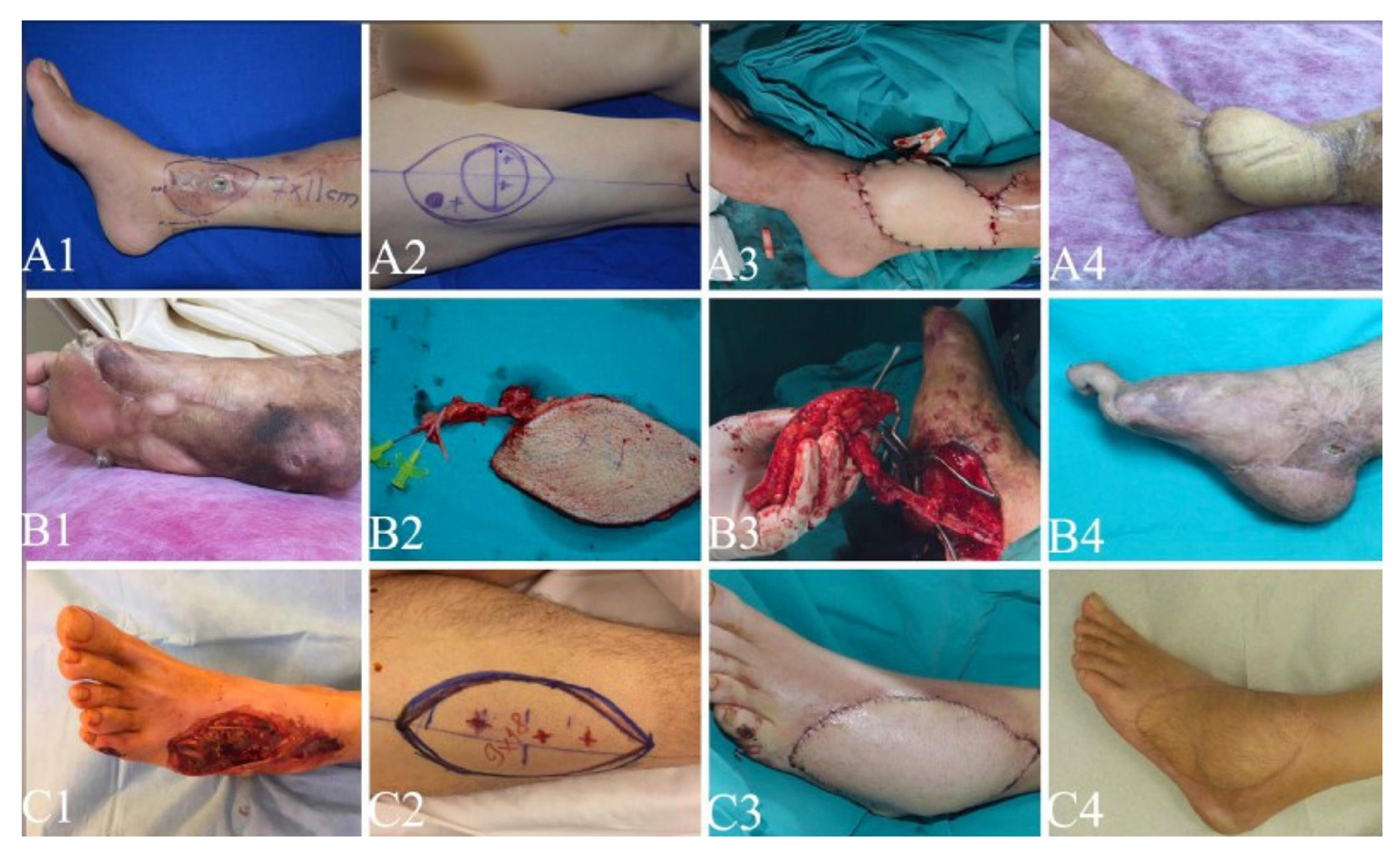
Figure 1 are reported herein in detail.
Case 1
A 61-year-old patient was operated on for a comminuted fracture of the tibia in another center after a traffic accident with open reduction and fixation with plate and screws. During the postoperative follow-up, he presented to our center with exposed plaque without local and/or systemic infection for 6 weeks. After radical debridement, a free ALT flap was transferred to the anterior tibial artery and vein with end-to-side anastomosis in a fasciocutaneous pattern (
Fig. 1 A1–A4).
Case 5
A 24-year-old patient developed localized tissue necrosis after an open reduction and fixation operation with plate and screws following a diagnosis of open calcaneal fracture after a traffic accident. Subsequently, after the plate and screws were removed, the defect was repaired with a skin graft and then osteomyelitis developed with fistula formation. For osteomyelitis, surgical treatment with previously carried out radical debridement was performed. A free ALT flap was transferred to the suprafascial posterior tibial artery and vein with end-to-side anastomosis in a suprafascial pattern (
Fig. 1 B1–B4).
Case 7
A 62-year-old patient was referred to our center with debridement after primary suturing in another center following a traffic accident. After repeated radical debridement and wound care, a free ALT flap was transferred to the anterior tibial artery and vein with end-to-side anastomosis in a fasciocutaneous pattern (
Fig. 1 C1–C4).
Results
The average patient age was 41.7 years (range, 16–62 years), and all the patients were male. Defect etiologies, localization and size after radical debridement, flap size and pattern, the type of dominant perforator artery of the flap, and the anastomosis applied artery in the recipient area are all shown in
Table 1. All flaps were free flaps applied to the lower extremity. The average area of the ALT flaps at inset was 95.7 cm
2 (range, 63–147 cm
2).
The average vein diameter, measured by the size of the venous coupler device at the level of the venous anastomosis, was 3.4 mm (range, 2.2–4.2 mm). On average, 1.8 venous anastomoses were performed. The descending branch of the lateral circumflex femoral artery almost always has two accompanying veins, one of which is slightly dominant in size and flow. The number of venous anastomoses performed is typically a function of recipient-site vein availability. There were no deaths, flap losses, or donor-site functional or cosmetic complications. There were two partial wound complications. The first was linear necrosis at the suturation borders of the flap because of flap recipient-site localization. The second was hematoma, which occurred at the recipient site in only one patient, patient 6, which was drained without affecting the flap.
Discussion
The two most important principles to be considered when choosing the most appropriate and feasible flap options to cover lower extremity tissue defects are the size and localization of the tissue defect [
7,
8]. The aim of surgical reconstruction of a lower extremity tissue defect regardless of etiologic factors should be to prevent loss of functionality in that limb and to preserve cosmetically acceptable appearance, with donor-site morbidity kept to a minimum. Although local fasciocutaneous and muscle flaps are often sufficient for defects of the lower extremity in the proximal third of the tibia midshaft, local flaps are often not sufficient to reconstruct defects in the distal third of the tibia and foot in particular. In addition, in cases in which there is excessive soft-tissue loss and extremity circulation in tissue defects secondary to trauma is adversely affected, these local tissues are often impossible to use in defect reconstruction. These types of defects nearly always require free flap transfers. In contrast, advances in microsurgery over the past 30 years have provided alternative reconstructive options. At the same time, a free flap minimizes the possible circulatory functional impairment of an already injured limb by protecting the local tissues. The free flap options in the literature to date containing the results of the first application include the rectus abdominis muscle [
9], the latissimus dorsi muscle [
10], the tensor fascia lata [
11], and scapular and parascapular flaps [
12] for lower extremity defects.
As indicated in the literature, the vascular pedicle of the ALT flap harvested can be either septocutaneous or musculocutaneous [
6,
7,
8]. A musculocutaneous pattern ALT flap could include the vastus lateralis muscle to cover a three-dimensional defect that did not require skeletonization of the perforator vessels when additional bulkiness was needed, as in case 5 (
Table 1). Moreover, a musculocutaneous pattern ALT flap could be trimmed to the fat layer subdermally, except for a small area around the pedicle entry to the flap, and it is appropriate for most superficial defects of the extremities. It is well known that flap elevation is slightly more difficult when the pedicle is musculocutaneous. However, when the musculocutaneous flap planned to be harvested includes part of the muscle, the harvesting is much easier, as dissection of the pedicle in the vastus lateralis is not necessary. In contrast, fasciocutaneous flaps may provide an opportunity to minimize bulkiness and to obtain an aesthetically more acceptable recipient site in the limb. Furthermore, it is easier to monitor fasciocutaneous flaps by simple clinical follow-up.
The ALT flap does not need positional changes during surgery, and flap elevation can be performed at the same time as the recipient site procedures and preparations. A large-caliber vascular pedicle facilitates microvascular anastomoses and provides a high anastomotic success rate. As in our series, vascular anastomoses can be successfully performed with a magnifying glass, except for in children. The flap can be used as a delicate flap by including the lateral femoral cutaneous nerve [
8,
9]. In our case series, sensory nerve reconstruction of all flaps was not performed. However, in our study, it is noteworthy that sensory recovery was achieved in all cases at 12 to 18 months postoperatively, which was sufficient to maintain the softtissue stability of flaps. Moreover, during the follow-up period, no patient was afflicted by ulceration on the weightbearing zones of flaps.
If the fascia lata is not removed with the flap, muscle herniation does not occur and primary closure can be easily attempted. A donor-site defect less than 8 cm in width can be closed primarily.
When the recipient site is on the lower extremity, the operation could be performed under epidural anesthesia, with peripheral blockage added for postoperative pain relief, as in all our cases. To harvest the anterolateral thigh flap as a flow flap, the distal end of the lateral circumflex femoral artery and the distal end of the descending branch can be placed or anastomosed into the recipient artery of the limb and receptive veins of the pedicles of other flaps. Therefore, it can be used to reconstruct softtissue defects while preserving and maintaining the vascular structure of the lower extremities.
To reduce the risk of thrombosis of recipient vessels during free flap transfer, microvascular anastomosis should be performed outside of the injured zones [
7,
9,
10]. Furthermore, recipient vessel selection is also vital to avoid free flap failure. Many authors suggested that the venous return of flaps was maintained well with only one accompanying vein anastomosis [
13,
14]. In our cases, because the ALT flaps were large, an average of 1.8 recipient veins, including an accompanying vein of the artery, were chosen for anastomosis to avoid congestion of the flap and postoperative complications. No flap congestion occurred postoperatively using this anastomosis method in our series. Despite the various advantages of this flap, the present study has some limitations, such as the small number of patients and the lack of investigation of sensory nerve coaptation of the ALT flap.
Conclusions
The ALT flap is an extremely versatile reconstruction option that can be easily applied to large soft-tissue defects, especially for the lower extremities, in addition to its applicability in different body regions. Because of the high reconstructive capacity of the ALT flap donor site, low donor-site morbidity, a long and large vascular pedicle with a reliable skin paddle, the possibility of use in a sensate and flow-through manner, and the possibility of performing the operation with two different surgical teams (which decreases the time of surgery), it should be considered as an ideal free flap reconstruction option for large tissue defects, especially those of the distal third of the tibia and foot.