Diabetes mellitus is a leading cause of morbidity and mortality in India. [
1] The prevalence of type 2 diabetes mellitus is huge both nationally and globally. Data from the International Diabetes Federation diabetes atlas (7th edition) suggested that the global prevalence of diabetes mellitus was 415 million (8.8%). It is expected to rise to 642 million (10.4%) in the next 25 years [
2]. In India, the International Diabetes Federation currently estimates 69.2 million people with diabetes mellitus, which is expected to exceed 123.5 million (78.5% increase) by 2040. [
3] The higher prevalence of type 2 diabetes in Asian Indians has been attributed to the so-called Asian Indian phenotype, which leads to unique clinical and biochemical changes in the body. [
1] The consequent changes lead to various alterations in microvasculature and macrovasculature, including foot complications. [
4] Diabetic peripheral neuropathy (DPN) is one the most common and severe complications of chronic hyperglycemia, affecting 50% of the diabetic population. [
5] The impact of these changes in the patient’s quality of life is significant. [
6] People with DPN have reported a severe burning and tingling sensation in their feet that initially keeps them from walking. Their physical activity gradually reduces. Poor physical fitness could potentially progress the disease. [
7] Also, retinopathy, nephropathy, dermopathy, neurologic pain, claudication, and diabetic foot could be other common manifestations of DPN. [
6] Diabetic foot syndrome is a severe complication characterized by the triad of neuropathy, ischemia, and infection. [
8] Therefore, the etiopathogenesis of diabetic foot complications includes vascular, neurologic, and musculoskeletal changes. [
9]
Foot complications are often ignored in diabetes management in Indian settings. Studies have shown that foot complications in Asians with type 2 diabetes mellitus have a major effect on health- related quality of life. [
10] Prevention and proper management of diabetic foot complications could be very effective in controlling the morbidity and mortality associated with the disease. Nevertheless, it is well-known that the biomechanical changes could cause a neuroischemic pressure ulcer in the presence of DPN. [
11] A nonhealing diabetic foot pressure ulcer could eventually lead to an amputation of the foot. [
12] Therefore, the neurovascular screening and biomechanical assessment of foot (kinetic and kinematic analysis) for risk factor prediction for foot ulcers could be an important part of diabetic foot care and management.
The biomechanical analysis mainly includes kinetic and kinematic parameters. Kinetic variables include plantar pressure, pressure-time integral, joint moment, and ground reaction force. Kinematic variables such as the joint angle, joint velocity, joint acceleration, and gait parameters have attained their importance in the recent past. [
13,
14,
15,
16,
17,
18,
19] In the presence of DPN, these changes could be even greater, causing a specific alteration in posture and gait biomechanics and leading to further foot complications. For example, some studies have compared gait velocity between participants with diabetes with peripheral neuropathy and those with diabetes without peripheral neuropathy. [
20,
21,
22,
23,
24,
25] Most of the studies suggested that participants with DPN have slower gait velocity compared with age-matched nonneuropathic patients with diabetes and healthy individuals. This finding was also supported by the meta-analysis by Hazari et al, [
26] who concluded that there was a significantly lower gait velocity in participants with DPN (–0.09; 95% CI, –0.13 to 0.05;
P , .0001). Similarly, the other spatiotemporal parameters, such as stride length and stance period, were reported to be significantly lower in participants with DPN. [
20,
25,
27,
28] Joint kinematics such as joint range of motion at the hip, knee, and ankle has been reported by various authors. [
22,
25,
29,
30] Studies by Gomes et al [
22] and Rasporvic [
25] suggested significantly higher maximum hip flexion in the neuropathy group compared with the nonneuropathy group, whereas the study by Yavuzer et al [
30] found it to be significantly lower. The study by Hazari et al [
26] concluded a significant difference at knee flexion and ankle dorsiflexion range of motion in diabetic participants with and without peripheral neuropathy compared with nondiabetic healthy participants. The maximum knee flexion angle was found to be significantly greater in nondiabetic participants compared with participants with diabetes with and without peripheral neuropathy (
P = .0008). [
26] The kinetic variable higher plantar pressure was reported as the most important etiopathogenic risk factor for the development of diabetic foot ulcer. [
19,
30] The study by Cavanagh and Ulbrecht [
31] reported that the plantar pressure measurement is the only dynamic component of the foot examination. It could provide information on the foot-ground interaction during walking in which plantar ulceration usually occurs in the diabetes population. [
32] A significantly higher average plantar pressure has been reported in individuals with DPN compared with diabetic patients without neuropathy and healthy participants in the previous studies. [
27,
29] Segmental plantar pressures, including forefoot and hindfoot pressures, have also been reported by a few authors. [
15,
16,
33,
34,
35] Ground reaction force and joint moment have also attained importance in the previous literature. [
25,
28,
35,
36] Pressure-time integral was reported by some authors to express the plantar loading in the diabetic foot. However, the study by Bus and Waaijman [
37] suggested that pressure-time integral would be more useful if it could predict ulceration, otherwise its value remains limited.
The kinematic and kinetic analysis of a joint segment or whole body requires sophisticated software and biomechanical tools. Previous studies have used some advanced biomechanical instruments and methods. For example, various marker- based motion analysis systems synchronized with force plates have been used by previous researchers. For example, Sawacha and colleagues [
19] used the BTS motion capture system (BTS Bioengineering Corp, Quincy, Massachusetts) with Bertec force plates (Bertec Corp, Columbus, Ohio) integrated with the Imago plantar pressure system, whereas Guldemond et al [
26] used a pressure-sensitive platform (EMED SF-4; Novel Electronics Inc, Munich, Germany) for barefoot pressure analysis. The other advanced and currently used 3-dimensional (3-D) motion analysis software include infrared cameras with a retroreflective marker capturing system, such as VICON (Vicon Oxford Metrics Ltd, Oxford, England), QUALISYS (Qualisys AB, Gothenburg, Sweden), and SIMI (SIMI Reality Motion Systems GmbH, Unterschleissheim, Germany), synchronized with Kistler force plates (Kistler Inc, Amherst, New York). These instruments are highly expensive and require an appropriate laboratory setup. Other instruments, such as the F-Scan (shoe-pressure) measure system and F-Scan mat system (Tekscan, Boston, Massachusetts), have also been used with greater efficacy in clinical practices. [
16,
29,
32] Biaxial electrogoniometers (models SG110/A and SG150; Biometrics Ltd, Newport, Wales) have been used to calculate joint kinematics. [
22] From the review of the literature, it could be suggested that a variety of biomechanical tools and methods are available to determine the kinetics and kinematics for a joint segment. Comprehensive information on various biomechanical tools and outcome variables suggested by previous researchers for determining the kinetics and kinematics of the foot in participants with diabetes can be seen in the review study by Hazari et al. [
26]
In a review of the literature, we found that there were a variety of studies reporting the altered kinetics and kinematics of the foot and its complications in participants with type 2 diabetes mellitus worldwide. However, there is a clear gap in the literature on biomechanical characteristics of the foot in type 2 diabetes mellitus in an Indian population. We believe that there could be multiple factors leading to the given biomechanical alterations, including kinetic and kinematic changes with the subsequent foot complications. Factors such as the living and work environment, quality of life, occupation, and socioeconomic conditions could have a great effect on the nervous, vascular, and musculoskeletal systems, which are the major determinants of the diabetic foot. Accordingly, the characteristic representation of diabetic foot complications in the Indian population could be different from that in Western countries. Because there is a dearth of literature for kinetic and kinematic analysis of the foot in type 2 diabetes mellitus with peripheral neuropathy in the Indian population, the given study could be very significant in Indian and Asian populations. These findings could be effectively used to understand the risk factors and clinical characteristics of foot complications in the Indian population. It could be altered biomechanics and musculoskeletal changes not only at the foot but also at other joints of the body leading to altered plantar pressure. It is also possible that the changes in the foot could be closely associated with the consequent changes in the musculoskeletal system, such as muscle weakness, muscle tightness, and neuropathic pain, that lead to altered gait patterns. Also, the postural changes in the body could lead to altered kinetics and kinematics in a closed chain. An overall observation, examination findings, and state-of-the- art advanced biomechanical motion analysis need to be reported in the Indian clinical setup. The main purpose of the study was to determine the diabetic foot kinetic and kinematic profile of the Indian population. It could also be important to understand the baseline difference and compare these changes from the developed countries and their possible reasons/factors. Therefore, the aim of this study was to determine the overall biomechanical changes in individuals with type 2 diabetes having peripheral neuropathy. The objectives of the study were 1) to determine the kinetics and kinematics of the foot in type 2 diabetes mellitus with peripheral neuropathy, 2) to determine the biomechanical changes in the musculoskeletal system and posture in type 2 diabetes mellitus with peripheral neuropathy in open and closed chains, and 3) to determine associations between the variables with risk factors for foot complications in type 2 diabetes mellitus with peripheral neuropathy.
Methods
Study Setting and Design
This cross-sectional study was conducted at the Diabetic Foot Clinic, Kasturba Hospital, Manipal University, Manipal, Karnataka, India.
Participant Recruitment
A total of 120 participants with type 2 diabetes mellitus and peripheral neuropathy without any previous ulceration or amputations of the lower limb were recruited under the purposive sampling method. Concerned physicians referred the participants from different medical units within Kasturba Hospital. Ethical approval and informed consent were obtained from all of the participants, followed by orientation about the procedure for data collection. A participant information sheet was given to all of the participants containing the detailed procedures and contact information for any further queries.
Instrumentation and Setup
Screening for DPN. All of the participants were thoroughly screened for the presence of DPN. The screening consisted of detailed vascular (ankle-brachial index, pedal pulse characteristics, infrared thermal imaging), neurologic (touch sensation, ankle reflex, 10-g Semmes-Weinstein monofilament, and vibration pressure threshold [VPT]), autonomic changes (skin texture, color, nail bed), and musculoskeletal (foot size, type, structural changes, navicular drop height, Foot Posture Index, quadriceps angle (Q-angle), muscle strength, muscle tightness, and postural analysis) assessment. The presence of DPN was confirmed by the absence of 10-g Semmes-Weinstein monofilament test and VPT greater than 25 V. [
33] Michigan Neuropathy Screening Instrument (MNSI) A and B were also incorporated as neuropathy screening tests.
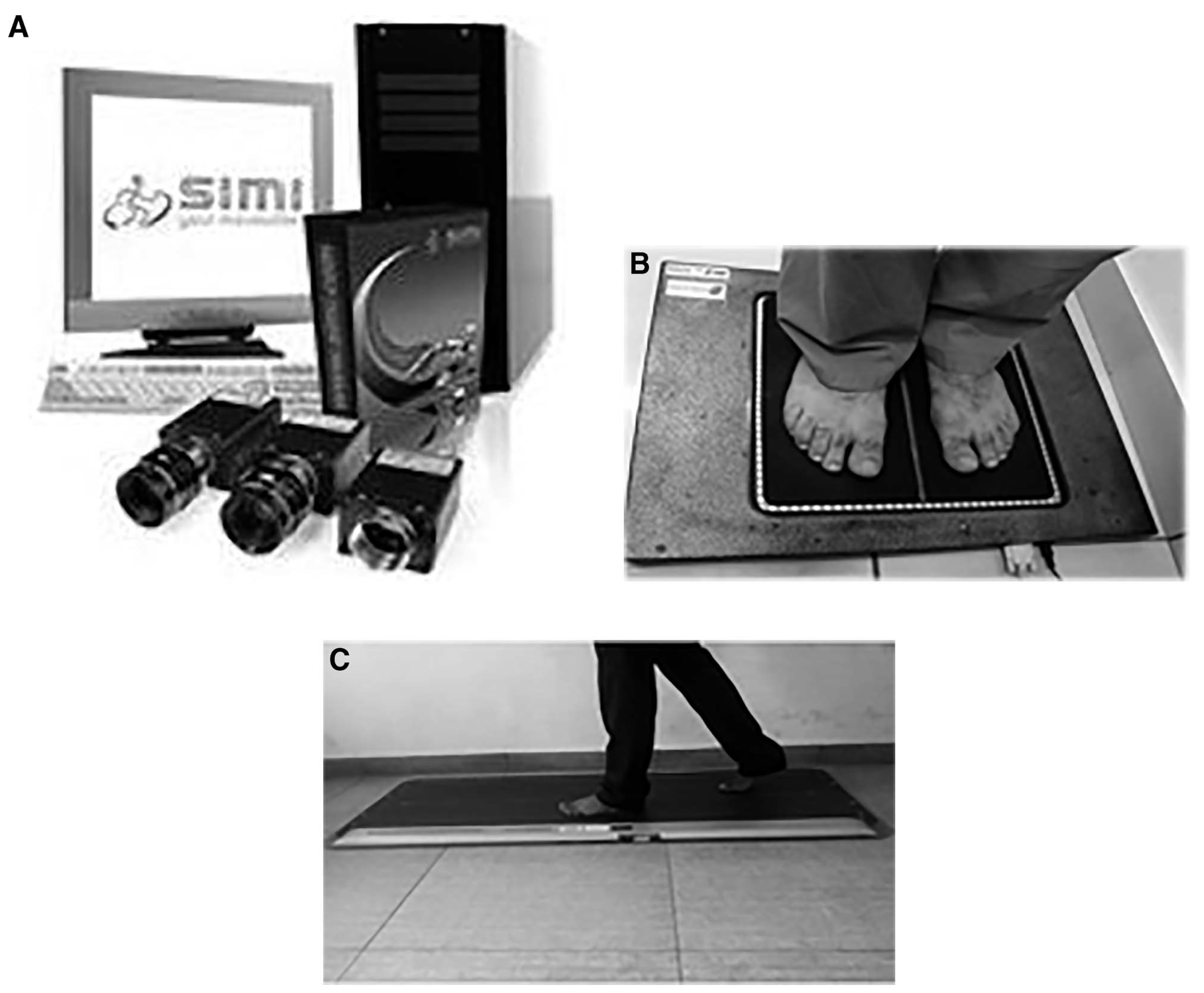
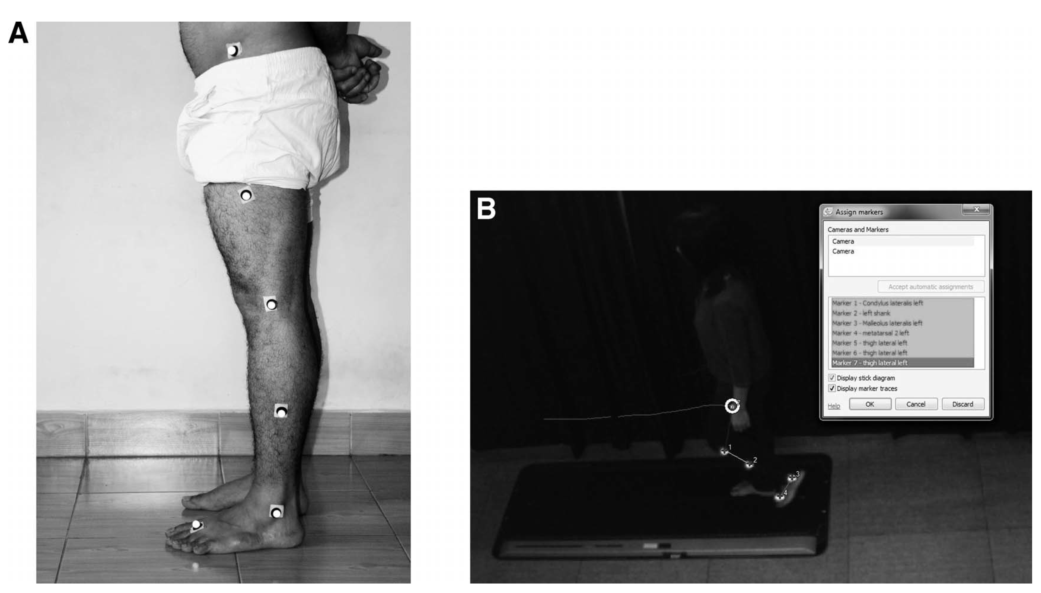
Motion Analysis. The 3-D kinematic data (joint angle, joint velocity, and joint acceleration) for the foot and knee joints were collected using SIMI 3-D motion analysis software with two high-frequency cameras (Basler 139a/b GigE, 100 frames per second at 1 megapixel [Basler AG, Ahrensburg, Germany]) (
Figure 1A). The current state of the art for gait analysis comprises marker-based systems such as the SIMI reality motion system. Marker-based systems, such as BTS, VICON, and SIMI, use retroreflective markers placed on the anatomical landmark to perform a 3-D analysis. The marker- based tracking system is considered the gold standard for clinical gait analysis. [
38] The SIMI motion system is a very reliable and valid tool used in gait analysis worldwide. A study by Frenken et al [
39] used the SIMI motion system as the gold standard for validating Timed Up and Go assesment in gait analysis. Although markerless techniques have gained popularity in the biomechanics in the past decade, their application in the clinical setting is a matter of debate. [
38]
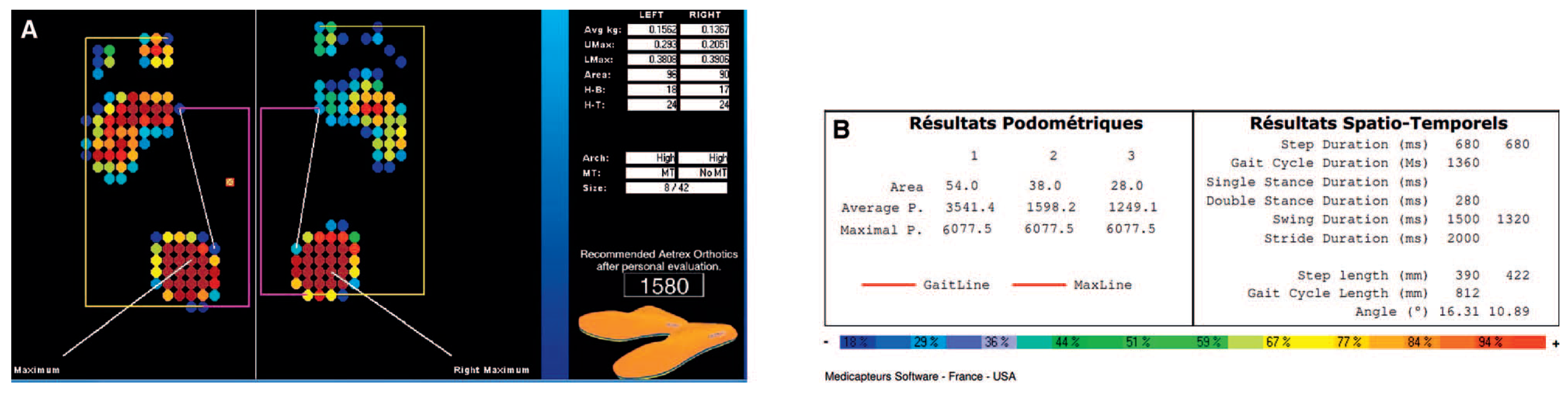
The kinetic variable average and maximum pressure was collected using an iStep static scanner (Aetrex Worldwide Inc, Teaneck, New Jersey). Spatiotemporal parameters of gait were calculated using WinTrack software (Medicapteurs France SAS, Balma, France (
Figure 1B and C). Increasing research work has demonstrated that portable systems based on body sensors are a promising method for clinical gait analysis regarding precision, comfortability, and usability. [
40]
Observational Analysis
This included observing structural changes such as toe deformities (clawing, hammer toes, hallux valgus), Charcot’s foot, changes in the nail bed, dryness/discoloration of the skin, tibial torsion, the position of the patella, femoral rotation, pelvic tilt, spine curvature, and head on shoulder position.
Other Biomechanical Analysis
These findings were reported from both open and closed kinematic chains as applicable. The examination of the foot included a standard procedure for the navicular drop test [
41] and the Foot Posture Index, [
42] plantar fascia tightness, and manual muscle testing of a major group of muscles in the lower limb, including foot muscles. At the knee, the (Q- angle was examined using a goniometer. Flexibility testing of the proximal and distal muscles was performed. A skilled physical therapist performed this clinical testing.
Statistical Analysis
SPSS for Windows, Version 16.0 (SPSS Inc, Chicago, Illinois) was used for descriptive statistics. The demographics and the kinematic and kinetic results are reported as mean 6 SD. Linear regression analysis was performed to determine the association between the variables and foot complications.
Results
The important kinetic and kinematic findings from the static and dynamic foot scans are shown in the figures and tables throughout. The outcome variables of interest from the static foot scan consisted of average foot pressure, maximum foot pressure, forefoot-to-hindfoot pressure ratio, metatarsal loading, and type of arch with foot size (
Figure 3A). The results of the dynamic foot scan included spatiotemporal gait parameters such as step length, stride length, gait cycle length, cadence, duration of double support, step duration, stride duration, swing duration, and toe-out angle (
Figure 3B). The results for knee and ankle joint kinematics from the SIMI motion system are presented herein, along with other important biomechanical findings. The mean age, height, weight, body mass index, and duration of diabetes are given in Table 1. Table 2 shows the clinical characteristics of the participants. The regression analysis showed statistical significance for maximum plantar pressure at the forefoot with age, weight, height, duration of diabetes, body mass index, knee and ankle joint angle at the toe-off phase of the gait cycle, pinprick sensation, and ankle reflex (
R = 0.71,
R2 = 0.55,
F12,108 = 521.9 kPa;
P = .002) (see Table 3 for associations of individual variables). For all of the variables, statistical significance was set at
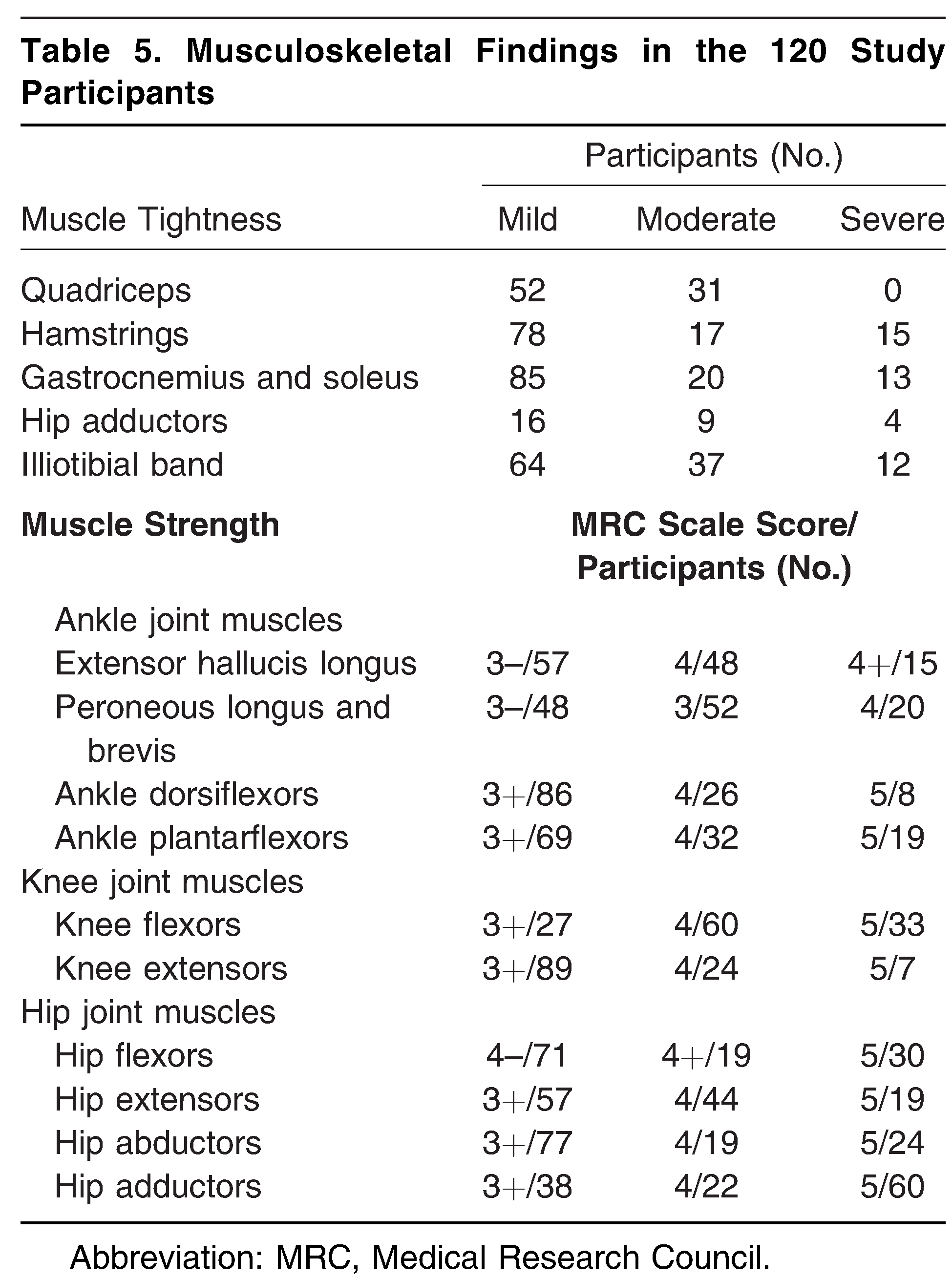
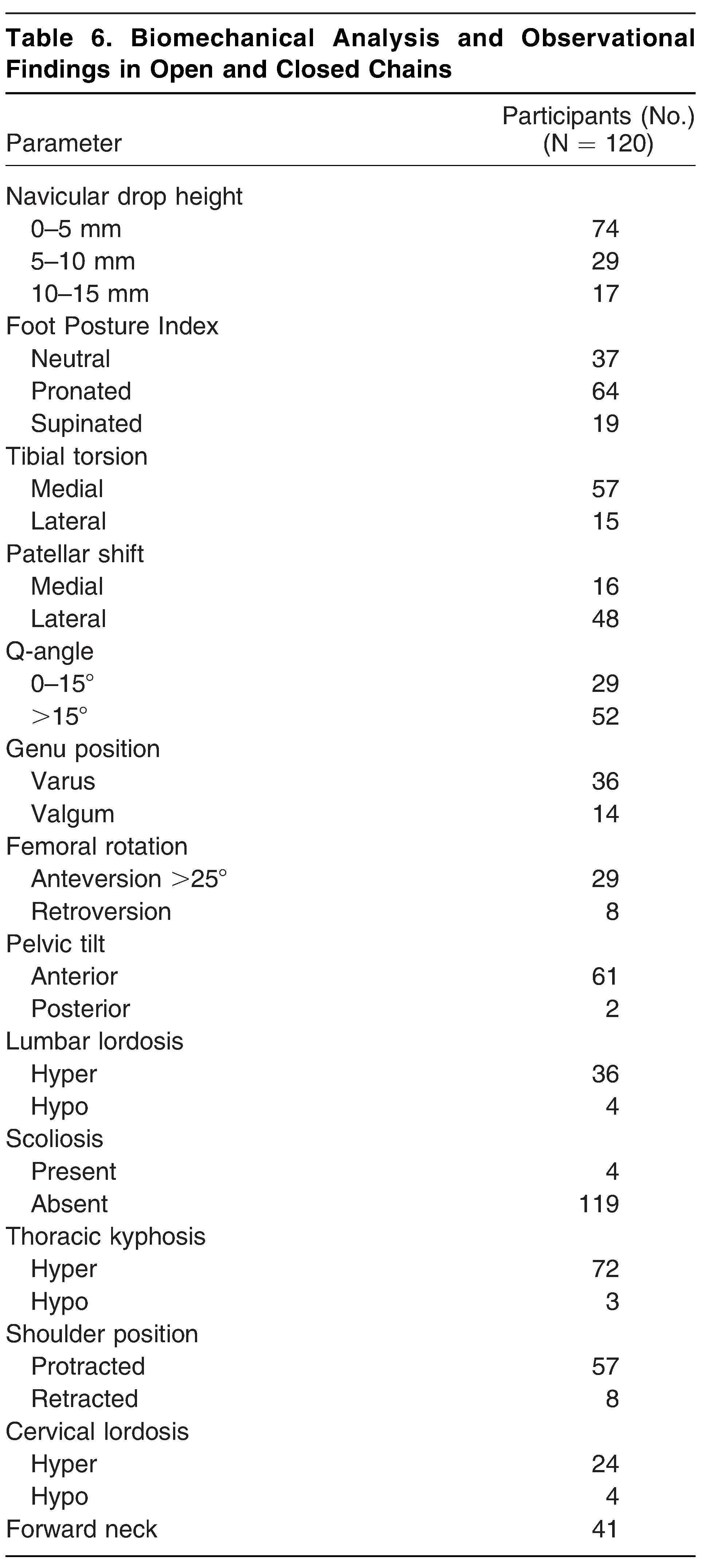
P , .05. Mean values for the kinematic variables joint angle, velocity, and acceleration at the ankle are given in Table 4. Musculoskeletal and other biomechanical findings are reported in Table 5 and Table 6, respectively. Kinetic variables such as plantar pressure are presented in Table 7. Knee joint kinematics are presented in Table 8, and spatiotemporal parameters of the gait are given in Table 9.


Discussion
Diabetic foot is a major complication and concern for poor prognosis of the disease. It leads to prolonged hospital stays. The forefoot, the base of the first metatarsal joint, and the heel are the most common area of foot ulcers in patients with type 2 diabetes. [
44] Peripheral neuropathy has been considered the most lethal risk factor for foot ulceration in diabetes mellitus. In the present study, many participants were diagnosed as having DPN and, thus, could be at higher risk for foot complications and ulcerations. The present study highlights various important clinically symptomatic and observational findings and their association with risk of foot complications in type 2 diabetes mellitus with peripheral neuropathy.
Sensory Neuropathy
In the present study, the presence of DPN was confirmed based on the results obtained from ankle reflex examination, the 10-g Semmes-Weinstein monofilament test, the VPT on a biothesiometer, and the MNSI. A study reported that considering nerve conduction study as the gold standard, ankle reflex had very high sensitivity and specificity (91.5% and 67.4%, respectively) in detecting DPN. [
45] In the present study, we found that 31 participants had diminished ankle jerk, and nine had an absent response suggestive of the absolute presence of DPN. The clinical inferences to these findings suggest that 33% of the population had sensory and motor, large and small nerve fiber damage (40 of 120). Also, DPN was confirmed using VPT on all of the participants. Also, based on the clinical findings and symptomatic observation, a vibration sense of 20 to 29 V was considered as mild, 30 to 39 V as moderate, and greater than 39 V as severe neuropathy; however, an established reference to this interpretation has not been suggested in the previous literature. Findings from previously herein and the results in Table 2 suggested that 16 participants had reduced touch sensation in the lower limb, whereas 21 participants could not feel intact sensation at all. The study performed by Driver et al [
46] concluded that lack of protective sensations due to sensory neuropathy could lead to ulceration repetitive microtrauma. The regression analysis results in Table 3 suggest that pinprick sensation, ankle reflex, MNSI score, and VPT showed a significant statistical association with peak/maximum plantar pressure at dynamic gait cycle (
P , .05). These findings could be supported with similar findings suggested by Barn et al [
47] and Fawzy et al. [
48] It could be seen that MNSI score and VPT were highly significant (
P = .001 and .000, respectively). This demonstrates that any increase in either of them could lead to an increase in the maximum forefoot pressure. In other words, the higher values of the MSNI and the VPT could be considered an important indicator for risk of diabetic foot ulcerations. Because the higher MNSI score and VPT could also stratify the participants with severity of neuropathy, it could also be suggested that the risk could be higher in severe neuropathy compared with moderate and mild neuropathy. Similarly, the regression analysis showed a strong association of peak pressure with pinprick sensation and ankle reflex (
P = .04 and .01, respectively). These findings further strengthen the possibility of diabetic foot syndrome in these participants. Among the blood variables, hemoglobin A
1c showed postive correlation with peak pressure, whereas foot deformities such as clawing, hammer toes, and hallux valgus also showed a significant increase in peak pressure (
P = .02, .004, .025, and .009, respectively). These results were similar to previous findings and are discussed in the subsequent section.
Vascular Findings and DPN
The mean local skin temperature at the dorsum of the foot using infrared thermal imaging was within the reference range of 32.8 86 6.1 8 C. Studies have shown that patients with diabetes could demonstrate an increased temperature at the lower extremity as a consequence of DPN. [
49] The study by Jayakumar et al [
49] reported that there was a significant positive correlation of mean foot temperature (32 8–35 8 C) with VPT greater than 20 V (
r = 0.301,
P = .001, n = 112) in individuals with peripheral neuropathy compared with those with- out neuropathy. However, in the present study, only four participants had an elevated temperature in the range of 378 to 388C. The elevated temperature of the foot could be suggestive of inflammatory changes, which could lead to further foot complications. An altered foot temperature may not directly affect the kinetic and kinematic parameters of the foot. However, care must be taken because higher plantar foot temperature in the presence of DPN should be considered as the high-risk diabetic foot, leading to neuroischemic changes. [
49]
Autonomic Neuropathy
The findings from the present study suggested that autonomic changes were the highest clinical manifestation among Indian participants with type 2 diabetes mellitus and peripheral neuropathy. These included findings such as skin texture, skin color, fissures on the plantar aspect and heel, and nail abnormalities. The present study found that of 120 participants, 98 presented with dryness of skin, 64 had discoloration (cyanotic, blackish, blister red, etc), 104 showed fissures, and 87 presented with ingrown nails and nail bed infections. Orthostatic hypotension was also noted in seven participants. These findings could be attributed to reduced blood supply in the periphery as a consequence of DPN. [
50] A study reported that autonomic neuropathy could present with increased peripheral blood pooling, leading to swelling in the foot. These changes could lead to an area of increased plantar pressure, resulting in foot ulceration. [
29]
Motor Neuropathy
Two components were assessed: muscle strength and muscle length.
The results of this study suggest that most of the participants had motor neuropathy regarding muscle weakness. The quadriceps and extensor hallucis longus muscles were the most significant to show lower grades on manual muscle testing. Other muscles, such as the tibialis anterior, gluteus, and foot everters, also showed a decrement in their strength but to a lesser extent (Table 5). A reduction of strength (30%–50%) has been shown in the previous study. [
51] The role of muscular contraction in shock absorption and energy conservation is an important aspect of efficient gait. Reduced muscular forces may increase joint reaction forces, leading to altered joint kinetics and kinematics. [
52] It is well- known that during the early stance phase of the gait cycle, 60% of the body weight is loaded abruptly on the ipsilateral limb in fewer than 20 sec. The abrupt impact is potentially attenuated by muscular con- traction of the lower-extremity joints. [
52] During loading response, the eccentric contraction of pretibial muscles helps to restrain the passive plantarflexion force. From the given study, it could be suggested that weakness of the extensor hallucis and tibialis anterior muscles may result in greater plantarflexion force at loading response with a consequent rise in ground reaction force. This finding suggests that participants with motor neuropathy may be at higher risk for foot ulceration due to muscular weakness. Similarly, the role of quadriceps strength is very crucial at initial contact and the toe-off phase of the gait cycle. It is well- known that the external ground reaction force has a tendency to produce knee flexion at initial foot contact, which must be encountered by knee extensors for mechanical stability with good shock absorption of the knee joint. In the given process, the balance between knee joint stability and shock absorption should be maintained by eccentric contractions of quadriceps. In the present study, we found that most of the participants had motor weakness of the quadriceps, and, thus, altered kinetics and kinematics at the knee could be seen. Because maximum knee flexion angle was found to be higher in participants with DPN, it may cause altered forces at the knee joint. Regarding the hip abductors, the impact of loading should be minimized at the hip during single-leg support through good hip abductor contraction. [
53] Because the results of the present study showed that strength of hip abductors in DPN was compromised, we assume that there might be consequent changes in the kinetics and kinematics of the hip joint and gait cycle.
Possible reasons for the reduction in muscle strength in patients with DPN have been reported in previous studies. Studies have shown that one of the most common reasons for the loss of muscle strength in patients with DPN was diabetes muscle infarction. A study has reported ischemic infarction of thigh muscles (vastus lateral, thigh adductors, and biceps femoris). [
54] In the present study, we found a similar result in hip joint abductor muscles; however, hip adductors showed relatively greater strength on manual muscle testing (50% of participants [n = 60] had normal strength) (Table 5). Similarly, a study reported diabetes muscle infarction of the extensor hallucis and tibialis anterior muscles. [
55] Again, the present results are similar to previous findings, with a significant strength reduction in these muscles as mentioned previously herein and seen in most of the participants (Table 5). However, note that both of these studies had participants with uncontrolled sugar levels. There- fore, minimal strength loss in the hip abductor and thigh muscles in the present study could be explained by the fact that none of the participants had uncontrolled sugar levels. While examining muscle length, tightness was predominantly seen in the posterior and lateral compartment of the foot compared with the anterior and middle compartments. Muscles such as hamstrings, gastrocnemius, soleus, and iliotibial band showed more participants with moderate-to-severe tightness. Tightness in this muscle could be seen as a result of an altered length–tension relationship, neural inhibition of the muscle group, or postural abnormalities. [
56] The association of calf muscle (gastrocnemius and soleus) tightness with kinetics and kinematics of the foot could be well explained by the split second effect suggested by a study. [
57] The study suggested that there were two types of forces (direct and indirect) created by the split second effect over a critical period of 120 msec during terminal mid- stance. Just before the heel-off phase, damaging forces are produced where both direct and indirect forces are created and magnified by the tightness of calf muscles restricting the tibial translation over the ankle joint. [
57] The tightness of calf muscles could also result in altered kinematics at the ankle such as decreased ankle dorsiflexion at terminal midstance and toe-off (,108), as seen in the present study (Table 4).
Clinical Biomechanical Findings
The postural analysis was included as an observational finding under this segment. These findings included shoulder position, spinal curvature, pelvic tilt, femoral rotation, tibial rotation, patellar shift, and calcaneal neutral position (Table 6). The other important clinical examinations for foot kinetics and kinematics included the navicular drop test, the Foot Posture Index, the first and fifth metatarsal lengths from the heel, and measurement of the Qangle. The results of the present study suggested that participants with type 2 diabetes mellitus and peripheral neuropathy showed some clinically significant differences in baseline posture. Hyper- lordosis of the cervical (n = 24) or lumbar (n = 36) spine and thoracic kyphosis (n = 72) were the major postural deviations seen at the spinal level. Changes in the posture could have led to altered closed kinetic and kinematic changes in all of the other joint segments, including the foot. A study reported that the lumbosacral angle was significantly correlated with the maximum forefoot plantar pressure in both static and dynamic analyses using pedobarography. [
58] The study results suggested that a decreased lumbosacral angle leads to lower maxi- mum pressure under the great toe. Conversely, in the present study, an increased lumbosacral angle due to excessive anterior pelvic tilt and lumbar hyperlordosis (Table 6) could result in increased pressure on the ball of the great toe. This could be an important biomechanical factor for risk of ulceration at the great toe in DPN. Forward neck and shoulder was found in one-third of the participants, suggesting that DPN could affect the scapular muscles significantly. [
59] The position of the patella was shifted more laterally in most of the participants (n = 48), leading to consequent changes in lower segments of the foot, resulting in a high- arched foot (n = 24). A high-arched foot could expose the metatarsal head to higher plantar pressure and ground reaction force, thereby in- creasing the risk of foot ulcerations. [
47] On the other hand, flatfoot (pes planus) was observed in 53 participants. The clinical confirmation of pes planus was evident from a positive navicular drop height of more than 10 mm in 17 participants. This finding suggested the collapse of the medial longitudinal arch while loading in a closed chain (standing). The study by Periyasamy and Anand [
60] concluded that a low-arched foot could be a high risk for midfoot collapse and Charcot’s foot deformity. A greater Q- angle, as seen in the present study, could also be attributed to flatfoot deformity (hyperpronation). The tibial and femoral rotation could be a reason for the greater Q-angle due to hyperpronation of the foot. Hyperpronation was clinically assessed by positive values of the Foot Posture Index in the studied population. From the given findings, it could be suggested that changes in the Q-angle could have a strong effect on patellofemoral and tibiofemoral kinetics and kinematics. [
59] Also, various foot deformities could be seen in participants with DPN in the present study. Hallux valgus (n = 36), claw toes (n = 29), and callus (n = 49) were common foot deformities. One study has shown a strong statistical correlation of foot deformities in DPN with peak plantar pressure (odds ratio = 8.7). [
48]
Kinetics and Kinematics at the Ankle and Knee
Plantar pressure is the most important kinetic variable and etiological factor for the development of foot ulcer in DPN. [
17,
19,
26] The results of the present study reported that the mean maximum pressure was 603.85 kPa, equivalent to 60.38 N/cm
2 (Table 7). A study [
26] reported that a pressure of 60 N/ cm
2 is the upper threshold for the development of an ulcer in DPN. In the other study, a cutoff point of 335 kPa (peak plantar pressure) was considered as a risk for ulceration at the forefoot. However, recent studies have been contradictory. The study by Bus et al [
61] suggested that plantar pressure threshold should not be considered as a suitable method for detecting the risk of foot ulceration in participants with diabetes. Nevertheless, the higher plantar pressure in the presence of sensory and motor neuropathy could be a potential risk of foot ulceration. [
61] Higher plantar pressure could be significantly associated with deformities and soft- tissue changes in the foot. [
61] In the present study, we also found that the participants had higher peak forefoot pressure compared with hindfoot pressure. The mean 6 SD ratio of the forefoot to the hindfoot was found to be 1.24 6 0.29 (Table 7). This finding suggests that people with DPN could be at higher risk for foot ulceration. [
16] Looking at the kinematic variables, we found that participants with DPN walked with less dorsiflexion at midstance (mean 6 SD: 106.19 6 7.58 at heel strike to 101.49 6 3.15 at midstance). Studies have reported a minimum dorsiflexion angle of 108 at midstance. [
62] Lower values could be attributed to plantar-tissue stiffness. Also, a higher knee flexion angle at midstance and toe-off (mean 6 SD: 169.968 6 2.718) was observed compared with healthy participants (mean 6 SD: 155.8486 8.928) (Table 8). [
63] As suggested previously herein, it could be seen due to the weakness of proximal group muscles, particularly the quadriceps. A strong eccentric contraction is required to maintain balance during the late stance phase of the gait cycle. Because the literature on joint velocity and joint acceleration in DPN is scarce, it is difficult to comment on these findings with great precision. However, based on other functionally related parameters, the results of this study suggested that angular velocity and angular acceleration could be lower in DPN and so would be joint power, as suggested in a previous study. [
64] In the present study, we used a marker-based tracking system, which has been considered the gold standard for motion analysis. The marker-based system has advantages over the markerless and noninfrared motion tracking systems. The study by Ceseracciu et al [
38] concluded that markerless systems had gained their position in the biomechanical analysis based on the precise anatomy of patients. However, the estimation of joint angles in the transverse plane was not precise enough to allow its application in the clinical setting. [
38]
Gait Characteristics
The results of gait parameters such as gait velocity, step length, stride length, and cadence are lower in participants with DPN (Table 9). These findings were similar to the results found in the previous study and reported in detail in the systematic review and meta-analysis study. [
26] The most noticeable gait variable in the present study was degree of toe-out. We found that the mean toe-out angle of all of the participants during the gait cycle was 13.28. A higher toe-out angle could be associated with a reduction in external adduction moment, predisposing the knee joint to osteoarthritis and menisci injuries. [
44] This could also be seen as a result of tight everter muscles at the ankle and iliotibial band at the knee and hip, as seen in the present study. In the present study, we found that most participants (113 of 120) have tightness of the iliotibial band.
Findings from the Indian Context
Findings such as dryness of the skin (n = 98), plantar fissures (n =104), and callus (n = 41) were commonly seen. It is possible that scoring based on these findings could hamper the results of the MNSI and other such questionnaires in the Indian population. The dryness of foot skin may be attributed to the type of footwear and the weather conditions rather than to consequent changes of DPN. Barefoot walking, agricultural work in the field, poverty, unawareness of diabetic footwear, lower socioeconomic conditions, and illiteracy could be other important factors for these findings in Indian clinical settings. These factors should be carefully assessed while scoring for neuropathy. Similarly, deep fissures could be seen as being occupation related (agriculture is the major occupation in India), and callus may be present on the lateral malleoli due to traditional sitting postures at dining and worship.
Conclusions
The findings from this study suggest that people with diabetes and peripheral neuropathy in India have various kinetic and kinematic changes at the foot. These changes are similar to changes seen in previous studies. There is a strong association between plantar pressure and anthropometric/demographic variables such as age, weight, height, body mass index, duration of diabetes, foot deformities, muscular strength, and muscle length. The increased plantar pressure, foot deformity, and DPN are a potential risk factor for foot complications and future ulcerations. Therefore, appropriate measures should be taken to control diabetes and neuropathy. Appropriate off-loading devices should be suggested in clinical settings. However, factors such as environment, living style, socioeconomic conditions, occupations, religious beliefs, and traditions should be carefully assessed for clinical presentations of DPN in the Indian population. They should also be considered a potential factor for causing altered kinetics and kinematics. From the results of the present study, we conclude that for early detection and prevention, a detailed biomechanical and neurologic assessment must be performed in every diabetic center and clinical setting in India. Foot complications are often ignored in India. Appropriate screening, assessment, and foot care are the need of the hour in the diabetes capital, India.
Future Scope
Opportunities to conduct future research on interventional studies in the given area are abundant. The present study is part of a PhD thesis, and the authors would like to extend their research toward developing a biomechanical model for ulcer prediction based on the results of the present study. Studies with larger sample sizes and more variables for risk factors of foot complications could be reported in the future. Early screening for neuropathy, comparison of biomechanical characteristics based on the duration of disease, body mass index, and other clinical parameters in diabetic patients with and without neuropathy followed by corrective interventional exercises and measures could be an area of high clinical significance.
Financial Disclosure
None reported.