Magnetic resonance imaging (MRI) is both sensitive (90%–96%) and specific (79%–87%) for osteomyelitis.[
1,
2] In addition, its value in preoperative planning has been described because it can delineate the extent of infection and, thus, how much bone would need to be resected.[
3-
7] Before ordering the MRI, however, plain radiographs are a simple diagnostic study to obtain. Because osseous changes have been reported to take 10 to 14 days to be seen on radiographs, the sensitivity is lacking.[
8] However, when apparent, osteomyelitis produces characteristic changes denoted by focal osteolytic lesions, cortical destruction, and periosteal new bone formation.[
2,
8]
When radiographic findings are suggestive of osteomyelitis, it is common to proceed to MRI. Not only is MRI the most accurate test,[
2] but it will also delineate the extent of infection, including a determination of which bones are and are not infected. In cases of contiguous spread osteomyelitis of the toes, the MRI can determine whether infection has spread from the radiographically involved phalanx to the next proximal bone.
The importance of delineating the extent of infection is not to be understated. Although it is important not to resect so much bone as to leave a biomechanically unstable foot, it is also generally accepted that all necrotic bone must be surgically resected. Residual osteomyelitis at the resection margin is associated with poor outcomes postoperatively.[
7,
9,
10] Furthermore, it has been noted that gross intraoperative appearance of the bone is not reliable for determining whether the resection margins are infected.[
9,
10] Whereas one early study found difficulty differentiating osteomyelitis from adjacent marrow edema on MRI,[
11] more recent works by Fujii et al[
6,
7] have demonstrated the utility of MRI in preoperative planning for the debridement of infected bone.
We sought to review the utility of radiographs and MRI ins determining which phalanges were affected by osteomyelitis by investigating occurrences wherein MRI detected infection spreading to more proximal bones than those already seen to be infected on radiographs. This was accomplished by retrospectively reviewing imaging of patients who have had radiographs and MRIs positive for osteomyelitis of one or more toes. Patients' radiographs were reviewed by a blinded investigator to identify frankly infected bones in each toe. This was then compared with the MRI to assess for osteomyelitis of the adjacent proximal bones. With the results, we determined the accuracy of radiographic analysis.
Materials and Methods
We performed a medical record review of patients seen by the podiatric medical service at Lenox Hill Hospital (New York, New York) from July 1, 2014, through February 28, 2016. Patients were included in the study if they presented with cellulitis overlying a clinically infected toe ulceration, as well as pedal radiographs and MRIs positive for osteomyelitis of at least one bone. Patients were excluded if they were younger than 18 years or pregnant. The institutional review board of Lenox Hill Hospital approved this study and waived the requirement to obtain consent.
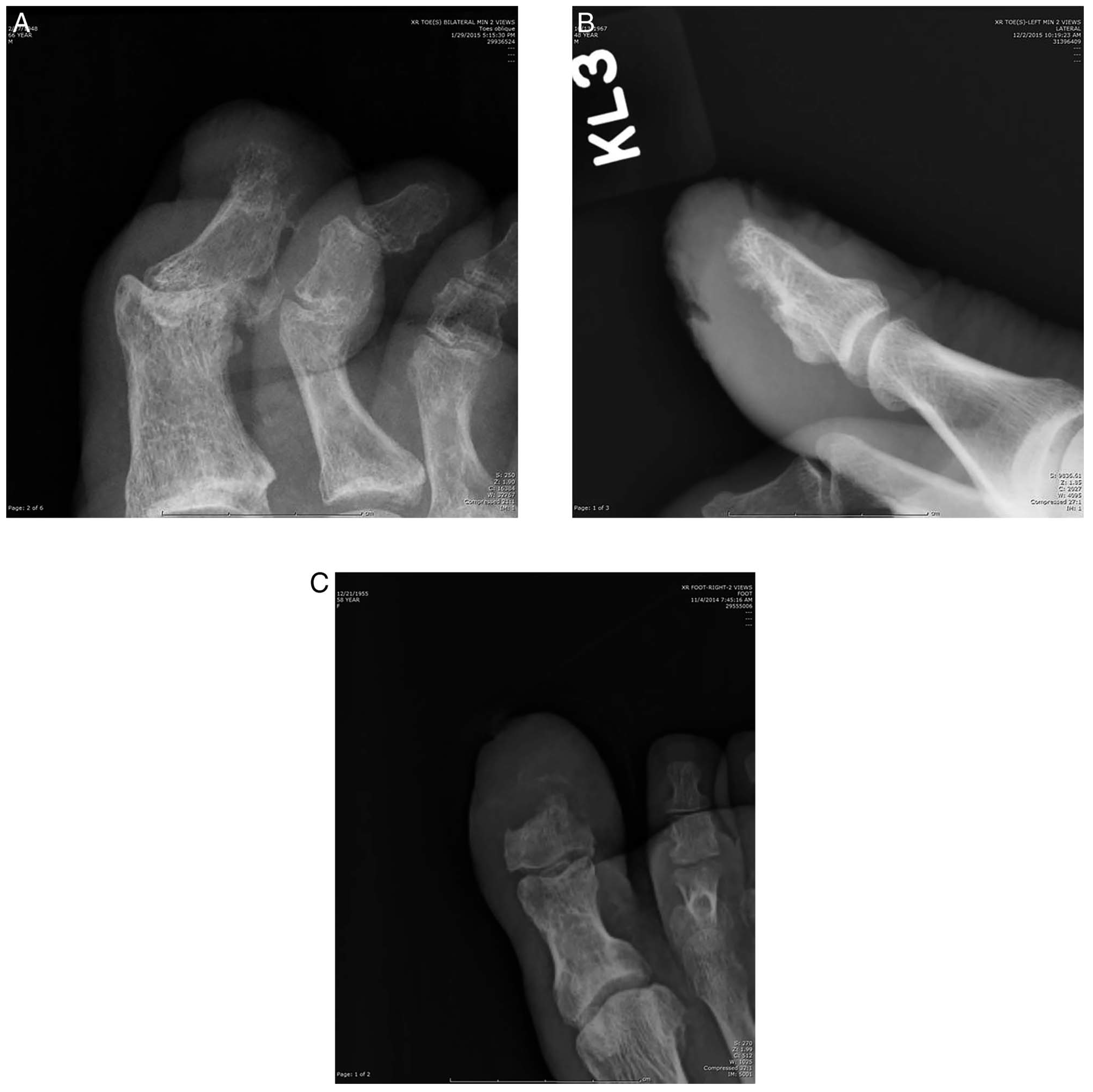
The senior author (W.S.), who is certified by the American Board of Foot and Ankle Surgery and has 28 years of experience, reviewed all of the radiographs and was blinded to the MRI findings. The senior author was apprised of which digit in each radiographic series had the infected wound to provide correlating clinical information. The senior author then reviewed each radiograph and evaluated each of the bones of the toe in question (distal, middle, and proximal phalanges and metatarsal) for one or more of the following radiographic signs of osteomyelitis: osteolysis, cortical destruction, and periosteal new bone formation (
Fig. 1). The radiographic findings for each bone were then compared with the associated MRI findings.
Figure 1.
A, Cortical destruction at the distal aspect of the distal phalanx of the hallux. B, Periosteal new bone formation at the plantar aspect of the distal phalanx. C, Osteolysis of the distal phalanx of the hallux represented by complete loss of radiographic density.
Figure 1.
A, Cortical destruction at the distal aspect of the distal phalanx of the hallux. B, Periosteal new bone formation at the plantar aspect of the distal phalanx. C, Osteolysis of the distal phalanx of the hallux represented by complete loss of radiographic density.
Results
Demographic and clinical data are summarized in
Table 1. Categorical variables are described using counts and percentages, and continuous variables are described using mean ± SD. Records from 14 patients (16 toes) were recovered. Of the 14 patients, 12 (85.7%) were men and ten (71.4%) had diabetes mellitus. No patients in the series had any history of gout or inflammatory arthropathies. In 13 of the 14 patients, clinical suspicion was based on the presence of neuropathic ulcers probing to bone. One case of a patient with a longstanding paronychia and radiographic evidence of osteomyelitis also satisfied the inclusion criteria. There were 11 patients (68.8%) with hallux osteomyelitis , four (25.0%) with second toe involvement, and one (6.3%) with fifth toe involvement. Data on inflammatory markers were available on 12 of 14 patients, with a mean erythrocyte sedimentation rate of 57.4 mm/h and a mean C-reactive protein level of 7.49 mg/L.
Table 1.
Descriptive Data for the 14 Study Patients
Table 1.
Descriptive Data for the 14 Study Patients
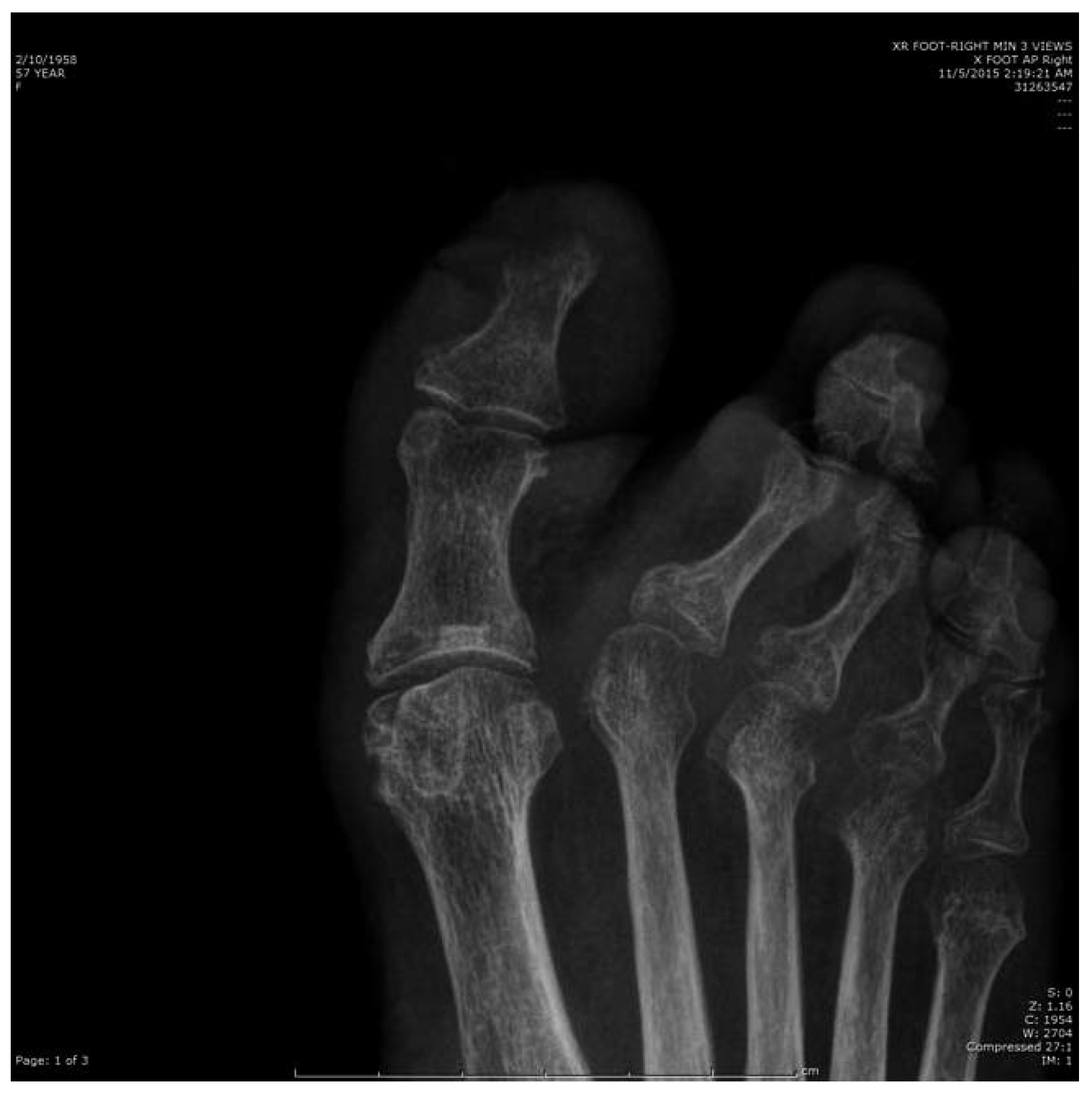
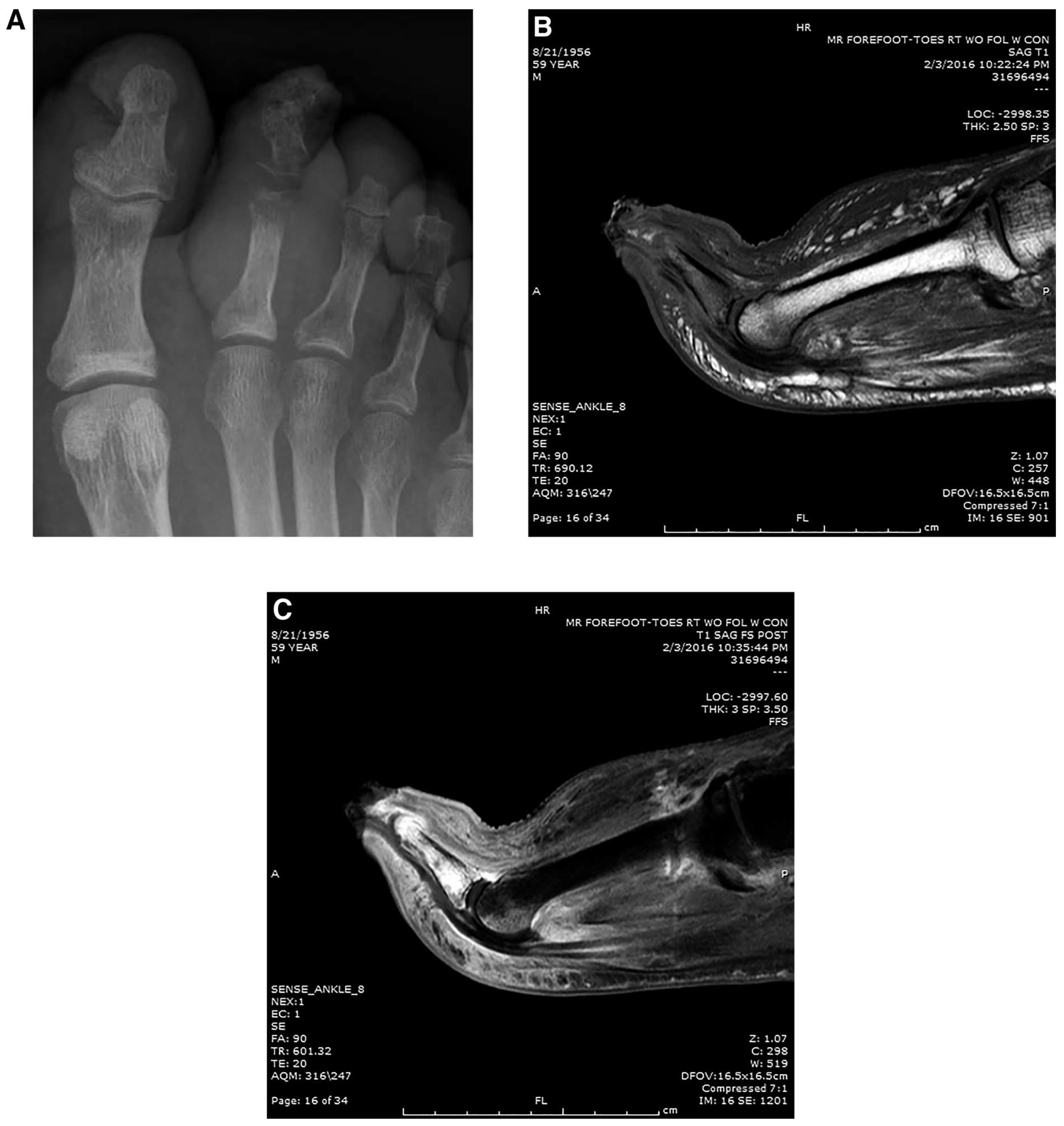
In 14 of 16 toes (87.5%), the ulcer was noted overlying the distal phalanx. In 2 of 16 toes (12.5%), osteomyelitis spread beyond the phalanx most directly underlying the ulceration to adjacent bones. In 14 of the 16 toes (87.5%), the blinded examiner was able to accurately determine which phalanges demonstrated radiographic changes consistent with osteomyelitis. There was one overread: a patient with osteomyelitis involving the distal phalanx of the hallux who also had severe osteopenia affecting the contiguous proximal phalanx and first metatarsal (
Fig. 2). There was also one underread: a patient with severe osteolysis of the phalanges of the second toe that had spread without obvious radiographic sign to the contiguous metatarsal (
Fig. 3).
Figure 2.
Demineralization seen in the proximal phalanx of the hallux and the first metatarsal head overread as osteomyelitis when infection was confined to the distal phalanx.
Figure 2.
Demineralization seen in the proximal phalanx of the hallux and the first metatarsal head overread as osteomyelitis when infection was confined to the distal phalanx.
Figure 3.
A, Osteomyelitis was interpreted to be confined to the phalanges of the second toe, but the second metatarsal head was found to be positive. B, T1-weighted image depicting loss of marrow signal in the phalanges and metatarsal head. C, T1-weighted fat-suppressed image with contrast enhancement depicting hyperintense signal in the phalanges and metatarsal head.
Figure 3.
A, Osteomyelitis was interpreted to be confined to the phalanges of the second toe, but the second metatarsal head was found to be positive. B, T1-weighted image depicting loss of marrow signal in the phalanges and metatarsal head. C, T1-weighted fat-suppressed image with contrast enhancement depicting hyperintense signal in the phalanges and metatarsal head.
Discussion
Although the sensitivity and specificity of plain radiographs in the diagnosis of osteomyelitis are imperfect,[
1,
2,
8] we have presented a case series of toe ulcerations in which plain radiographs in combination with strong clinical suspicion were sufficient to determine which phalanges and metatarsals were affected by osteomyelitis in 87.5% of toes.
Study weaknesses include its retrospective design and relatively small sample size. In addition, no differential diagnosis, including gout or inflammatory arthropathies as a cause for radiographic osteolysis, has been described in these patients, although each patient's history and physical examination findings pointed overwhelmingly toward an infectious etiology. This study is strengthened by its inclusion of a blinded examiner to assess which phalanges showed signs of osteomyelitis using radiographic and clinical information alone.
Many of the original research studies on imaging osteomyelitis were published 20 to 30 years ago,[
2-
4,
8,
11,
12] with some subsequent investigations into the characteristics of osteomyelitis on T1-weighted imaging.[
13,
14] In the intervening years, an increasing number of review articles have been produced,[
15-
22] warranting reconsideration of the original papers on which these reviews are based. Since the turn of the millennium, the landscape of health care has shifted sufficiently that increasing confidence can be placed in the radiographic diagnosis of osteomyelitis. In 1983, Bonakdar-pour and Gaines[
8] first made the observation that radiographic changes require 10 to 14 days to become apparent because that is the time it takes for bony cortex to undergo 30% to 50% destruction. This was noted before the present diabetes epidemic in an era when hematogenously seeded osteomyelitis was more common. That paper's only example cited of contiguous spread infection was in a paraplegic patient with an ischial decubitus ulcer. Yet, this 30% to 50% figure continues to be repeated in the diabetic foot literature.[
16-
18,
21] In the more commonly encountered contiguous spread osteomyelitis of present-day diabetic foot infections, cortical destruction would be the first step leading to marrow involvement, compared with a hematogenous marrow infection that breaks through the bony cortex over time. One may hypothesize that radiographic changes ought to occur more rapidly than 10 to 14 days in this patient population because cortical breakthrough is the necessary first step for bone infection.
To our knowledge, no reference exists estimating the incidence of occurrences in which osteomyelitis of a suspected phalanx spreads to an adjacent uninfected bone. In this series, only 12.5% of toe ulcerations with underlying osteomyelitis demonstrated spread of infection to more proximal bones. It is possible that this low incidence and not the sensitivity of radiographs is what obviates the need for advanced imaging. Although estimating the true incidence of bone-to-bone spread of osteomyelitis was not a goal of this study, the observation that it is low nonetheless merits further inquiry into the necessity of MRI in these patients.
Operative techniques differ in the debridement of infected phalanges. A recent report, using a Delphi method, reached a consensus that a bone margin specimen should be sent for pathology and microbiology in addition to a sample of frankly necrotic bone.[
23] Although this recommendation was made in reference to osseous debridement in general and not to osteomyelitis of the toes specifically, it would seem to suggest that a sample from the more proximal phalanges or metatarsal head would be warranted. However, in the process of obtaining the clear margin specimen, the medullary cavity of unaffected bones will be exposed, carrying the risk of intraoperative contamination. At our institution, we routinely perform simple phalangectomy/terminal Symes amputations based off of preoperative imaging findings without additional sampling. However, an approach involving a proximal margin specimen is even less reliant on MRI and other imaging than in the approach we use because it uses microbiology and not imaging to determine clear margins.
Conclusions
The importance of MRI in the diagnosis and management of osteomyelitis is well-established in the literature. However, we presented a subset of patients in whom infection was detected radiographically in one of the phalanges, and, in most cases, no additional information was gained from the MRI beyond what was already seen on the radiograph. Further research is warranted to replicate these results in a larger series, and an algorithm may then be suggested wherein the MRI is bypassed and operative planning is based solely on the radiographic presentation. This will reduce patient inconvenience and save health-care dollars.