Spiral fractures of the tibia are often the result of torsion trauma. They happen because the mid-distal third of the tibia head shifts from a triangle into a quadrangle; and because it is the thinnest site of the bone, it is the most common place for fractures to occur.[
1,
2] If the torsion trauma is violent enough, this can lead to proximal fibula fractures.[
3,
4] Spiral fractures of the distal third of the tibia are common fractures that are thus often complicated with a proximal fibula fracture.[
1,
2] In clinical practice, this type of fracture is also frequently complicated by posterior malleolus fractures, with an incidence of ankle fractures concurrent with tibial fractures ranging from 0.6% to 88%, and this complication is difficult to visualize on plain film radiography.[
5-
11]
Recent studies have shown a higher incidence of these concurrent fractures, and this, in part, may be due to increased detection with improving imaging methods.[
12] Radiography and computed tomography (CT) examinations are conventional tools for the diagnosis of bone fractures,[
13] and some clinicians now use magnetic resonance imaging (MRI) as well.[
12,
14,
15] However, in some hospitals, diagnostic CT is not available (eg, in many rural hospitals throughout China), and using radiography examination alone could lead to missed diagnoses.[
16]
Despite the increased detection of posterior malleolus fractures, there is a possibility that their incidence is still underestimated.[
16] The pain experienced during tibial fracture may mask the posterior malleolar fracture.[
12] When these ankle fractures go undiagnosed or are not effectively treated, this can lead to incongruity and cartilaginous damage, resulting in posttraumatic arthritis,[
17] and undiagnosed posterior malleolus fractures may displace during reduction maneuvers or surgical treatment of the tibial fracture.[
18] Thus, it is suggested that anteroposterior, mortise, and lateral views are obtained during radiography,[
10] and a communication line may also be observed that suggests posterior malleolus fracture.[
12] Preoperative CT can then assist in diagnosis and lead to prompt fracture stabilization.[
16]
Imaging and Diagnosis
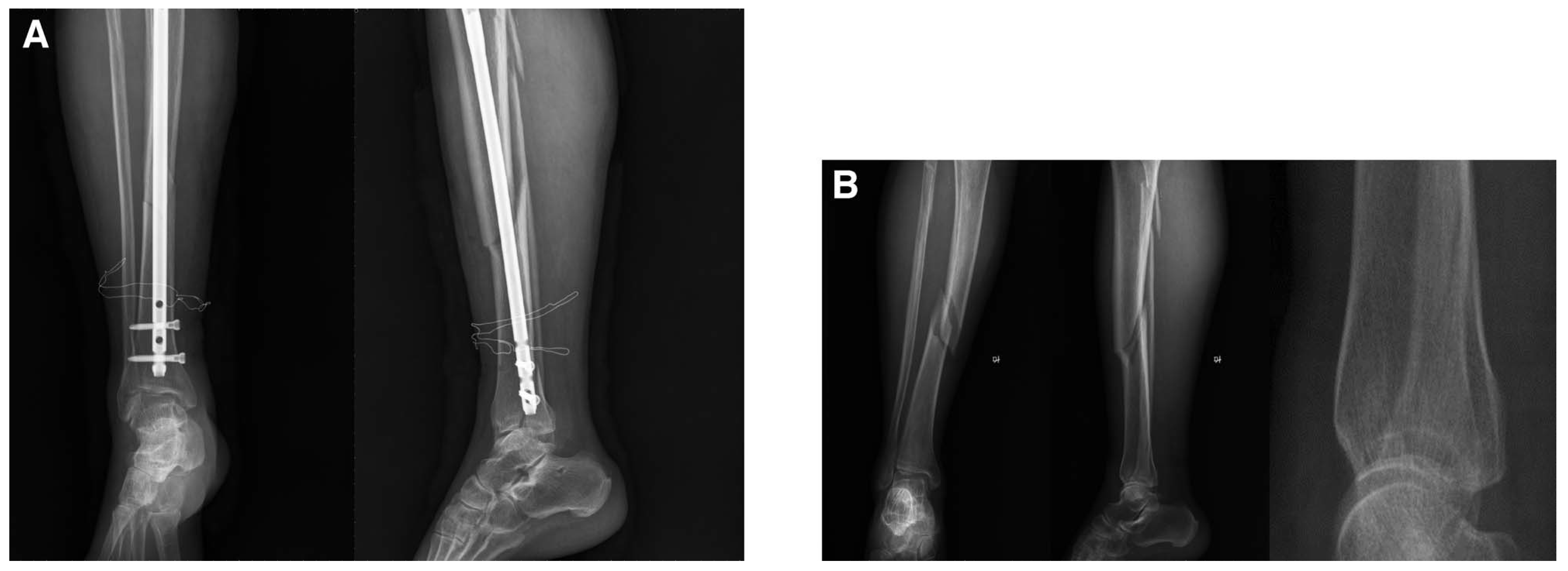
The 28 patients in the spiral tibia group were admitted 1.5 to 8 hours after injury. They all had pain and swelling of the injured limbs. Two patients in the early study period underwent radiographic examination only, one of whom had a displaced posterior malleolus fracture after surgical fixation (
Fig. 2A). After careful interpretation, the preoperative radiograph did in fact indicate a nondisplaced posterior malleolus fracture (
Fig. 2B). The radiographic examination alone did not show any posterior malleolus fractures in one patient in the early study period, and nine subsequent patients underwent both radiography and CT, which revealed the complicated fractures.
Figure 2.
A 57-year-old woman who had right spiral mid-distal third tibia fractures with posterior malleolus fractures. A, Postoperative radiograph indicating displaced posterior malleolus fractures: no broken line is shown in the anterior radiograph (left), clear posterior malleolus fractures in the lateral radiographs (middle, right). B, Fracture lines of the posterior malleolus are unclear in the radiographic images in the anterior (left), posterior (middle), and lateral (right) positions, which were taken before surgical treatment.
Figure 2.
A 57-year-old woman who had right spiral mid-distal third tibia fractures with posterior malleolus fractures. A, Postoperative radiograph indicating displaced posterior malleolus fractures: no broken line is shown in the anterior radiograph (left), clear posterior malleolus fractures in the lateral radiographs (middle, right). B, Fracture lines of the posterior malleolus are unclear in the radiographic images in the anterior (left), posterior (middle), and lateral (right) positions, which were taken before surgical treatment.
Table 1 presents the diagnostic accuracy of radiography and CT for the patients who underwent both (n = 26). Using CT as the gold standard, radiography had a sensitivity of 33.3%, a false negative rate of 66.7%, specificity of 100.0%, a misdiagnosis rate of 0%, a positive predictive value of 100%, and a negative predictive value of 73.9%.
Table 1.
Diagnostic Accuracy of Radiography and CT for Spiral Fractures of the Distal Third of the Tibia Complicated by Posterior Malleolus Fractures.
Table 1.
Diagnostic Accuracy of Radiography and CT for Spiral Fractures of the Distal Third of the Tibia Complicated by Posterior Malleolus Fractures.
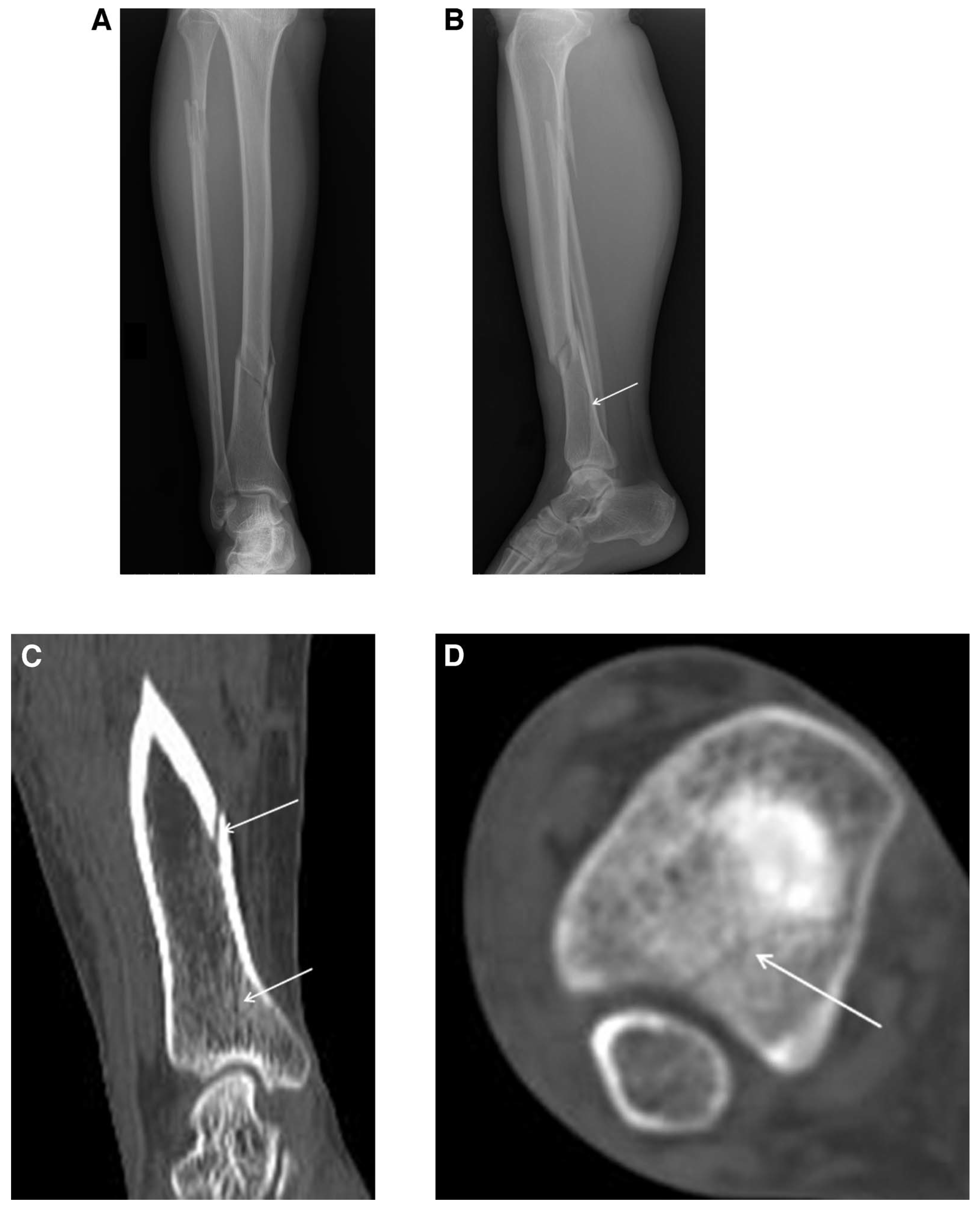
All ten patients in the concurrent group had proximal fibula fractures. All of the fracture lines were oblique (higher laterally and lower medially), with mild lateral displacement (0.05 cm or so). The distance between the distal end of the tibia fracture lines and the articular surfaces at the ankle level was within 3.5 to 9.5 cm (mean, 6.5 cm). All of the patients with combined posterior malleolus fracture showed involvement of one-quarter to one-half of the ankle joint surface of the distal tibia, which resulted from coronal fractures of the distal tibia (
Fig. 3).
Figure 3.
A 31-year-old woman with a right spiral distal third tibia fracture and a proximal fibula fracture. For this patient, the initial radiograph did not show any malleolus fracture, but the computed tomographic (CT) scan showed the fracture. The radiography was reviewed, and the fracture line could be visualized, but with difficulties. The surgical approach was then changed. A, The original radiograph showing no posterior malleolus fracture lines from the anterior radiographic image. B, After review, unclear fracture lines can be seen from the lateral position radiographic image (arrow). C, Mild posterior malleolus fractures from the lateral CT scan (arrows). D, Obvious posterior malleolus fractures from the coronal CT scan (arrow).
Figure 3.
A 31-year-old woman with a right spiral distal third tibia fracture and a proximal fibula fracture. For this patient, the initial radiograph did not show any malleolus fracture, but the computed tomographic (CT) scan showed the fracture. The radiography was reviewed, and the fracture line could be visualized, but with difficulties. The surgical approach was then changed. A, The original radiograph showing no posterior malleolus fracture lines from the anterior radiographic image. B, After review, unclear fracture lines can be seen from the lateral position radiographic image (arrow). C, Mild posterior malleolus fractures from the lateral CT scan (arrows). D, Obvious posterior malleolus fractures from the coronal CT scan (arrow).
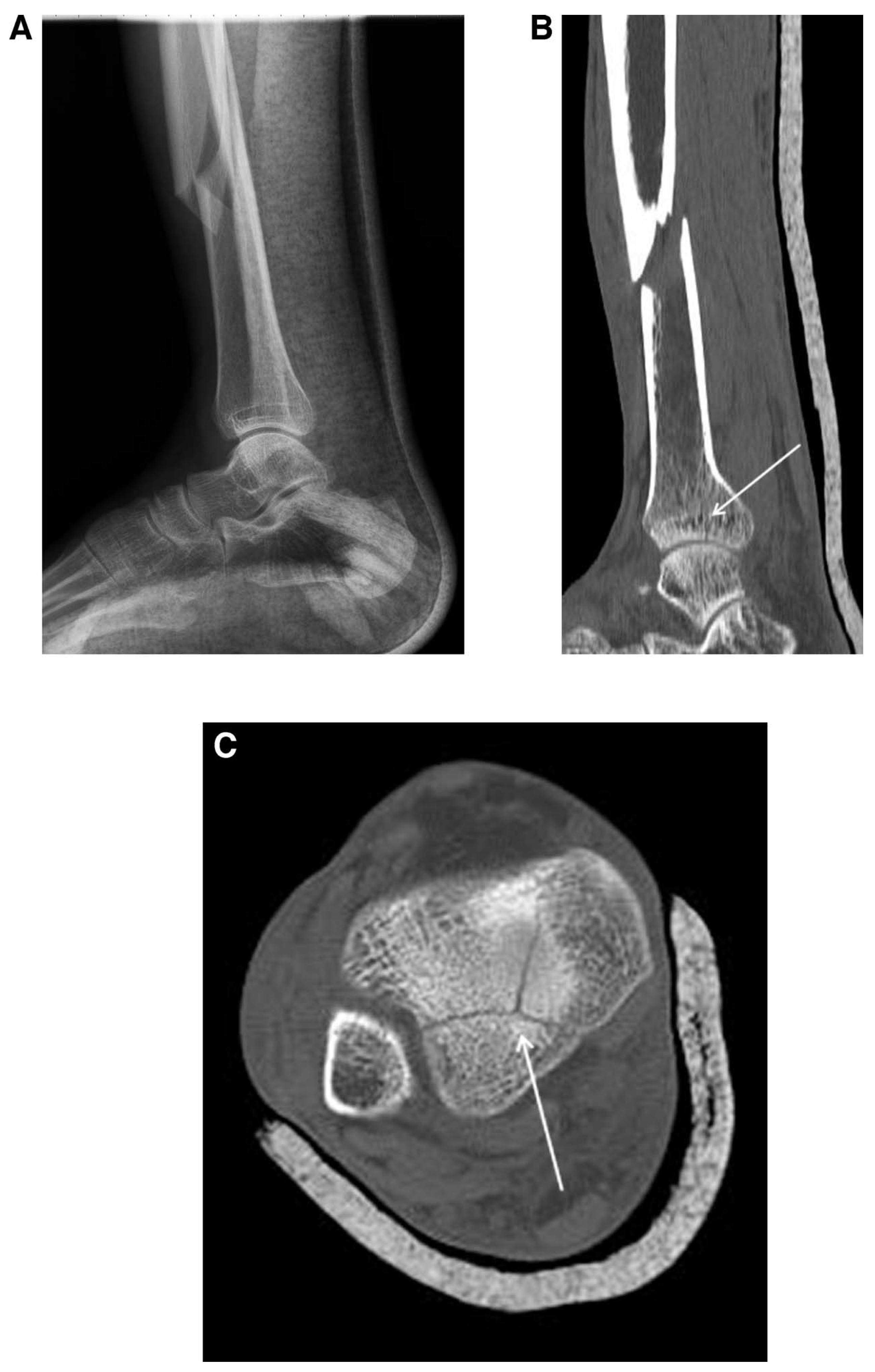
Seven of the ten patients had no displaced posterior malleolus fractures. The remaining three patients had their fractures displaced by approximately 2 cm. The CT showed that in five of the nine patients, the fractures were individual fractures and discontinuous with the tibia fracture lines (
Fig. 4).
Figure 4.
A 46-year-old man with spiral distal third tibia fractures. A, Lateral radiograph. B, Computed tomographic (CT) scan does not clearly indicate posterior malleolus fracture lines (arrow). C, Coronal CT scan showing two clear fracture lines from the posterior malleolus (arrow).
Figure 4.
A 46-year-old man with spiral distal third tibia fractures. A, Lateral radiograph. B, Computed tomographic (CT) scan does not clearly indicate posterior malleolus fracture lines (arrow). C, Coronal CT scan showing two clear fracture lines from the posterior malleolus (arrow).
All of the fractures included in the concurrent group were low-energy indirect violence traumas, and all of the tibial fractures were spiral fractures. The tibias of seven patients formed forward mild angular deformities, and the remaining three did not. Eight of the posterior malleolus fractures were coronal fractures of the distal tibias. One case was complicated by both the medial and lateral malleolus fractures at the same time. The fracture line of the lateral malleolus was from anteroinferior to posterosuperior.
Discussion
The aim of this study was to retrospectively evaluate the diagnosis and treatment of posterior malleolus fracture with spiral fracture of the distal third of the tibia in a Chinese hospital. The information gained from this study should help provide better diagnosis of these fractures and effective treatment. We found that spiral fractures of the distal third of the tibia were often complicated by posterior malleolus fractures, with a concurrent rate of 35.7% (ten of 28) and that radiographic diagnosis was not sufficient to fully diagnose the ankle injury.
In the present series, there were seven men and three women, with a mean age of 50.9 years (range, 23–75 years). Men were more often affected than women, which may be related to practicing more at-risk activities. In addition, two patients were older than 60 years, which may be related to loss of balance and dexterity with aging. Nevertheless, the sample size of patients with spiral fractures of the distal third of the tibia complicated by posterior malleolus fractures could be too small to reach any firm conclusions about the epidemiology of this fracture type.
The incidence of concurrent fractures of the tibial distal third and posterior malleolar during this study was quite high but lower than that found by Boraiah et al,[
9] at 48%, or that at 88% found by Hou et al[
12] using both CT and MRI, although higher than the upper levels of approximately 11% found using radiography alone.[
8] In the present study, the posterior malleolus fractures were usually coronal fractures. The displacements were not obvious, with a low positive rate in examinations (three of ten). The fracture lines were seldom continuous with the fracture lines of the tibia (five of ten). Most of the orthopedists had an inadequate understanding of this fracture type; it was easily missed and could, therefore, cause displaced posterior malleolus fractures postoperatively.[
18] One patient in the present study was the victim of inadequate diagnosis during the early period of the study. The posterior malleolus fracture was not identified on the radiographs, resulting in a missed diagnosis. However, after intramedullary nailing, the distal segments of the screws caused a displaced posterior malleolus fracture. The patient then went on to experience ankle pain and arthritis, which was not the case for the nine patients correctly diagnosed as having concurrent fractures from the start. Of course, the surgical approach is not the same in the presence of a malleolus fracture. The misdiagnosed patient first received an intramedullary nail, but the intramedullary nail resulted in a displacement of the posterior malleolus, leading to poor outcomes. In the patients presented in
Figures 3 and
4, the original surgical plan (based on radiographic findings alone) was simple intramedullary nail fixation, but after CT, the surgical procedure was changed to screw fixation of the posterior malleolus first, followed by intramedullary nail fixation. Based on our experience, an intramedullary nail should be the first choice. If the patient is showing combined posterior malleolus fracture, then the surgical approach could be changed. Plate fixation could be selected, but the trauma is often more important. This sequence of fixation is supported by a previous study that showed similar outcomes to the ones observed in the present study.[
19]
There are a wide range of options for the treatment of distal tibia fractures.[
20] Spiral fracture of the distal third of the tibia was an indication for intramedullary nailing. For simple tibial shaft fracture, fixation with intramedullary nailing is the primary choice and has many advantages. However, because of the complications of malleolus fractures, its application has been restrained and the result has been the chance of severe soft-tissue damage from plate fixation.[
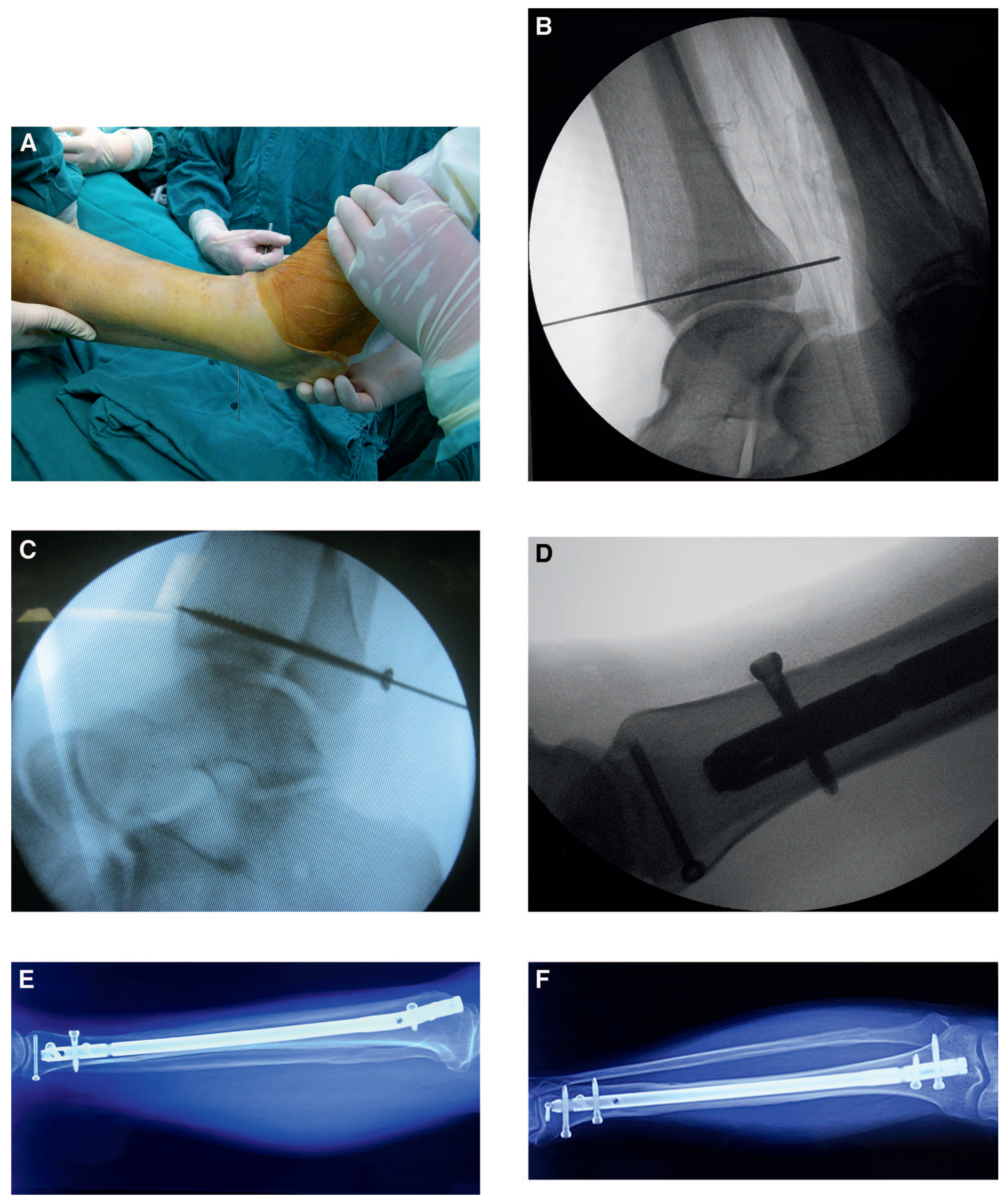
21] Patients with posterior malleolar fracture can still undergo intramedullary nail fixation after screw fixation. For relatively large fragments, it is possible to use anterior to posterior screw fixation, but the fracture pattern studied here often had small fragments, so that treatment was inadequate. Furthermore, it was also not conducive for compression because open internal posterior to anterior fixation required the patient to be in either the lateral or prone position. However, intramedullary nailing of the tibia requires a supine position. Our innovative method was easy and feasible, while it fixed the posterior malleolus fragments and compressed them without changing their positions. The patients were placed in the supine position, the guide pin was inserted from anterior to posterior until the guide pin exited from the posterior side, and then a hollow screw was used from posterior to anterior through the guide pin. Therefore, this novel method allowed us to fix the small bone block to the bigger bone block in the supine position. The advantage of this surgical procedure included minimally invasive fixation, small trauma, short surgical time, and reliable fixation, which may be the innovation of this method.
The optimal treatment of posterior malleolus fractures is also the subject of much debate.[
22] A recent survey of surgeons in the United States showed that there was wide variation in the range of treatments,[
23] and there is no consensus on the size of fracture that should be fixed.[
24] Even minimal displacement may cause posttraumatic arthritis.[
25] Therefore, anatomical reduction of all displaced posterior malleolus fractures should be considered.[
26] The method used in our center for posterior malleolus screw fixation gave good results when the posterior malleolus fractures were identified before the tibial fracture was treated, emphasizing the importance of correct diagnosis at the outset of treatment.
A possible mechanism of injury for these fractures could be a static position of the foot and ankle during exercise, with the injured leg kept moving forward and rotating outward due to inertia.[
3,
4] In this condition, the rotating force could continue to act on the shaft of the tibia, and, thus, could result in spiral fracture of the mid-distal third tibia. The fracture of the posterior malleolus might have been caused by a sudden stop of the foot and ankle during exercise, so while the tibia continued to move forward, it struck the tail. However, in our analysis of the imaging data, the fractures of the posterior malleolus were individual fragments (five of ten) and discontinuous with the tibial fracture lines, in contrast to most of those seen by Hou et al.[
12] It could be that in the patients presented herein the posterior malleolus fracture was related to the tractions of the posterior tibiofibular ligament.
In reference to the published literature,[
9-
11] along with the present data, spiral fractures of the distal third of the tibia could often be complicated by posterior malleolus fractures, which were sometimes misdiagnosed. The reasons for misdiagnoses included 1) insufficient knowledge of the high chance of posterior malleolus fracture concurrent with spiral fracture of the distal third of the tibia; 2) obviously displaced spiral tibia fractures were diagnosed but slight posterior malleolus fractures were sometimes undetected; 3) most of the fracture lines were coronal and, therefore, difficult to identify on the anterior radiographs; 4) the fracture lines were also obscured by the fibula in the posterior radiographs; and 5) the fractures were mainly nondisplaced or invisible, thus the examinations of the radiographs were negative. These reasons for misdiagnosis present important arguments to always perform CT or MRI when patients present with spiral fracture of the distal third of the tibia. Based on the features of the fractures, we think that certain relationships could be present between the causing mechanism of the fracture pattern and the occurrence of the ankle fractures (supination-external rotation type). Whether there is any correlation between the positions of these fracture patterns and the position of the knee or the ankle at the time of injury or the angle between the tibial shaft and the ground will require further investigation.
Of course, the present study is not without limitations. The sample size was small and from a single center, which could introduce a bias due to the treatment methods. In addition, no biomechanical experiment was performed, which would be useful to better understand the formation of posterior malleolus fractures with spiral fractures of the distal third of the tibia. Additional studies are necessary to improve the diagnosis and treatment of the fractures.
In conclusion, despite previous studies extolling the virtues of CT in addition to radiography for the successful diagnosis of posterior malleolus fractures with spiral fractures of the distal third of the tibia, misdiagnosis can still occur. This study emphasizes the need for CT scanning of spiral fractures of the distal third of the tibia and presents a successful method of treating these fractures. The present study offers some clues for better diagnosis and treatment of these fractures.