Antibiotic-impregnated nonabsorbable beads, also known as polymethylmethacrylate (PMMA) beads, have historically been used as adjunctive treatment with surgical debridement and as void filler, and they are well established in North America and Europe [
1-
4]. Absorbable cement antibiotic carriers have been widely reported in the adjunctive treatment of osteomyelitis [
5,
6]. There is an abundant amount of in vitro, in vivo, and human reports of successful combination use of calcium sulfate, hydroxyapatite, and iohexol with antibiotics [
7-
13]. A calcium sulfate and hydroxyapatite drug carrier vehicle has been successfully used by the author in the adjunctive surgical management of osteomyelitis. This calcium sulfate and hydroxyapatite carrier vehicle is a biocompatible, bioabsorbable, sterile, injectable, moldable, nonexothermic, radiopaque, and nontoxic ceramic bone void filler that acts as an osteoconductive scaffold that, due to its biodegradability, does not need to be removed after implantation [
14].
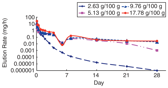
An antibiotic carrier vehicle should be flowable to allow complete contact of the antibiotic with the bacteria adhering to the bone, to allow significant drug elusion compared with PMMA drug elusion, and to have adequate minimal inhibitory concentration (MIC) levels. X. Yang, PhD, of the Hospital for Special Surgery in New York, New York, reported excellent calcium sulfate and hydroxyapatite carrier vehicle vancomycin elusion rates at three separate concentrations recorded over 10 days compared with PMMA vancomycin elusion rates for the initial burst and subsequent tail (
Fig. 1) [
15]. The calcium sulfate and hydroxyapatite carrier vehicle vancomycin elusion concentration initial release was at 10 mg/h or more versus approximately 0.25 mg/h for the PMMA vancomycin elusion rates. The tail concentration plateau elusion rates for the calcium sulfate and hydroxyapatite carrier vehicle was maintained at 0.5 mg/h for 4 weeks versus 0.05 mg/h for the PMMA [
15]. An in vitro study of zones of inhibition created by a mixture of the calcium sulfate and hydroxyapatite carrier vehicle with either single or multiple antibiotics versus methicillin-resistant
Staphylococcus aureus or Pseudomonas aeruginosa was compared with the Clinical Laboratory Standards Institute's criteria for MIC. The results demonstrated consistent significant inhibitory effects of the delivered antibiotics versus
S aureus or
P aeruginosa well beyond the MIC susceptibility for all of the antibiotics tested [
15].
Figure 1.
Vancomycin elusion from the calcium sulfate and hydroxyapatite carrier vehicle.
Figure 1.
Vancomycin elusion from the calcium sulfate and hydroxyapatite carrier vehicle.
The percutaneous antibiotic delivery technique (PAD-T) was first used by the author in 2009 as a means to deliver antibiotics with a flowable carrier vehicle into an area of bone infection with complete bone penetration and contact with the adherent surface bacteria/biofilm, obtaining significant drug elusion and drug levels well above the MIC in the bone defect. This technique avoids a more invasive bone surgery, such as placing and removing nonabsorbable antibiotic beads. The PAD-T minimizes time away from work, does not require external fixation, and generally requires no change in weightbearing status. The PAD-T involves a small percutaneous skin incision followed by a small bone cortex incision into the infected bone. Irrigation and spongiosum bone cultures can be performed through the bone cortex incision. Through this small bone cortex incision, the calcium sulfate and hydroxyapatite carrier vehicle in the liquid state mixed with 1.2 g of tobramycin and 1.0 g of vancomycin is then delivered under fluoroscopy into the area of bone infection.
Technique
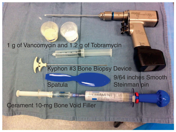
The setup for this procedure is fairly simple (
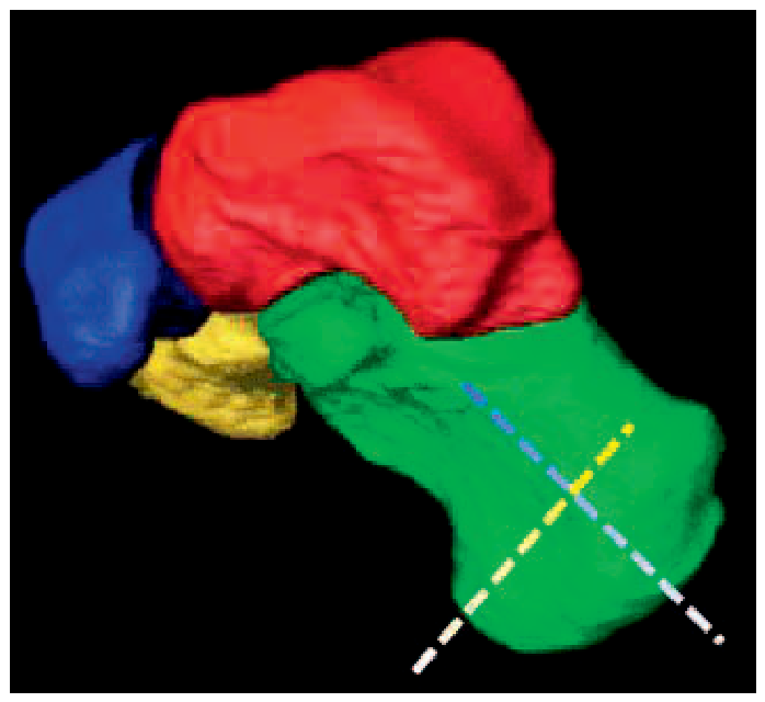
Fig. 2). A tourniquet should be used to minimize medullary bleeding. Any dry antibiotic or antifungal drug can be used. There is significant broad-spectrum bacterial coverage when vancomycin and tobramycin are used together with the bone void filler. A simple percutaneous skin incision is completed superficial to the area of osteomyelitis. Under intraoperative fluoroscopy, an incision is made into the near bone cortex and is then completed with a smooth 9/64-inch Steinman pin into the area of osteomyelitis. The Steinman pin is then advanced to the far bone cortex without far bone cortex violation (
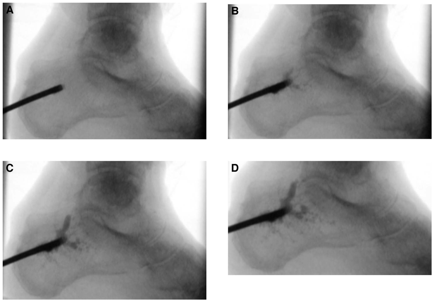
Fig. 3). Usually a single plane pass of the Steinman is sufficient. Multiple passes of the Steinman pin in long bones can unnecessarily increase the risk of stress risers and should be avoided. Because the bone void filler is being delivered in a liquid medium, the bone void filler will move along the path of least resistance along the micro channels and cavities created by the bone infection. After the bone cortex incision, spongiosum biopsies for bacteria can be completed with a curved curette, and irrigation can also be completed. The calcium sulfate and hydroxyapatite carrier vehicle is mixed per protocol and then added to the antibiotics. This mixture is placed in a 20-mL syringe and is then connected to the size 3 Kyphon bone biopsy device (Medtronic, Minneapolis, Minnesota). Under intraoperative fluoroscopy, the bone biopsy device is then advanced through the guide hole at the near bone cortex incision and advanced to the far cortex. There should be a nice filling effect seen under intraoperative fluoroscopy as the calcium sulfate and hydroxyapatite carrier vehicle and antibiotics fill the damaged bone (
Fig. 4). Once the calcium sulfate and hydroxyapatite carrier vehicle is activated with iohexol (Omnipaque; GE Healthcare, Chicago, Illinois), the workable time of the product is approximately 10 min, with a final set time of approximately 45 min.
Figure 2.
Instrumentation required for the percutaneous antibiotic delivery technique.
Figure 2.
Instrumentation required for the percutaneous antibiotic delivery technique.
Figure 3.
Two approaches to the calcaneus. The blue dashed line is the preferred approach. Note that neither approach violates the far cortex.
Figure 3.
Two approaches to the calcaneus. The blue dashed line is the preferred approach. Note that neither approach violates the far cortex.
Figure 4.
A, Bone cortex incision with bone biopsy device needle delivery. B, Initial antibiotic delivery. C, Continued antibiotic delivery. D, Completed antibiotic delivery.
Figure 4.
A, Bone cortex incision with bone biopsy device needle delivery. B, Initial antibiotic delivery. C, Continued antibiotic delivery. D, Completed antibiotic delivery.
Case Presentation
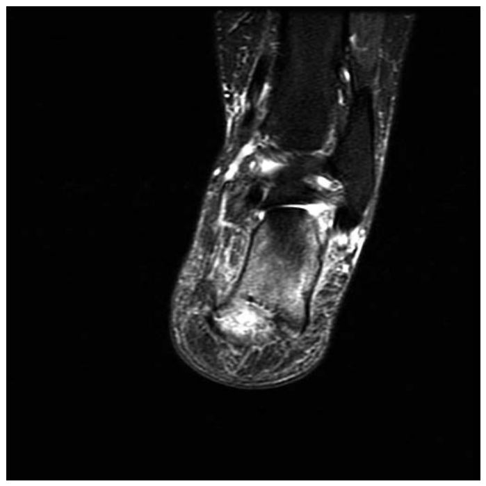
A 54-year-old diabetic woman presented with a right heel ulcer to the calcaneus with underlying acute calcaneal osteomyelitis following plantar fascia release and calcaneal exostectomy for chronic plantar fasciitis by a different surgeon. She had been treated with intravenous vancomycin for 1 month and continued to have significant right heel pain (8 on a visual analog scale from 1 to 10). Her initial erythrocyte sedimentation rate was 62 mm/h, white blood cell count was 9.0×10 [9]./L, and magnetic resonance imaging findings were consistent with osteomyelitis of the body of the calcaneus (
Fig. 5). The patient's diabetes was well controlled, and there was no peripheral arterial disease of that lower extremity. Preoperative deep-tissue biopsy cultures were positive for methicillin-resistant
S aureus.
Figure 5.
Preoperative magnetic resonance image of the foot.
Figure 5.
Preoperative magnetic resonance image of the foot.
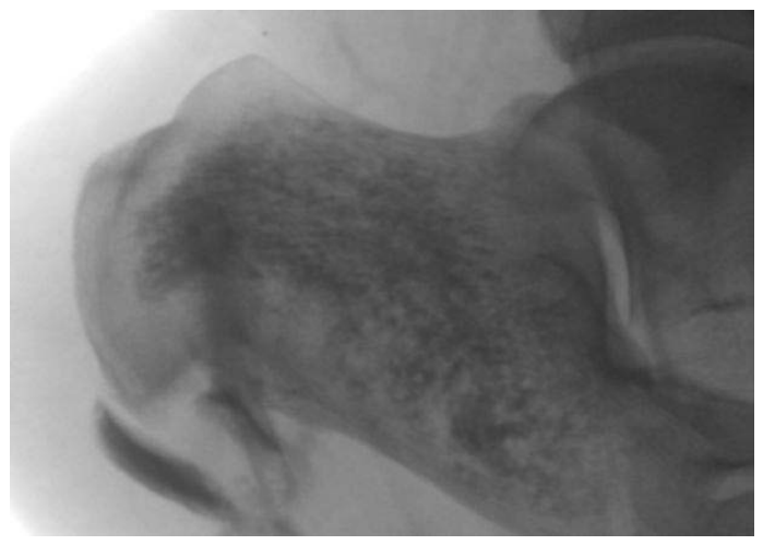
The PAD-T was used immediately after seeing the patient, 7 weeks after the original surgery, with delivery of calcium sulfate and hydroxyapatite bone void filler carrier vehicle with 1.2 g of tobramycin and 1.0 g of vancomycin under fluoroscopy directly into the area of bone infection (
Fig. 6). Extravasation of the carrier vehicle into soft tissue, as seen on the intraoperative image, can be hand-milked out of the soft tissue and elicits no soft-tissue reaction. There was no bone debridement, but the wound was debrided. The bone cultures were also positive for methicillin-resistant
S aureus. After surgery, the infectious disease physician placed the patient on 4 weeks of oral linezolid therapy. Five days after surgery, the heel wound was healed, her pain was considerably decreased, and she began to weightbear approximately 50% with crutches and a surgical shoe. At 3 weeks, the patient was weightbearing completely without restriction. One month later, the erythrocyte sedimentation rate was 17 mm/h, down from 62 mm/h. Four years after treatment, the patient is without wound, pain, or osteomyelitis reoccurrence and is weightbearing without restriction. Plain radiographs in the postoperative period did not demonstrate any calcaneal bone erosion or loss of trabeculation.
Figure 6.
Intraoperative fluoroscopy image of the antibiotic delivery.
Figure 6.
Intraoperative fluoroscopy image of the antibiotic delivery.
Discussion
The process of osteomyelitis is one of bone destruction, osseous vascular destruction and congestion, and adherent bacteria generation. Osteomyelitis causes the bone to be destroyed, leaving a path of least resistance throughout the affected bone. This leaves large and small voids of compromised bone harboring free-floating and adherent bacteria. All of these areas of bone infection are interconnected by macro and micro defects from the bone destruction. These areas of micro bone destruction can very easily be missed or not be accessible on a bone debridement and can serve as a nidus for reinfection after traditional bone debridement and intravenous antibiotic management. It is this devitalized and compromised bone that harbors adherent bacteria that contain pathogens that are resilient and serve as the nidus for continued infection. Even when bone debridement and irrigation may remove the nonadherent pathogens, the sessile biofilm remains. The successful treatment of this adherent bacterium is further diminished by the microvascular destruction and congestion in the infected bone, thus interfering with the body's ability to deliver antibiotics to the area of bone infection.
Furthermore, it is this dysvascular and avascular environment from the bone infection that also contains adherent bacteria, which significantly contributes to the difficulty in treating a bone infection with oral or parental antibiotics alone, relying on the patient's circulation to deliver the antibiotics in sufficient concentration to have an inhibitory effect on the bone infection. It is within this dysvascular and avascular bone that the surface-adhering bacteria survives and allows the chronicity of the bone infection to become established.
The PAD-T can be an effective adjunctive therapy to successfully treat osteomyelitis owing to the liquid nature of the drug carrier vehicle, the drug elusion levels obtained, and the significant drug MIC levels obtained [
5,
6,
15]. It is this fluid nature of the drug carrier vehicle that allows it to move along the path of least resistance created by the bone infection to penetrate into all of the microchannels, cavities, and abscesses in complete contact with all bone surfaces and adherent bacteria.
Conclusions
The PAD-T for the adjunctive management of osteomyelitis allows easy, direct placement of either concentrated antibiotic or antifungal medication into the area of osteomyelitis. There has not been a previously reported percutaneous antibiotic delivery technique using a simple bone cortex incision and flowable bone void filler in the adjunctive treatment of osteomyelitis. There is no restriction to either antibiotic or antifungal medication to be added to the calcium sulfate and hydroxyapatite carrier vehicle. Because of the simple skin and bone cortex incision, there is no required change in weightbearing in the postoperative period. The patient in this case presentation was kept 50% weightbearing initially after the author's surgery because of the previous calcaneal surgery by a different surgeon. The resolution of bone infection in the case presentation could have been obtained because the PAD-T is a potential adjunctive therapy at this time and further research is warranted. Tetsworth and Cierny [
16]. advocate an atraumatic surgical approach to the surgical management of osteomyelitis, which this percutaneous approach allows.
Financial Disclosure: None reported.
Conflict of Interest: None reported.