If midfoot dislocations are uncommon injuries, tarsal fractures or fracture-dislocations are even more so [
1]. The incidence of navicular fracture-dislocations has been estimated to be approximately 0.26% of all fractures [
2].
The tarsal navicular is the keystone of the longitudinal medial arch. It is both intrinsically and extrinsically stable, the former due to the high degree of congruity of the midfoot joints and the latter to its powerful dorsal and plantar ligamentous support, which explains the fact that fractures or fracture-dislocations associated with other important foot injuries are more likely to occur than are isolated fractures or fracture-dislocations [
3,
4]. Bearing this in mind, it is impossible that isolated dislocations occur without a simultaneous bone or ligament injury to the adjacent column. This associated injury can be either transient or permanent. Therefore, for a navicular fracture-enucleation to happen, an extensive dorsal, plantar, medial, and lateral ligament and capsular injury, as well as a dislocation or mediotarsal subluxation, must occur.
Regarding fracture-dislocations, the literature is confined to case reports, and they report only navicular fractures with dorsal displacement of the dorsal fragment in which the integrity of the talonavicular joint is partially maintained. According to the radiographic findings, this case seems closer to the published case reports on isolated navicular dislocations occurring with a total disruption of the talonavicular joint, as in the case report by Mathesul et al [
5]. Regarding pure dislocations, since 1924 [
6] fewer than 20 cases have been published [
4,
7-
12].
The most frequent cause of fracture-dislocations as described in the literature is high-energy trauma, mainly crush fractures, where the foot gets trapped [
7], and the pathophysiologic mechanism of this type of fracture remains unclear.
Navicular fractures can be classified into tuberosity, avulsion, overuse, and body fractures. The most extended classification for body fractures is that of Sangeorzan et al [
13]: fractures that divide the navicular into dorsal and plantar fragments (type I), fractures that divide it into dorsomedial and plantar-lateral segments (type II), and axial load comminuted fractures (type III).
The goal in dislocation and fracture-dislocation is restoring the medial longitudinal arch and the talonavicular joint congruity [
1]. Given the low incidence of this type of injury, the appropriate treatment is not clear, but surgical treatment seems to be necessary to achieve an acceptable functional outcome [
3,
5-
14]. Many options have been published, such as closed reduction with external fixation and percutaneous pinning [
5,
15], open reduction using a double approach [
1,
3,
7,
9,
14], partial or total excision [
5], and naviculocuneiform arthrodesis [
5,
7].
The long-term evolution of this kind of injury is uncertain. The most frequently reported complication is avascular necrosis [
3,
4,
7,
9,
12,
13], but post-traumatic flatfoot with no increase of forefoot adduction has also been described [
11].
In the present case we chose a single medial approach and little aggressive osteosynthesis (percutaneous pinning) with the aim of preserving bone vascularization and avoiding avascular necrosis.
Case Report
We report the case of a 20-year-old man with no significant medical history who came to the emergency department with left midfoot pain and deformity after falling while riding a bicycle, with pain at passive and active mobilization of the ankle. He could not specify the position of his foot when the accident took place.
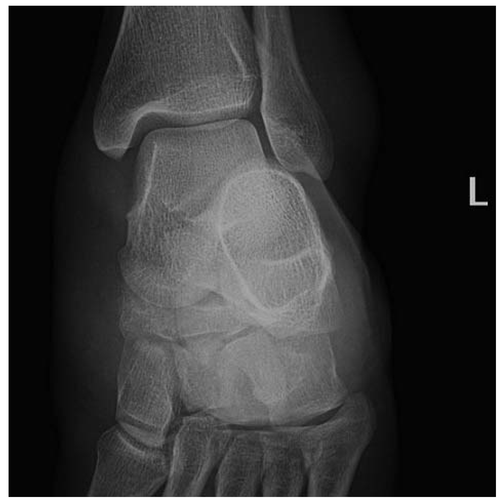
Initial radiographic examination with anteroposterior (
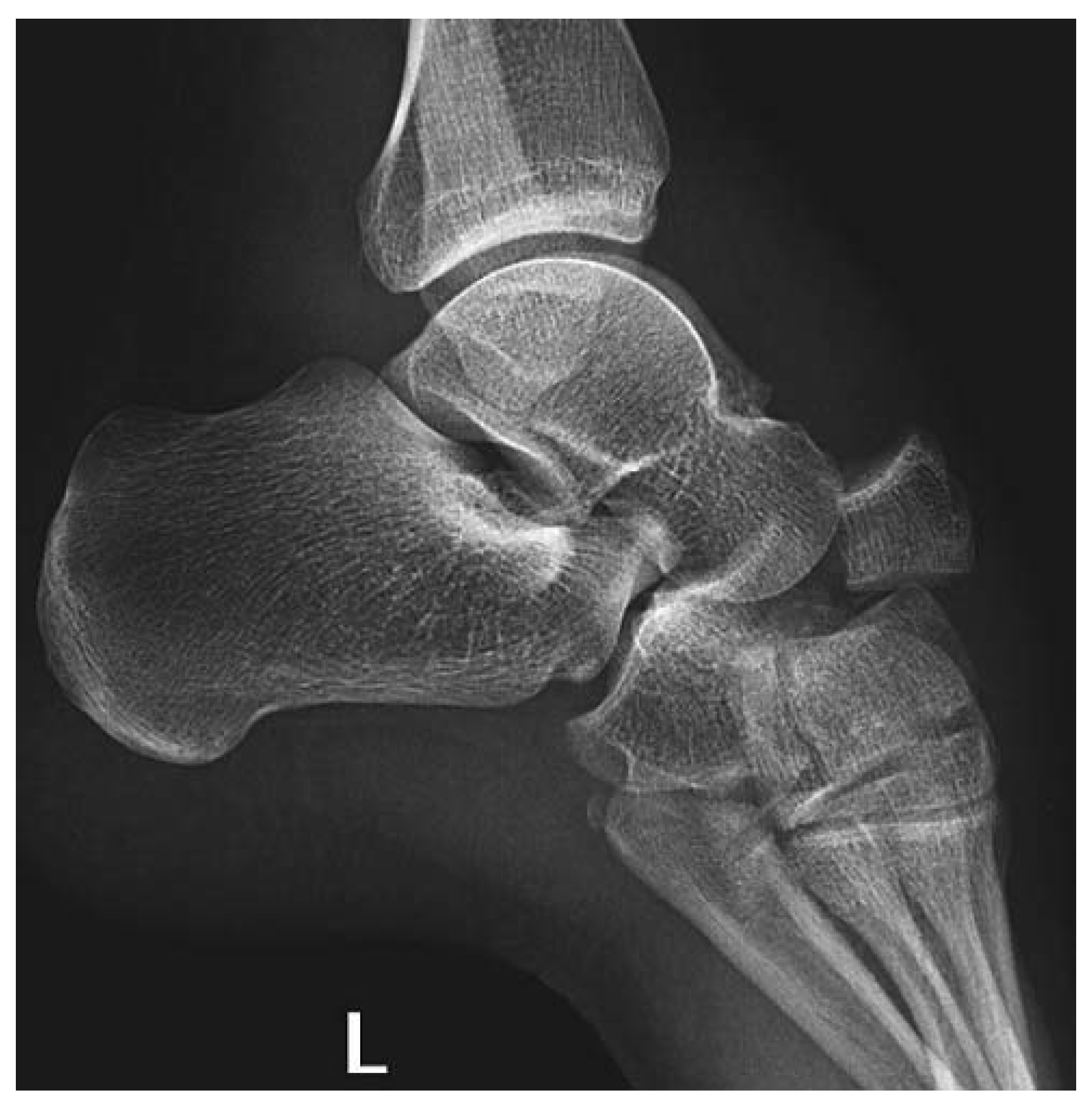
Fig. 1), lateral (
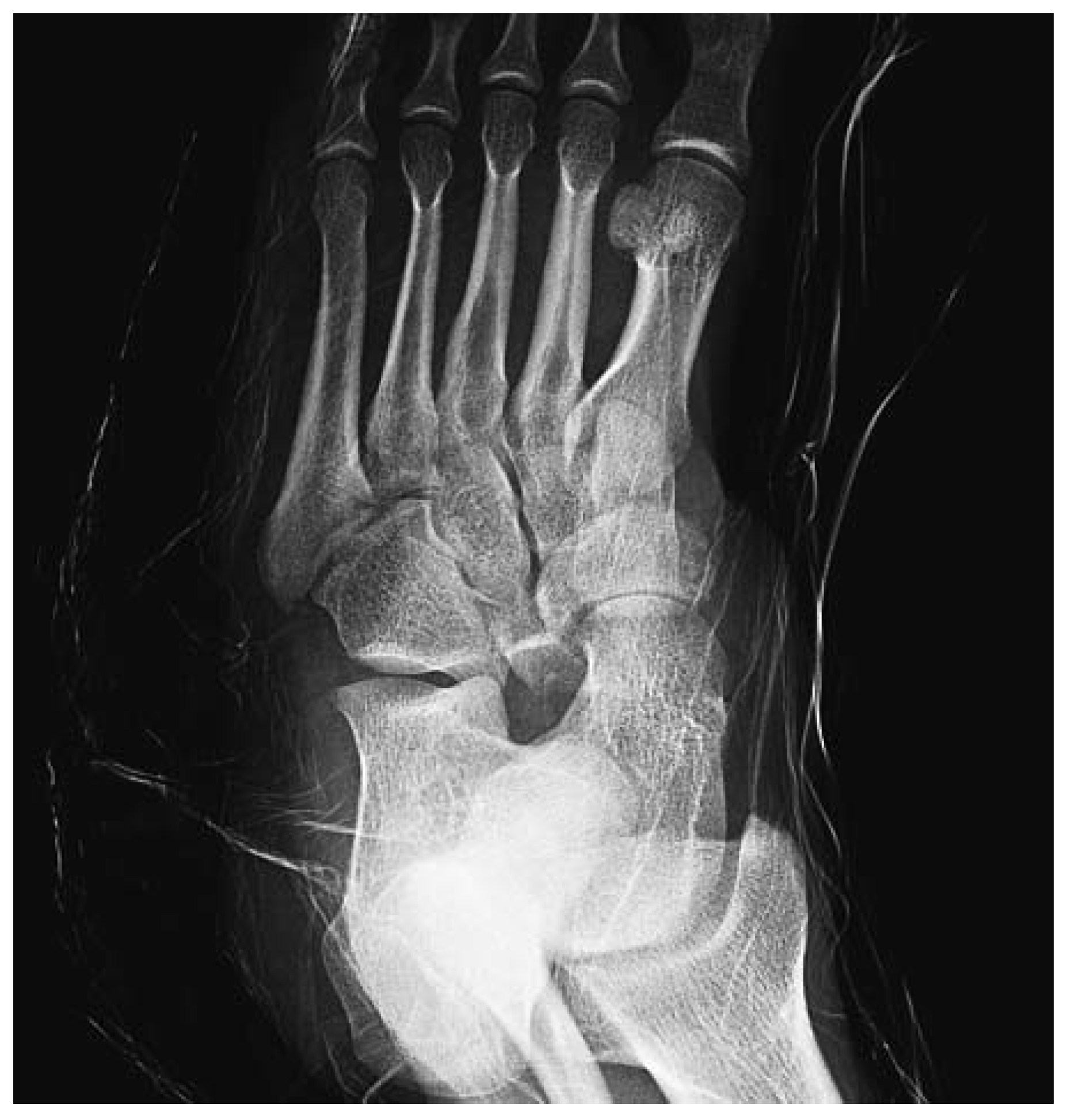
Fig. 2), and oblique (
Fig. 3) radiographs in the emergency department led to the diagnosis of dorsal fracture-dislocation of the tarsal navicular with total disruption of the talonavicular joint and medial swivel rotation of Chopart's joint. The calcaneocuboid joint remained intact. The final diagnosis was Sangeorzan type II [
13] fracture-dislocation of the navicular and Orthopaedic Trauma Association type 74-C2 [
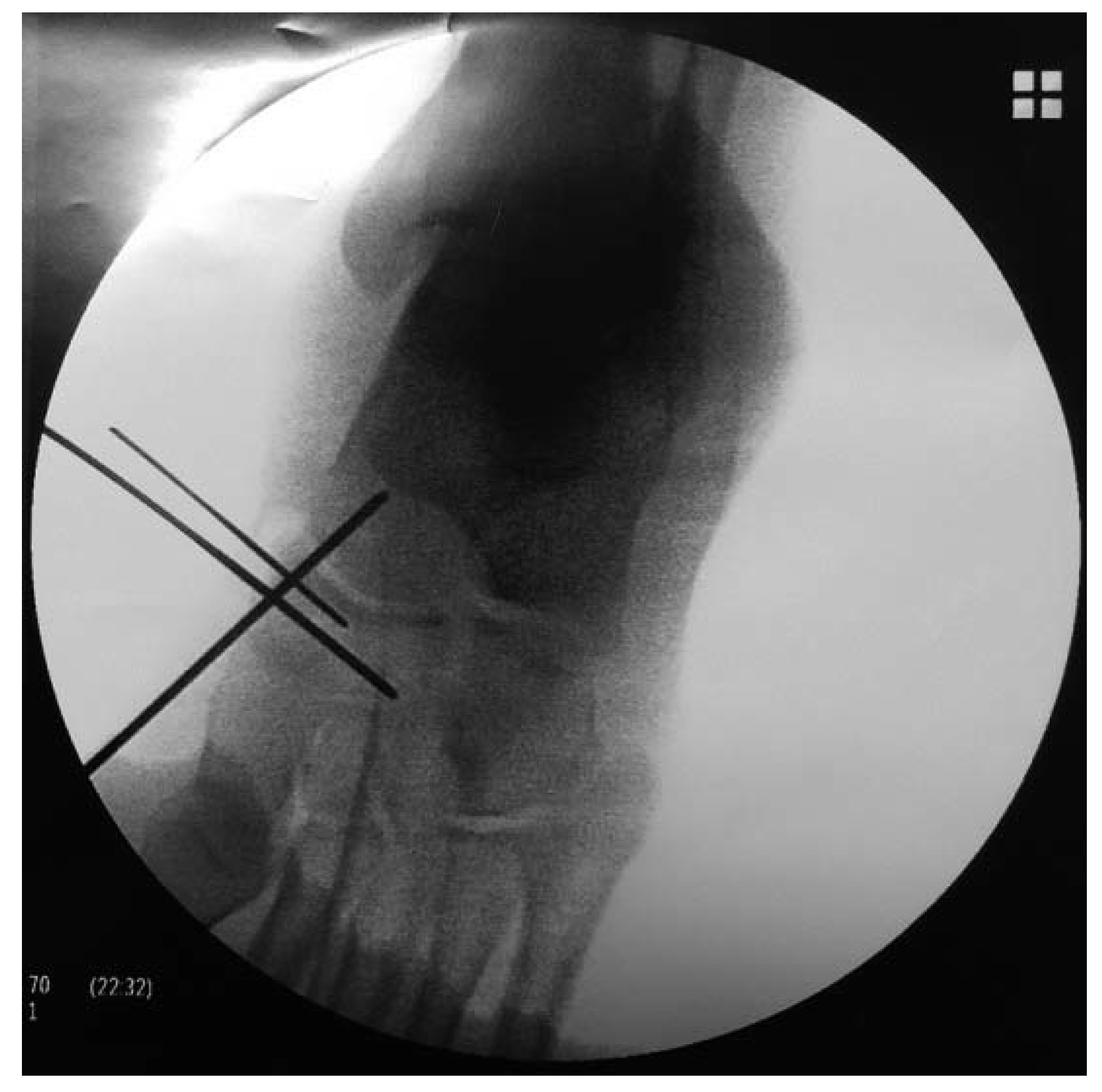
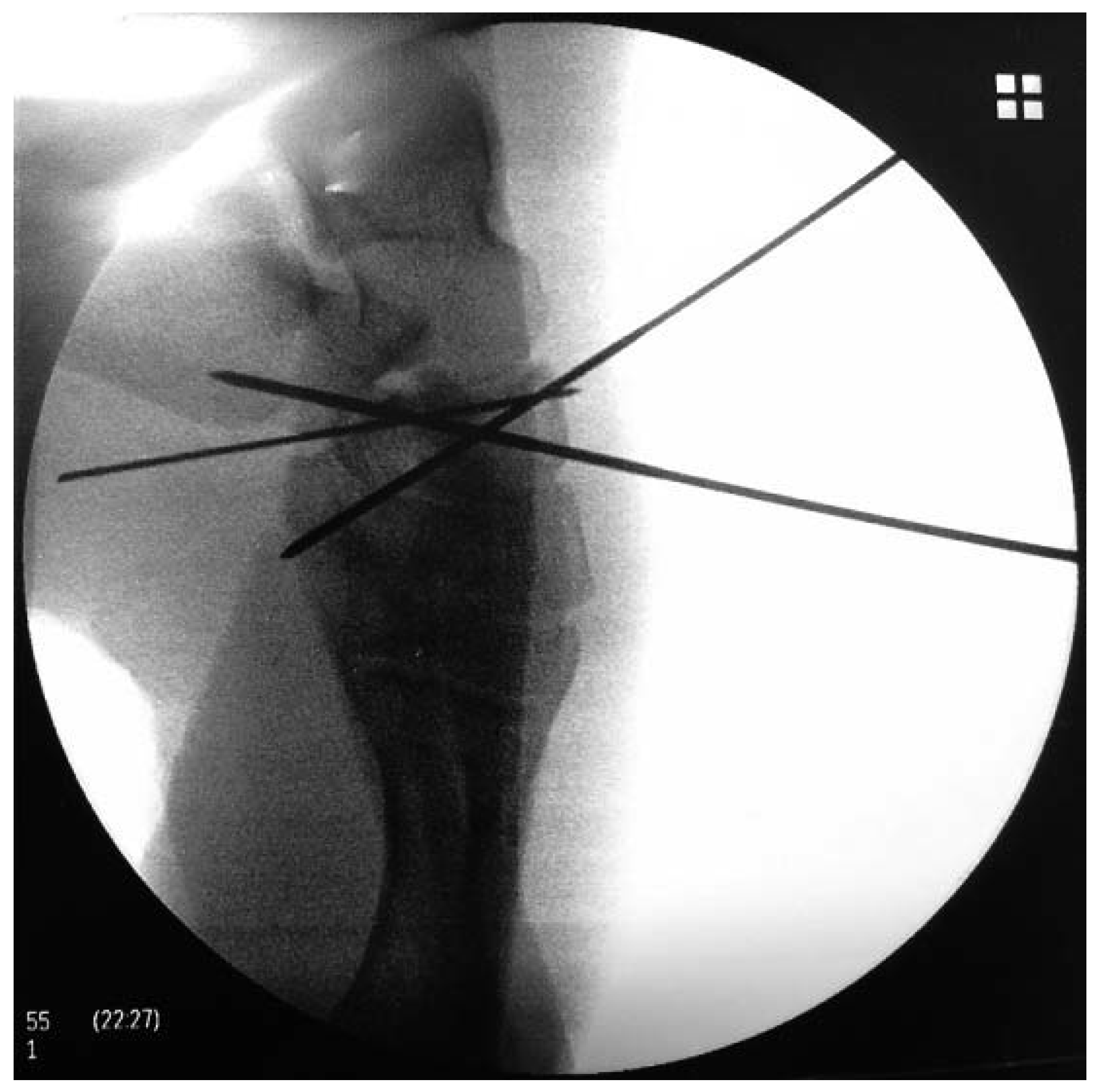
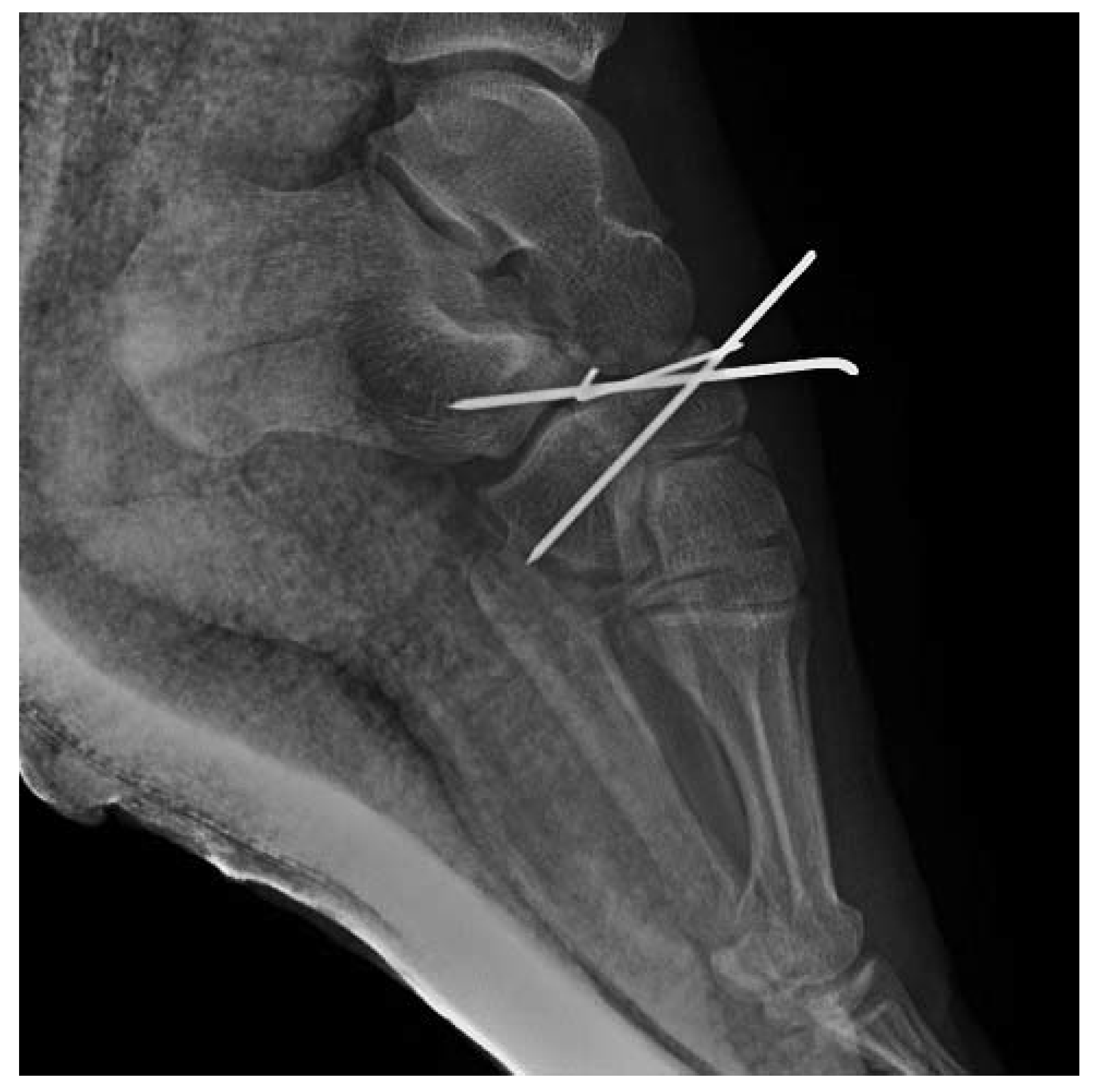
1], with dorsal enucleation of the dorsal fragment and a medial swivel dislocation of Chopart's joint. Under spinal anesthesia, open reduction and percutaneous pinning was performed through a medial approach. Three Kirschner wires were inserted, avoiding the talonavicular and the naviculocuneiform joints. The first Kirschner wire was inserted from medial to lateral, and the other two Kirschner wires were inserted diverging to plantar (
Figs. 4 and
5). This fixed the fracture and also worked as joint stops, thus avoiding recurrence of the dislocation as well as injury to the joint surface while trying not to be aggressive in order to avoid avascular necrosis. Immobilization in a plaster cast was used for 6 weeks (
Fig. 6).
Figure 1.
Anteroposterior radiograph taken at presentation to the emergency department.
Figure 1.
Anteroposterior radiograph taken at presentation to the emergency department.
Figure 2.
Lateral radiograph taken at presentation to the emergency department.
Figure 2.
Lateral radiograph taken at presentation to the emergency department.
Figure 3.
Oblique radiograph taken at presentation to the emergency department.
Figure 3.
Oblique radiograph taken at presentation to the emergency department.
Figure 4.
Intraoperative anteroposterior radiograph showing insertion of Kirschner wires in a medial to lateral direction.
Figure 4.
Intraoperative anteroposterior radiograph showing insertion of Kirschner wires in a medial to lateral direction.
Figure 5.
Intraoperative lateral radiograph showing insertion of Kirschner wires in a plantar direction.
Figure 5.
Intraoperative lateral radiograph showing insertion of Kirschner wires in a plantar direction.
Figure 6.
Postoperative lateral radiograph.
Figure 6.
Postoperative lateral radiograph.
Nonweightbearing ambulation was started on the second day. The Kirschner wires and the plaster cast were removed after 6 weeks, and the fracture remained adequately reduced. After 8 weeks, the patient reported no pain, and progressive partial weightbearing was permitted. Four months after the injury, the patient regained complete ankle mobility and partial subtalar joint mobility and could return to physical activity, experiencing only minor discomfort and swelling.
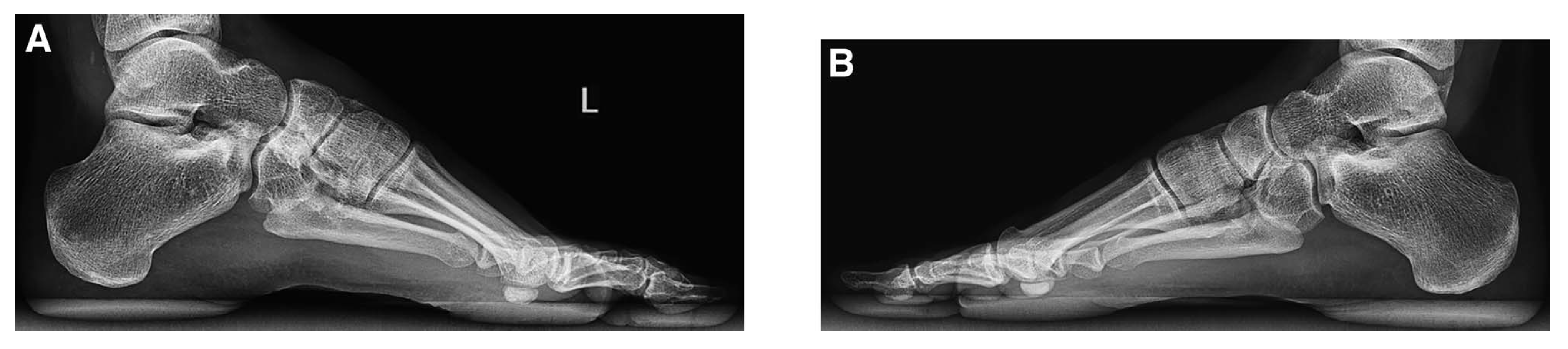
Eighteen months after the accident, the mobility of the ankle and foot was completely restored and pain free. The patient reported some discomfort and light swelling after intense exercise and on walking long distances on irregular surfaces. Lateral control radiographs showed minor differences in midfoot architecture compared with the contralateral foot: there was a difference of 2° in the Moreau-Costa-Bartani angle (constituted by the line formed from the lower point of first metatarsal head to the lower point of the talonavicular joint and the line formed from the lower point of the talonavicular joint to the lower point of the posterior calcaneal tuberosity) [
16] (114° in the left foot and 112° in the healthy right foot); the Meary-Tomeno angle (the line corresponding to the diaphyseal axis of the first metatarsal and the line corresponding to the axis of the talar neck overlap normally, so this angle is usually a line, 0°) [
16] was 7° in the left foot and 4° in the right; and, finally, the calcaneal pitch angle (formed by the horizontal and a line from the base of the heel to the inferior cortex of the calcaneus) [
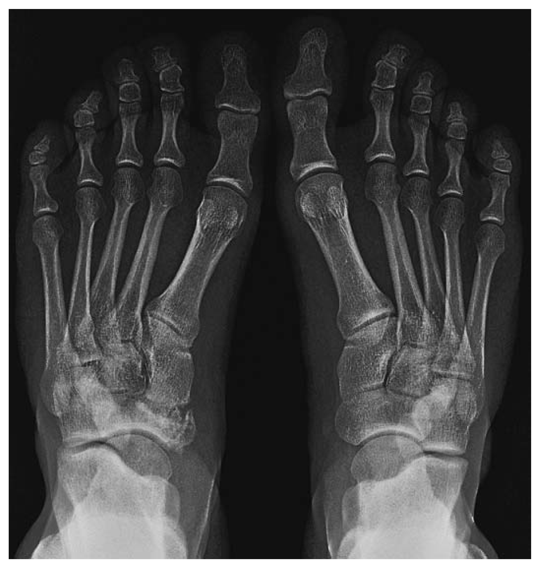
16] was 40° in the injured left foot and 39° in the right foot. The dorsoplantar view (
Fig. 7) revealed signs of post-traumatic arthritis of the naviculocuneiform joint. However, the medial column length (distance from the medial edge of the proximal joint surface of the navicular to the medial edge of the distal articular surface of the first cuneiform) remained unchanged (
Fig. 8). After 18 months, the patient's American Orthopaedic Foot and Ankle Society score reached 85 points (on a scale from 0 to 100).
Figure 7.
Dorsoplantar weightbearing radiograph 18 months postoperative revealed signs of post-traumatic arthritis of the naviculocuneiform joint. However, the medial column length remained unchanged.
Figure 7.
Dorsoplantar weightbearing radiograph 18 months postoperative revealed signs of post-traumatic arthritis of the naviculocuneiform joint. However, the medial column length remained unchanged.
Figure 8.
Lateral weightbearing radiographs 18 months postoperative showed minor differences in the injured left (A) midfoot architecture compared with the contralateral foot (B): there was a difference of 2° in the Moreau-Costa-Bartani angle, 3° in the Meary-Tomeno angle, and 1° in the calcaneal pitch angle.
Figure 8.
Lateral weightbearing radiographs 18 months postoperative showed minor differences in the injured left (A) midfoot architecture compared with the contralateral foot (B): there was a difference of 2° in the Moreau-Costa-Bartani angle, 3° in the Meary-Tomeno angle, and 1° in the calcaneal pitch angle.
Discussion
Different hypotheses have been published trying to clarify the pathophysiologic mechanism for this injury [
5,
13,
17-
19], eg, plantarflexion with hindfoot inversion [
20], dorsiflexion, or isolated forced foot rotation [
14], but it probably consists of a more complex mechanism based on the combination of different displacements. In all of the published case reports in which a tarsal bone dislocation is described [
10,
11], we believe that there probably are other associated bone or capsular-ligamentous injuries that have gone unobserved on imaging studies, and, therefore, after reviewing the published literature, we consider that in these types of injuries a computed tomographic scan is essential to understand the lesion, although in the present case report it was not performed.
Microangiographic studies have revealed that the medial and lateral thirds of the navicular are rich in anastomotic networks but that the central third is avascular [
20], which probably explains the risk of nonunion and avascular necrosis after fracture-dislocation of the navicular [
3,
4,
7,
9,
12,
13,
21]. Many treatment options have been reported. We believe that preserving bone vascularization and avoiding avascular necrosis should be mandatory, and that is why we chose a single medial approach and a less aggressive osteosynthesis such as Kirschner wire fixation avoiding the talonavicular and naviculocuneiform joints. The two Kirschner wires, inserted diverging to plantar, fixed the fracture and also worked as joint stops, thus avoiding recurrence of the dislocation and injury to the joint surface.
In summary, navicular dislocation and fracture-dislocation are highly complex injuries that involve the whole midfoot. After a careful literature review, we think it would be advisable to perform a computed tomographic scan to correctly define the injury. Surgical treatment seems to be mandatory, but it is important not to be too aggressive to avoid bone devascularization and avascular necrosis. Accordingly, we observed that an excellent clinical evolution is possible with a single medial approach as well as using Kirschner wires for fixation and as joint stops at the same time.