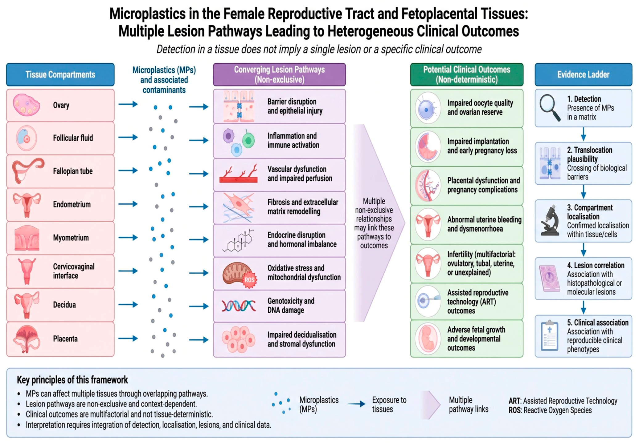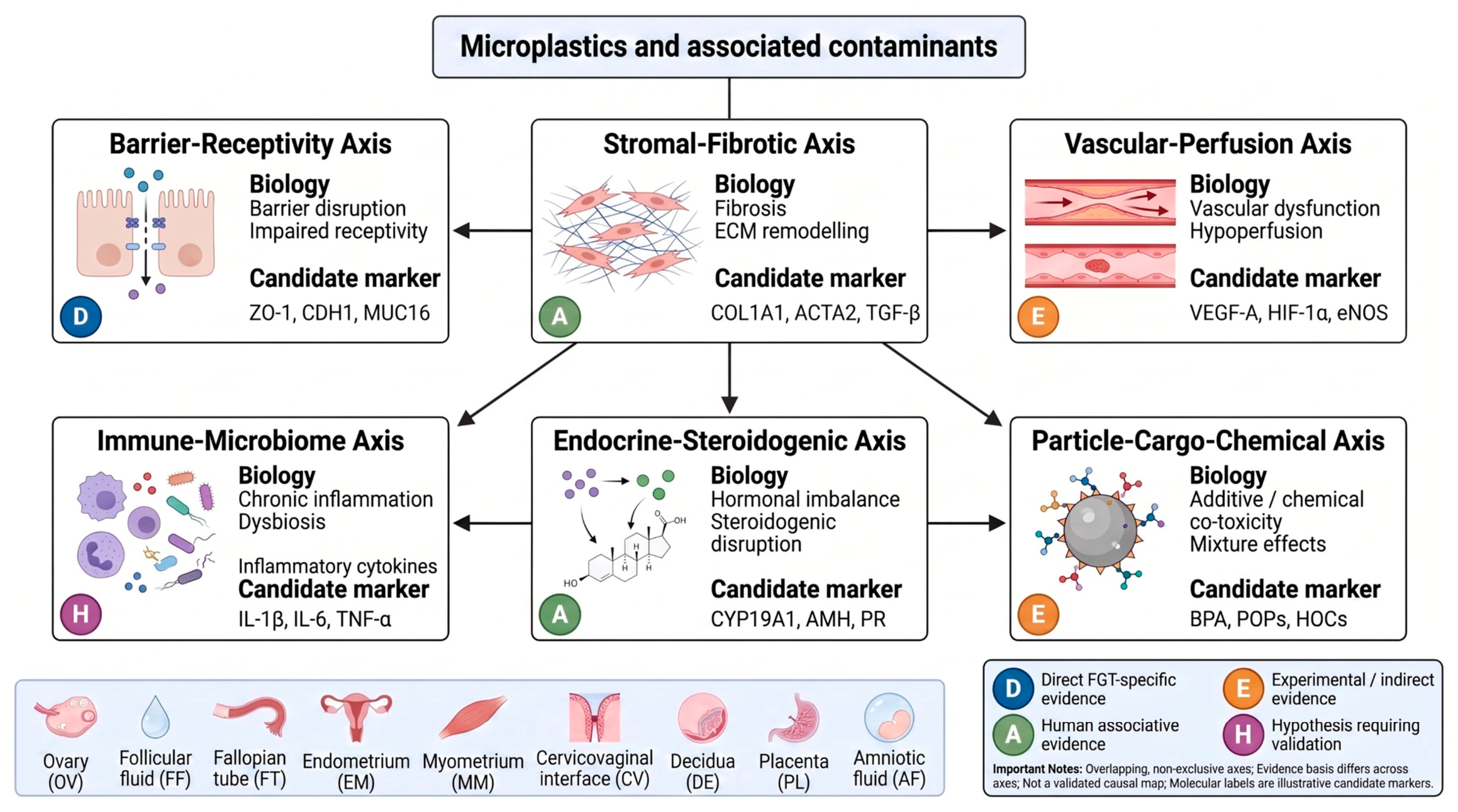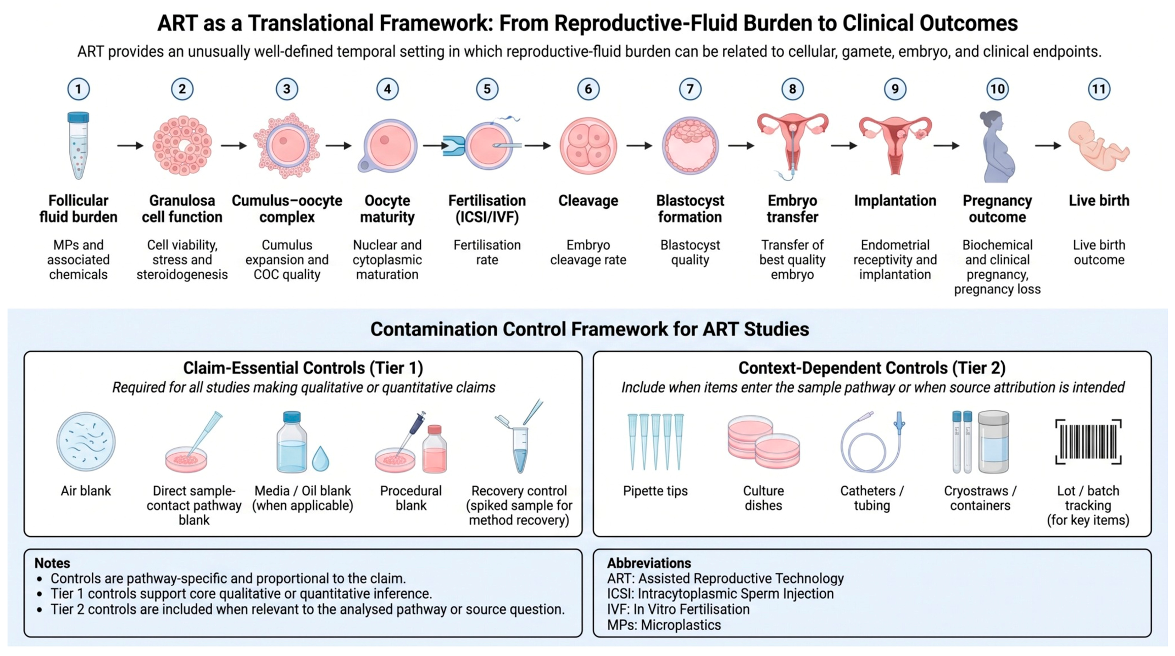

Microplastics in the Female Genital Tract and Fetoplacental Continuum: A Lesion-Based Framework for Pathophysiological Interpretation

 ,
,  , , and
Abstract
1. Introduction
2. Review Design and Evidence Interpretation
Claim-Calibrated Recommendation Grading and Feasibility
3. Exposure Pathways to FGT Compartments
3.1. Systemic Exposure
3.2. Local Mucosal Exposure
3.3. Clinical and Procedural Exposure
4. Reproductive Fluids as Biological Microenvironments
4.1. Functional Relevance of Reproductive Fluids

| Fluid Compartment | Biological Role | Current Primary Evidence | Permissible Interpretation | Principal Limitation |
|---|---|---|---|---|
| Follicular fluid | Oocyte-granulosa-cell microenvironment, steroidogenesis, and oxidative balance. | Human detection and selected clinical or experimental associations [33,38,39]. | Target-compartment exposure with potential for temporally ordered assisted reproductive technology analysis. | Assisted reproductive technology selection, stimulation, small cohorts, and laboratory contamination. |
| Uterine/endometrial fluid | Embryo-endometrial dialogue, receptivity, and implantation signalling. | Indirect support from endometrial-fluid biology and uterine-tissue microplastic research [23,51]. | Implantation-window exposure context when fluid is sampled directly. | Effusion, cycle timing, disease, and the collection procedure may alter the sample. |
| Tubal fluid | Sperm selection, fertilisation, ciliary transport, and early embryo support. | Direct human microplastic-fluid evidence is minimal; diseased tubal-tissue detection has been reported [35,50]. | A justified but currently hypothesis-generating target compartment. | Human sampling is difficult; infection, endometriosis, and surgery are strong confounders. |
| Cervicovaginal fluid/lavage | Lower-tract mucosal, immune, and microbiome interface. | Preliminary human lavage detection [34]. | Local lower-female-genital-tract exposure signal. | Does not establish epithelial uptake, persistence, or upper-tract ascent. |
| Menstrual effluent | Endometrial shedding, uterine-cavity contents, cervicovaginal passage, and product contact. | Direct MP evidence is limited to an exploratory three-sample menstrual-blood series; other studies establish biomarker-matrix feasibility rather than MP occurrence [29,47,48]. | Repeatable non-invasive sampling opportunity. | Very high product, device, clothing, air, and reagent contamination risk. |
| Intervillous blood | Maternal side of placental exchange. | No direct primary intervillous-blood MP study was identified; placental studies provide indirect exposure context [30,31,32,36,42]. | Hypothesis-generating maternal-side exposure context only; direct fluid occurrence remains unestablished. | Delivery-room and procedural contamination and uncertain tissue incorporation. |
| Amniotic fluid | Foetal-interface fluid. | Dual-method detection in 39 of 48 samples with no significant association with immediate outcomes, plus an exploratory five-sample report [29,37]. | Foetal-interface exposure signal. | Does not establish foetal-tissue deposition, developmental harm, or long-term outcome. |
4.2. Follicular Fluid
4.3. Uterine, Endometrial, and Cervicovaginal Fluids
4.4. Tubal Fluid
4.5. Menstrual Blood
4.6. Amniotic Fluid and Intervillous Blood
5. Compartment-Specific FGT Lesion Atlas
5.1. Ovary and Follicular Microenvironment
5.2. Endometrium
5.3. Endometrial Polyps
5.4. Myometrium, Leiomyoma, and Adenomyosis
5.5. Fallopian Tube
5.6. Cervicovaginal Interface
5.7. Decidua and Placenta
6. FGT Lesionome
6.1. Integrated Mechanistic Pathway and Terminology
6.2. Axis-Specific Evidence and Predictions
7. ART as a Human Translational Model
7.1. Value of ART Cohorts
7.2. ART Endpoints
7.3. ART Contamination Controls
8. Claim-Calibrated Methods for Pathology-Ready MP Studies
8.1. Contamination Controls
8.2. Analytical Confirmation
8.3. Spatial Localisation
8.4. Lesion Scoring and Clinical Annotation
9. Target-Tissue Interpretation and Prioritised Research Agenda
10. Conclusions
Supplementary Materials
Author Contributions
Funding
Institutional Review Board Statement
Informed Consent Statement
Data Availability Statement
Acknowledgments
Conflicts of Interest
Abbreviations
References
- Thompson, R.C.; Courtene-Jones, W.; Boucher, J.; Pahl, S.; Raubenheimer, K.; Koelmans, A.A. Twenty years of microplastic pollution research: What have we learned? Science 2024, 386, eadl2746. [Google Scholar] [CrossRef] [Scilit] [PubMed]
- European Commission. Commission Regulation (EU) 2023/2055 of 25 September 2023 amending Annex XVII to Regulation (EC) No 1907/2006 as regards synthetic polymer microparticles. Off. J. Eur. Union 2023, L238, 67–88. [Google Scholar]
- Karak, P.; Parveen, A.; Modak, A.; Adhikari, A.; Chakrabortty, S. Microplastic pollution: A global environmental crisis impacting marine life, human health, and potential innovative sustainable solutions. Int. J. Environ. Res. Public Health 2025, 22, 889. [Google Scholar] [CrossRef] [Scilit] [PubMed]
- Napper, I.E.; Thompson, R.C. Plastic debris in the marine environment: History and future challenges. Glob. Chall. 2020, 4, 1900081. [Google Scholar] [CrossRef] [Scilit] [PubMed]
- Osman, A.I.; Hosny, M.; Eltaweil, A.S.; Omar, S.; Elgarahy, A.M.; Farghali, M.; Yap, P.-S.; Wu, Y.-S.; Nagandran, S.; Batumalaie, K.; et al. Microplastic sources, formation, toxicity and remediation: A review. Environ. Chem. Lett. 2023, 21, 2129–2169. [Google Scholar] [CrossRef] [Scilit] [PubMed]
- Campanale, C.; Massarelli, C.; Savino, I.; Locaputo, V.; Uricchio, V.F. A detailed review study on potential effects of microplastics and additives of concern on human health. Int. J. Environ. Res. Public Health 2020, 17, 1212. [Google Scholar] [CrossRef] [Scilit] [PubMed]
- Guo, M.; Noori, R.; Abolfathi, S. Microplastics in freshwater systems: Dynamic behaviour and transport processes. Resour. Conserv. Recycl. 2024, 205, 107578. [Google Scholar] [CrossRef] [Scilit]
- Issac, M.N.; Kandasubramanian, B. Effect of microplastics in water and aquatic systems. Environ. Sci. Pollut. Res. 2021, 28, 19544–19562. [Google Scholar] [CrossRef] [Scilit] [PubMed]
- Ivleva, N.P. Chemical analysis of microplastics and nanoplastics: Challenges, advanced methods, and perspectives. Chem. Rev. 2021, 121, 11886–11936. [Google Scholar] [CrossRef] [Scilit] [PubMed]
- Pironti, C.; Ricciardi, M.; Motta, O.; Miele, Y.; Proto, A.; Montano, L. Microplastics in the environment: Intake through the food web, human exposure and toxicological effects. Toxics 2021, 9, 224. [Google Scholar] [CrossRef] [Scilit] [PubMed]
- Smith, M.; Love, D.C.; Rochman, C.M.; Neff, R.A. Microplastics in seafood and the implications for human health. Curr. Environ. Health Rep. 2018, 5, 375–386. [Google Scholar] [CrossRef] [Scilit] [PubMed]
- World Health Organization. Dietary and Inhalation Exposure to Nano- and Microplastic Particles and Potential Implications for Human Health; World Health Organization: Geneva, Switzerland, 2022. [Google Scholar]
- Hyman, S.; Kahn, L.G.; Cowell, W. Microplastics and reproductive health: Implications for human health and areas for future research. Hum. Reprod. 2026, 41, 1040–1049. [Google Scholar] [CrossRef] [Scilit] [PubMed]
- Ali-Hassanzadeh, M.; Arefinia, N.; Ghoreshi, Z.-A.S.; Askarpour, H.; Mashayekhi-Sardoo, H. The effects of exposure to microplastics on female reproductive health and pregnancy outcomes: A systematic review and meta-analysis. Reprod. Toxicol. 2025, 135, 108932. [Google Scholar] [CrossRef] [Scilit] [PubMed]
- Medley, E.A.; Spratlen, M.J.; Yan, B.; Herbstman, J.B.; Deyssenroth, M.A. A systematic review of the placental translocation of micro- and nanoplastics. Curr. Environ. Health Rep. 2023, 10, 99–111. [Google Scholar] [CrossRef] [Scilit] [PubMed]
- Balali, H.; Morabbi, A.; Karimian, M. Concerning influences of micro/nanoplastics on female reproductive health: Focusing on cellular and molecular pathways from animal models to human studies. Reprod. Biol. Endocrinol. 2024, 22, 141. [Google Scholar] [CrossRef] [Scilit] [PubMed]
- Hunt, K.; Davies, A.; Fraser, A.; Burden, C.; Howell, A.E.; Buckley, K.; Harding, S.; Bakhbakhi, D. Exposure to microplastics and human reproductive outcomes: A systematic review. BJOG 2024, 131, 675–683. [Google Scholar] [CrossRef] [Scilit] [PubMed]
- Doroftei, B.; Savuca, A.; Cretu, A.-M.; Maftei, R.; Anton, N.; Ilea, C.; Doroftei, M.; Puha, B. Microplastics and human fertility: A comprehensive review of their presence in human samples and reproductive implication. Ecotoxicol. Environ. Saf. 2025, 303, 118939. [Google Scholar] [CrossRef] [Scilit] [PubMed]
- Fitch, S.; Rogers, J.; Marty, S.; Norman, J.; Schneider, S.; Rushton, E.; Wikoff, D.; Ellis-Hutchings, R. Systematic review of potential developmental and reproductive toxicity of microplastics. Toxicol. Sci. 2025, 207, 289–305. [Google Scholar] [CrossRef] [Scilit] [PubMed]
- Inam, Ö. Impact of microplastics on female reproductive health: Insights from animal and human experimental studies: A systematic review. Arch. Gynecol. Obstet. 2025, 312, 77–92. [Google Scholar] [CrossRef] [Scilit] [PubMed]
- Carvajal, B.; Vivero Sun, R.; Piderit, F.; Zazueta, A.; Tapia, C.V.; Gotteland, M.; Magne, F. Microplastics in female reproductive and pregnancy organs: A systematic review. Life 2026, 16, 746. [Google Scholar] [CrossRef] [Scilit] [PubMed]
- Kazmi, S.S.U.H.; Batool, S.M.; Pastorino, P.; Nunes, L.M.; Li, G. Microplastic trafficking in maternal-fetal system: A systematic review and quantitative profiling linking particle characteristics to developmental biology. Crit. Rev. Toxicol. 2026. online ahead of print. [Google Scholar] [CrossRef] [Scilit] [PubMed]
- Qin, X.; Cao, M.; Peng, T.; Shan, H.; Lian, W.; Yu, Y.; Shui, G.; Li, R. Features, potential invasion pathways, and reproductive health risks of microplastics detected in human uterus. Environ. Sci. Technol. 2024, 58, 10482–10493. [Google Scholar] [CrossRef] [Scilit] [PubMed]
- Arcuri, S.; Pennarossa, G.; Bebbere, D.; Gandolfi, F.; Ledda, S.; Brevini, T.A.L. Microplastic exposure induces epithelial barrier alterations and increases collagen deposition in a 3D human endometrial model in vitro. J. Assist. Reprod. Genet. 2025, 42, 3065–3077. [Google Scholar] [CrossRef] [Scilit] [PubMed]
- He, S.; Zhang, Y. Detection and quantification of microplastics in endometrial polyps and their role in polyp formation. Reprod. Toxicol. 2025, 132, 108757. [Google Scholar] [CrossRef] [Scilit] [PubMed]
- Kim, N.; Lee, J.H.; Lee, I.; Park, J.H.; Jung, G.S.; Lee, M.J.; Im, W.; Cho, S.; Choi, Y.S. Investigation of potential toxic effects of nano- and microplastics on human endometrial stromal cells. Reprod. Toxicol. 2025, 132, 108848. [Google Scholar] [CrossRef] [Scilit] [PubMed]
- Xu, H.; Dong, C.; Yu, Z.; Hu, Z.; Yu, J.; Ma, D.; Yao, W.; Qi, X.; Ozaki, Y.; Xie, Y. First identification of microplastics in human uterine fibroids and myometrium. Environ. Pollut. 2024, 360, 124632. [Google Scholar] [CrossRef] [Scilit] [PubMed]
- Xu, H.; Dong, C.; Zhang, B.; Jin, Y.; Cheng, J.; Yu, Z.; Ozaki, Y.; Yu, J.; Ma, D.; Xie, Y. The role of microplastics in elevated risk and tissues metabolic alterations in uterine fibroid of female patients. Environ. Pollut. 2025, 374, 126251. [Google Scholar] [CrossRef] [Scilit] [PubMed]
- Sharma, G.C.; Kumar, N.; Devrukhkar, D.; Joshi, R.M.; Debnath, S.; Chaturvedi, B.; Rajotiya, S.; Mishra, S.; Sharma, A.; Singh, M.; et al. Human reproductive exposure to microplastics: A multi-technique analytical study of menstrual and amniotic fluids. NanoImpact 2026, 42, 100617. [Google Scholar] [CrossRef] [Scilit] [PubMed]
- Wang, P.; Li, G.; Shou, H.; Huang, X.; Xu, H.; Zhang, S.; Zhu, H. Microplastic abundance in placental chorionic villi detected by pyrolysis-gas chromatography/mass spectrometry in cases of spontaneous miscarriage during early pregnancy. eBioMedicine 2025, 120, 105918. [Google Scholar] [CrossRef] [Scilit] [PubMed]
- Feng, Y.; Wu, D.; Guan, Q.; Wang, Q.; Xu, Q.; Li, R.; Tu, C.; Pan, X.; Inman, J.L.; Snijders, A.M.; et al. Multiple types of exogenous microparticles coexist in the early human decidua and placental villi. Environ. Int. 2026, 212, 110283. [Google Scholar] [CrossRef] [Scilit] [PubMed]
- Shen, Y.; Ning, Y.; Chen, F.; Wang, X.; Zheng, D.; Sun, Y.; Zhang, X.; Zhang, X. Impact of placental microplastics on birth anthropometrics: A cross-sectional study. Ecotoxicol. Environ. Saf. 2026, 309, 119572. [Google Scholar] [CrossRef] [Scilit] [PubMed]
- Ni, D.; Yu, K.; Yan, N.; Chen, X.; Xie, Q.; Yang, Y.; Jiang, W.; Yang, Y.; Zhang, J.; Ling, X. Characterization of microplastics in human follicular fluid and assessment of their potential impact on mouse oocyte maturation in vitro. Ecotoxicol. Environ. Saf. 2025, 291, 117796. [Google Scholar] [CrossRef] [Scilit] [PubMed]
- Shim, Y.; Min, H. Identification and characterization of microplastics in human cervicovaginal lavage fluids using Raman spectroscopy: A preliminary study. Life 2025, 15, 357. [Google Scholar] [CrossRef] [Scilit] [PubMed]
- Dong, C.; Xu, H.; Lin, Y.; Zhang, B.; Yu, Z.; Xie, Y.; Yu, J.; Ma, D. Microplastics detected in three types of female reproductive organs using micro-Raman spectroscopy. Ecotoxicol. Environ. Saf. 2024, 285, 117099. [Google Scholar] [CrossRef] [Scilit] [PubMed]
- Garcia, M.A.; Liu, R.; Nihart, A.; El Hayek, E.; Castillo, E.; Barrozo, E.R.; Suter, M.A.; Bleske, B.; Scott, J.; Forsythe, K.; et al. Quantitation and identification of microplastics accumulation in human placental specimens using pyrolysis gas chromatography mass spectrometry. Toxicol. Sci. 2024, 199, 81–88. [Google Scholar] [CrossRef] [Scilit] [PubMed]
- Tian, J.; Liang, L.; Li, Q.; Li, N.; Zhu, X.; Zhang, L. Association between microplastics in human amniotic fluid and pregnancy outcomes: Detection and characterization using Raman spectroscopy and pyrolysis GC/MS. J. Hazard. Mater. 2025, 482, 136637. [Google Scholar] [CrossRef] [Scilit] [PubMed]
- Montano, L.; Raimondo, S.; Piscopo, M.; Ricciardi, M.; Guglielmino, A.; Chamayou, S.; Gentile, R.; Gentile, M.; Rapisarda, P.; Conti, G.O.; et al. First evidence of microplastics in human ovarian follicular fluid: An emerging threat to female fertility. Ecotoxicol. Environ. Saf. 2025, 291, 117868. [Google Scholar] [CrossRef] [Scilit] [PubMed]
- Si, M.; Xiong, X.; Yun, C.; Chen, Y.; Niu, H.; Qu, Y.; Liu, M.; Wang, Y.; Huang, L.; Long, X.; et al. Microplastics and nanoplastics in follicular fluid are associated with diminished ovarian reserve: Clinical and molecular insights. J. Adv. Res. 2026, in press. [Google Scholar] [CrossRef] [Scilit] [PubMed]
- Munno, K.; Lusher, A.L.; Minor, E.C.; Gray, A.; Ho, K.; Hankett, J.; Lee, C.-F.T.; Primpke, S.; McNeish, R.E.; Wong, C.S.; et al. Patterns of microparticles in blank samples: A study to inform best practices for microplastic analysis. Chemosphere 2023, 333, 138883. [Google Scholar] [CrossRef] [Scilit] [PubMed]
- Gruber, E.S.; Karl, V.; Duswald, K.; Bhamidipalli, M.S.; Schlederer, M.; Limberger, T.; Kopatz, V.; Teleky, B.; Kenner, L.; Brandstetter, M. Method for label-free and non-destructive detection of microplastics in human formalin-fixed paraffin-embedded tissue sections. Sci. Rep. 2025, 15, 42637. [Google Scholar] [CrossRef] [Scilit] [PubMed]
- Weingrill, R.B.; Lee, M.-J.; Benny, P.; Riel, J.; Saiki, K.; Garcia, J.; Oliveira, L.F.A.d.M.; Fonseca, E.J.d.S.; de Souza, S.T.; D’AMato, F.d.O.S.; et al. Temporal trends in microplastic accumulation in placentas from pregnancies in Hawai'i. Environ. Int. 2023, 180, 108220. [Google Scholar] [CrossRef] [Scilit] [PubMed]
- Marroquin, J.; Kiomourtzoglou, M.A.; Scranton, A.; Pollack, A.Z. Chemicals in menstrual products: A systematic review. BJOG 2024, 131, 655–664. [Google Scholar] [CrossRef] [Scilit] [PubMed]
- Upson, K.; Shearston, J.A.; Kioumourtzoglou, M.-A. Menstrual products as a source of environmental chemical exposure: A review from the epidemiologic perspective. Curr. Environ. Health Rep. 2022, 9, 38–52. [Google Scholar] [CrossRef] [Scilit] [PubMed]
- Marcelis, Q.; Deconinck, E.; Rogiers, V.; Demaegdt, H.; Di Mario, M.; Vanhaecke, T.; Desmedt, B. Chemical characterization of menstrual and intimate care products: An extractables and leachables investigation. Environ. Int. 2025, 198, 109401. [Google Scholar] [CrossRef] [Scilit] [PubMed]
- Shearston, J.A.; Upson, K.; Gordon, M.; Do, V.; Balac, O.; Nguyen, K.; Yan, B.; Kioumourtzoglou, M.-A.; Schilling, K. Tampons as a source of exposure to metal(loid)s. Environ. Int. 2024, 190, 108849. [Google Scholar] [CrossRef] [Scilit] [PubMed]
- Whitbread, A.L.; Mittelmeier, L.; Rao, R.P.; Mittelmeier, W.; Osmanski-Zenk, K. Menstrual blood as a non-invasive alternative for monitoring vitamin levels. J. Clin. Med. 2024, 13, 7212. [Google Scholar] [CrossRef] [Scilit] [PubMed]
- Warren, L.A.; Shih, A.; Renteira, S.M.; Seckin, T.; Blau, B.; Simpfendorfer, K.; Lee, A.; Metz, C.N.; Gregersen, P.K. Analysis of menstrual effluent: Diagnostic potential for endometriosis. Mol. Med. 2018, 24, 1. [Google Scholar] [CrossRef] [Scilit] [PubMed]
- Kouakou, F.; Denizot, A.-L.; L’Hostis, A.; Colet, J.; Jacques, S.; Sallem, A.; Ziyyat, A.; Vaiman, D.; Wolf, J.-P. Plastic used in in vitro fertilization procedures induces massive placental gene expression alterations. eBioMedicine 2023, 91, 104572. [Google Scholar] [CrossRef] [Scilit] [PubMed]
- Pérez-Cerezales, S.; Ramos-Ibeas, P.; Acuña, O.S.; Avilés, M.; Coy, P.; Rizos, D.; Gutiérrez-Adán, A. The oviduct: From sperm selection to the epigenetic landscape of the embryo. Biol. Reprod. 2018, 98, 262–276. [Google Scholar] [CrossRef] [Scilit] [PubMed]
- Alves, A.R.; Dias, M.F.; Silvestre, M. Endometrial fluid biomarkers and their potential as predictors of successful embryo implantation. Biomedicine 2023, 13, 1. [Google Scholar] [CrossRef] [Scilit] [PubMed]
- Redline, R.W.; Ravishankar, S.; Bagby, C.M.; Saab, S.T.; Zarei, S. Four major patterns of placental injury: A stepwise guide for understanding and implementing the 2016 Amsterdam consensus. Mod. Pathol. 2021, 34, 1074–1092. [Google Scholar] [CrossRef] [Scilit] [PubMed]
- Khong, T.Y.; Mooney, E.E.; Ariel, I.; Balmus, N.C.M.; Boyd, T.K.; Brundler, M.-A.; Derricott, H.; Evans, M.J.; Faye-Petersen, O.M.; Gillan, J.E.; et al. Sampling and definitions of placental lesions: Amsterdam Placental Workshop Group consensus statement. Arch. Pathol. Lab. Med. 2016, 140, 698–713. [Google Scholar] [CrossRef] [Scilit] [PubMed]
- ESHRE Guideline Group on Good Practice in IVF Laboratories. Revised guidelines for good practice in IVF laboratories (2015). Hum. Reprod. 2016, 31, 685–686. [CrossRef] [Scilit] [PubMed]
- Rauert, C.; Charlton, N.; Bagley, A.; Dunlop, S.A.; Symeonides, C.; Thomas, K.V. Assessing the efficacy of pyrolysis–gas chromatography–mass spectrometry for nanoplastic and microplastic analysis in human blood. Environ. Sci. Technol. 2025, 59, 1984–1994. [Google Scholar] [CrossRef] [Scilit] [PubMed]
- Qian, N.; Gao, X.; Lang, X.; Deng, H.; Bratu, T.M.; Chen, Q.; Stapleton, P.; Yan, B.; Min, W. Rapid single-particle chemical imaging of nanoplastics by SRS microscopy. Proc. Natl. Acad. Sci. USA 2024, 121, e2300582121. [Google Scholar] [CrossRef] [Scilit] [PubMed]
- Ao, J.; Xu, G.; Wu, H.; Xie, L.; Liu, J.; Gong, K.; Ruan, X.; Han, J.; Li, K.; Wang, W.; et al. Fast detection and 3D imaging of nanoplastics and microplastics by stimulated Raman scattering microscopy. Cell Rep. Phys. Sci. 2023, 4, 101623. [Google Scholar] [CrossRef] [Scilit]
- European Food Safety Authority. EFSA Scientific Colloquium 25: A coordinated approach to assess the human health risks of micro- and nanoplastics in food. EFSA Support. Publ. 2021, 18, EN-6815. [Google Scholar] [CrossRef] [Scilit]
- EFSA Panel on Contaminants in the Food Chain. Presence of microplastics and nanoplastics in food, with particular focus on seafood. EFSA J. 2016, 14, e04501. [CrossRef] [Scilit] [PubMed]


| Evidence Level | Claim-Specific Minimum Evidentiary Requirement | Supported Inference | Remaining Limit |
|---|---|---|---|
| 1. External exposure opportunity | A source and credible route are documented (ingestion, inhalation, local contact, or procedure-related contact) [2,3,10,11]. | Exposure is plausible. | Does not establish internal access, target-compartment dose, lesion, or disease. |
| 2. Confirmed matrix detection | Polymer identity is confirmed in a defined biological matrix with appropriate blanks and spectral criteria [23,29,33,34,35,36,37,38]. | The polymer signal is present in the sampled matrix. | Does not establish persistent tissue retention, spatial localisation, or injury. |
| 3. Internal or target-compartment burden | Burden is quantified by number, mass, size, and polymer where feasible, with recovery and blank correction [23,27,28,29,30,31,32,33,34,35,36,37,38,39,40]. | Biological access or exposure within a reproductive compartment is supported. | Does not establish cellular uptake or a lesion. |
| 4. Spatial localisation | Polymer-confirmed particles are co-registered with a named anatomical compartment using a validated spatial method [23,35,41,42]. | Anatomical context distinguishes luminal, surface, stromal, vascular, cellular, and lesion-adjacent findings. | Localisation alone is not injury and does not establish clinical causation. |
| 5. Lesion association | Burden or localisation is linked to a prespecified, reproducible histological, cellular, or molecular response in the same compartment [24,25,26]. | A lesion-relevant association is supported. | Causality still requires temporality, exposure-response, alternative-explanation control, and replication. |
| 6. Clinical association | A prespecified, clinically annotated reproductive or pregnancy outcome is analysed with effect estimates, temporal information, and confounder control; prospective design strengthens temporality but is not a universal prerequisite for association [32,37,38,39]. | The finding is clinically associated with the measured endpoint. | Selection, multiple testing, reverse causation, residual confounding, analytical heterogeneity, and absent independent replication may remain. |
| 7. Causal inference | Exposure precedes outcome; target dose, spatial concordance, lesion response, exposure-response, contamination control, confounder control, and independent replication converge. | A causal interpretation may become supportable. | This level has not been established for human female genital tract or fetoplacental disease. |
| Compartment | Candidate Lesion Axis (Non-Exhaustive; Evidence Status Varies) | Potential Clinical Endpoints (Hypothesis-Specific) | Main Confounders or Limitations | Claim-Contingent Priority and Feasibility |
|---|---|---|---|---|
| Ovary, follicular fluid, granulosa/cumulus cells | Oxidative and mitochondrial stress, steroidogenic dysfunction, altered cumulus-oocyte signalling, and impaired oocyte maturation. | Anti-Müllerian hormone, antral follicle count, follicle-stimulating hormone, oocyte yield, metaphase-II rate, fertilisation, cleavage, blastocyst formation, implantation, miscarriage, and live birth. | Infertility diagnosis, age, stimulation protocol, embryo selection, and assisted-reproductive-technology laboratory particles. | Tier 1 occurrence: polymer confirmation, matched assisted-reproductive-technology blanks, size-resolved burden, and recovery for quantitative comparisons. Tier 2: paired fluid/cells and one prespecified oocyte or embryo endpoint. Tier 3: broad molecular panels or the full sequence to live birth [33,38,39]. |
| Endometrium | Barrier dysfunction, impaired receptivity or decidualisation, inflammation, and stromal remodelling. | Implantation failure, pregnancy loss, abnormal bleeding, and unexplained infertility. | Cycle phase, infection, hormonal exposure, bulk analysis, and absent spatial co-localisation. | Tier 1 lesion claim: spatial confirmation, cycle and hormonal context, and a prespecified blinded lesion score. Tier 2: one hypothesis-specific receptivity, decidualisation, or fibrosis measure. Tier 3: broad marker and outcome panels [23,24,26]. |
| Endometrial polyp | Stromal proliferation and migration, PI3K/AKT signalling, vascular change, inflammation, and matrix remodelling. | Bleeding, polyp recurrence, and implantation outcome. | Cross-sectional association, age, hormones, and disease-related particle trapping. | Tier 1 disease comparison: matched polyp, adjacent endometrium, and control tissue with identical processing, mapping, and lesion scoring. Tier 2: bleeding or recurrence. Tier 3: implantation and broad pathway panels [25]. |
| Myometrium, leiomyoma, adenomyosis | Smooth-muscle or stromal oxidative stress, extracellular-matrix remodelling, fibrosis, inflammation, and vascular change. | Bleeding, pain, lesion burden, and fertility outcomes. | Diseased tissue, fibrosis-related trapping, prior treatment, and procedural contamination. | Tier 1 disease comparison: identically processed lesional, adjacent, and independent controls with procedural blanks. Tier 2: matrix or vascular scoring. Tier 3: longitudinal symptom or fertility outcomes [27,28,35]. |
| Fallopian tube | Ciliary and secretory-cell injury, mucosal inflammation, hydrosalpinx-wall remodelling, fibrosis, and altered transport. | Tubal-factor infertility, hydrosalpinx, chronic salpingitis; ectopic pregnancy remains a hypothesis. | Infection, pelvic inflammatory disease, endometriosis, surgery, and under-sampling. | Tier 1 occurrence or disease comparison: exact compartment, surgical blanks, and infection or endometriosis data. Tier 2: ciliary and lesion scoring and paired fluid where feasible. Tier 3: transport or ectopic-pregnancy outcomes [35,50]. |
| Cervicovaginal interface | Barrier alteration, epithelial inflammation, microbiome disturbance, and mucosal immune activation. | Local inflammation, altered microbiome, and product-related exposure. | Lavage dilution, recent product or sexual exposure, device composition, and no tissue mapping. | Tier 1 occurrence: dilution, device composition, exposure history, and matched blanks. Tier 2: epithelium and pH. Tier 3: microbiome and immune profiling [34]. |
| Decidua and fetoplacental interface | Trophoblast stress, decidual arteriopathy, maternal/foetal vascular malperfusion, Hofbauer-cell response, inflammation, and fibrin deposition. | Miscarriage, foetal growth, preterm birth, hypertensive disorders, placental insufficiency, and neonatal outcome. | Multifactorial lesions, delivery contamination, gestational age, maternal disease, and uncertain localisation. | Tier 1 incorporation or lesion claim: delivery blanks, compartment mapping, and recognised sampling and lesion terminology. Tier 2: paired matrices and one prespecified pregnancy outcome. Tier 3: broad mechanistic or long-term neonatal panels [29,30,31,32,36,37,42,52,53]. |
| Reporting Domain | Claim-Specific Priority and Feasibility | Purpose and Interpretive Value |
|---|---|---|
| Specimen and anatomical site | Tier 1 for all claims; routine. Specify tissue or fluid, exact anatomical compartment, sampling route, and whether material is lesional, adjacent non-lesional, control, luminal, surface, or fluid. | Defines the biological target and prevents dissimilar matrices from being interpreted as equivalent [23,35,53]. |
| Physiological and clinical context | Tier 1 for lesion or clinical claims; routine. Record only hypothesis-relevant variables, including age, menstrual phase or gestational age, assisted reproductive technology setting, diagnosis, hormonal treatment, and procedure. | Allows the burden and lesion to be interpreted in the correct biological and clinical state. |
| Sampling and contamination control | Core air or field, container or reagent, and pathway-specific procedural blanks are Tier 1 for occurrence claims; routine to specialised. Recovery is Tier 1 for quantitative burden or exposure-response claims. Context-specific assisted-reproductive-technology, delivery, surgery, or formalin-fixed paraffin-embedded controls are added only when that pathway is used; correction or exclusion rules are prespecified. | Identifies ambient, material, procedural, and analytical contributions and prevents post hoc handling of contamination [40]. |
| Analytical platform and polymer confirmation | Tier 1 for all microplastic claims; specialised. Use a polymer-specific method with spectral criteria and library details. Orthogonal confirmation is Tier 2 when spectra are ambiguous or claims are high consequence; visual microscopy alone is insufficient. | Establishes that the signal is polymeric and defines the method’s size, mass, and spatial capabilities [9,41,55,56,57]. |
| Particle characteristics and dose | Number or concentration, size, morphology, polymer, and unit normalisation are Tier 1 for quantitative comparisons; mass and recovery are added where the platform permits. Weathering, surface chemistry, and cargo are Tier 3 unless the mechanism specifically depends on them. | Permits comparison, exposure-response analysis, and distinction between particle-count and polymer-mass evidence [27,28,29,30,31,32,33,34,35,36,37,38,39]. |
| Spatial localisation | Tier 1 for localisation or lesion claims, but not for descriptive matrix occurrence; specialised or research-intensive. State spatial resolution and registration method. | Converts bulk presence into anatomical context; localisation remains distinct from injury [23,41,42,53]. |
| Lesion scoring and biological response | Tier 1 for lesion claims; routine to specialised once tissue is available. Use a limited prespecified, preferably blinded lesion score. Molecular markers are Tier 2 or Tier 3 unless directly supported by the stated hypothesis. | Tests whether burden or localisation aligns with a reproducible response rather than an assumed mechanism [24,25,26,52]. |
| Clinical outcomes, temporality, and confounders | Tier 1 for clinical or causal claims; feasibility varies from routine annotation to research-intensive longitudinal follow-up. Prespecify the primary outcome, effect estimate, timing, missing data, and a limited confounder set. Prospective design strengthens temporality but is not a universal prerequisite for association. Broad outcome panels are exploratory and require multiplicity control. | Supports bounded clinical inference and makes residual confounding, selection, temporality, and causal limitations explicit [30,32,37,38,39]. |
Disclaimer/Publisher’s Note: The statements, opinions and data contained in all publications are solely those of the individual author(s) and contributor(s) and not of MDPI and/or the editor(s). MDPI and/or the editor(s) disclaim responsibility for any injury to people or property resulting from any ideas, methods, instructions or products referred to in the content. |
© 2026 by the authors. Licensee MDPI, Basel, Switzerland. This article is an open access article distributed under the terms and conditions of the Creative Commons Attribution (CC BY) license.
Share and Cite
Das, S.K.; Karak, P.; Parveen, A.; Das, S.N.; Nerune, S.M. Microplastics in the Female Genital Tract and Fetoplacental Continuum: A Lesion-Based Framework for Pathophysiological Interpretation. Pathophysiology 2026, 33, 66. https://doi.org/10.3390/pathophysiology33030066
Das SK, Karak P, Parveen A, Das SN, Nerune SM. Microplastics in the Female Genital Tract and Fetoplacental Continuum: A Lesion-Based Framework for Pathophysiological Interpretation. Pathophysiology. 2026; 33(3):66. https://doi.org/10.3390/pathophysiology33030066
Chicago/Turabian StyleDas, Sayandeep K., Prithviraj Karak, Afsona Parveen, Swastika N. Das, and Savitri M. Nerune. 2026. "Microplastics in the Female Genital Tract and Fetoplacental Continuum: A Lesion-Based Framework for Pathophysiological Interpretation" Pathophysiology 33, no. 3: 66. https://doi.org/10.3390/pathophysiology33030066
APA StyleDas, S. K., Karak, P., Parveen, A., Das, S. N., & Nerune, S. M. (2026). Microplastics in the Female Genital Tract and Fetoplacental Continuum: A Lesion-Based Framework for Pathophysiological Interpretation. Pathophysiology, 33(3), 66. https://doi.org/10.3390/pathophysiology33030066