

MRI Voxel Morphometry Shows Brain Volume Changes in Breast Cancer Survivors: Implications for Treatment

 ,
,  ,
,  , , ,
, , , 
Abstract
1. Introduction
2. Materials and Methods
2.1. Patients and Healthy Volunteers
2.2. MRI Study
2.3. Statistical Analysis
3. Results
3.1. Patient Characteristics
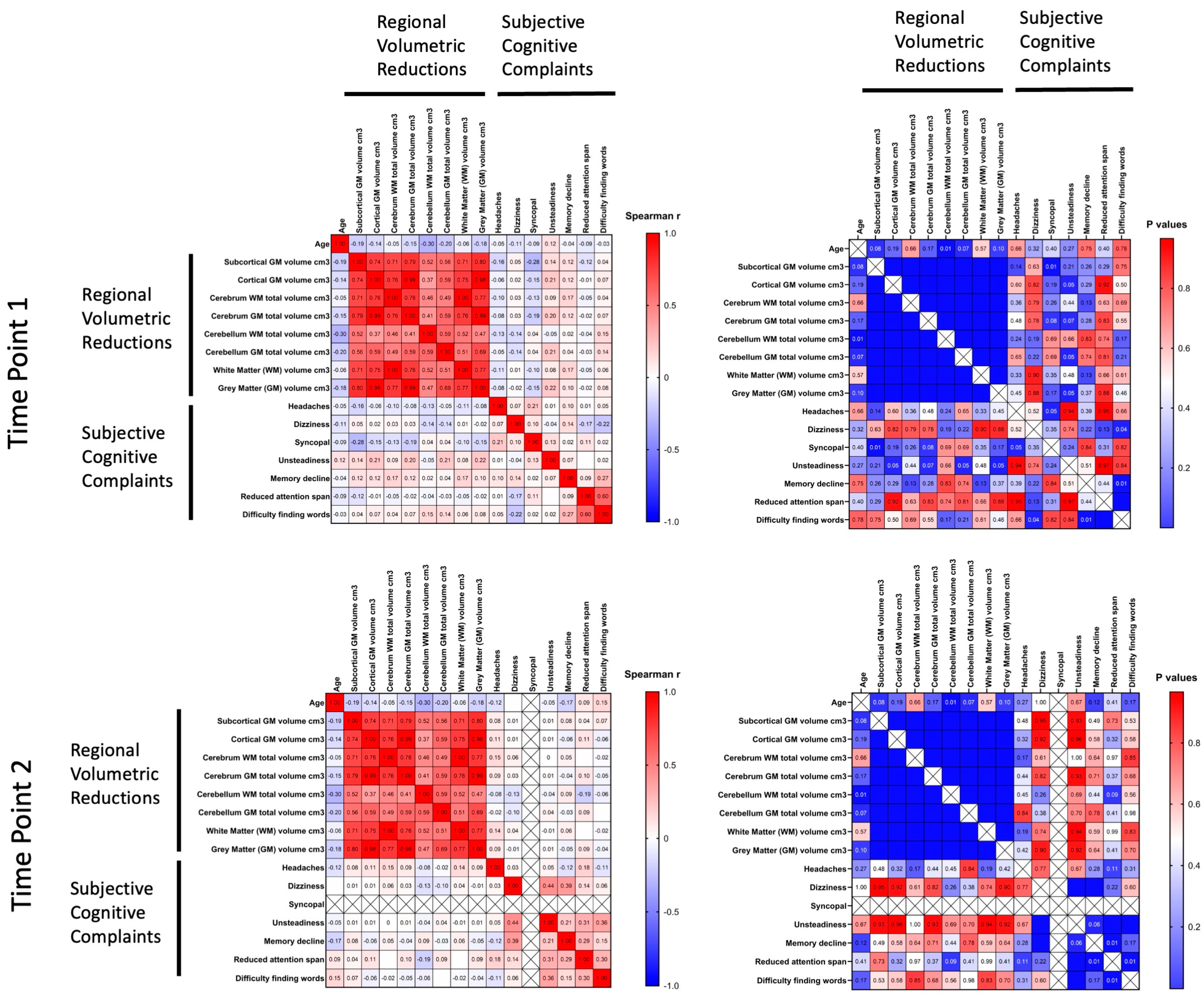
3.2. MRI Voxel Morphometry
4. Discussion
5. Conclusions
Author Contributions
Funding
Institutional Review Board Statement
Informed Consent Statement
Data Availability Statement
Conflicts of Interest
Abbreviations
| AC | Cyclophosphamide and Adriamycin (chemotherapy regimen) |
| CAF | Cyclophosphamide, Adriamycin, and Fluorouracil (chemotherapy regimen) |
| CAP | Cyclophosphamide and Adriamycin (chemotherapy regimen) |
| CNS | Central Nervous System |
| DOC | Docetaxel/Paclitaxel (chemotherapy agents) |
| DWI | Diffusion-Weighted Imaging |
| FAC | Fluorouracil, Adriamycin, and Cyclophosphamide (chemotherapy regimen) |
| GM | Gray Matter |
| ICAM-1 | Intercellular Adhesion Molecule 1 |
| MPRAGE | Magnetization Prepared Rapid Acquisition Gradient Echo |
| MRI | Magnetic Resonance Imaging |
| PECAM-1 | Platelet Endothelial Cell Adhesion Molecule 1 |
| PMES | Post-Mastectomy Syndrome |
| T1WI | T1 weighted image |
| TIRM | Turbo Inversion Recovery Magnitude |
| VolBrain | Automated MRI Brain Volumetric System |
| WM | White Matter |
References
- Cancer Today. Available online: https://gco.iarc.who.int/today/en/dataviz/pie?mode=population&group_populations=0&cancers=20&populations=100_112_191_196_203_208_233_246_250_276_300_348_352_372_380_40_428_440_442_470_498_499_528_56_578_616_620_642_643_688_70_703_705_724_752_756_8_804_807_826 (accessed on 13 August 2024).
- Breast Cancer Treatment (PDQ®)–NCI. Available online: https://www.cancer.gov/types/breast/hp/breast-treatment-pdq (accessed on 4 October 2024).
- Lee, E.Q. Neurologic Complications of Cancer Therapies. Curr. Neurol. Neurosci. Rep. 2021, 21, 66. [Google Scholar] [CrossRef]
- Fleming, B.; Edison, P.; Kenny, L. Cognitive Impairment after Cancer Treatment: Mechanisms, Clinical Characterization, and Management. BMJ 2023, 380, e071726. [Google Scholar] [CrossRef]
- Onzi, G.R.; D’Agustini, N.; Garcia, S.C.; Guterres, S.S.; Pohlmann, P.R.; Rosa, D.D.; Pohlmann, A.R. Chemobrain in Breast Cancer: Mechanisms, Clinical Manifestations, and Potential Interventions. Drug Saf. 2022, 45, 601–621. [Google Scholar] [CrossRef]
- Pinter, N.K.; Fritz, J.V. Neuroimaging for the Neurologist: Clinical MRI and Future Trends. Neurol. Clin. 2020, 38, 1–35. [Google Scholar] [CrossRef]
- Yousaf, T.; Dervenoulas, G.; Politis, M. Advances in MRI Methodology. Int. Rev. Neurobiol. 2018, 141, 31–76. [Google Scholar] [CrossRef]
- Deelchand, D.K.; Henry, P.G.; Joers, J.M.; Auerbach, E.J.; Park, Y.W.; Kara, F.; Ratai, E.M.; Kantarci, K.; Öz, G. Plug-and-Play Advanced Magnetic Resonance Spectroscopy. Magn. Reson. Med. 2022, 87, 2613. [Google Scholar] [CrossRef]
- Yao, S.; Zhang, Q.; Yao, X.; Zhang, X.; Pang, L.; Yu, S.; Cheng, H. Advances of Neuroimaging in Chemotherapy Related Cognitive Impairment (CRCI) of Patients with Breast Cancer. Breast Cancer Res. Treat. 2023, 201, 15–26. [Google Scholar] [CrossRef]
- Bukkieva, T.; Pospelova, M.; Efimtsev, A.; Fionik, O.; Alekseeva, T.; Samochernykh, K.; Gorbunova, E.; Krasnikova, V.; Makhanova, A.; Nikolaeva, A.; et al. Microstructural Properties of Brain White Matter Tracts in Breast Cancer Survivors: A Diffusion Tensor Imaging Study. Pathophysiology 2022, 29, 595–609. [Google Scholar] [CrossRef]
- Nikolaeva, A.; Pospelova, M.; Krasnikova, V.; Makhanova, A.; Tonyan, S.; Krasnopeev, Y.; Kayumova, E.; Vasilieva, E.; Efimtsev, A.; Levchuk, A.; et al. Elevated Levels of Serum Biomarkers Associated with Damage to the CNS Neurons and Endothelial Cells Are Linked with Changes in Brain Connectivity in Breast Cancer Patients with Vestibulo-Atactic Syndrome. Pathophysiology 2023, 30, 260–274. [Google Scholar] [CrossRef]
- Goto, M.; Abe, O.; Hagiwara, A.; Fujita, S.; Kamagata, K.; Hori, M.; Aoki, S.; Osada, T.; Konishi, S.; Masutani, Y.; et al. Advantages of Using Both Voxel- and Surface-Based Morphometry in Cortical Morphology Analysis: A Review of Various Applications. Magn. Reson. Med. Sci. 2022, 21, 41–57. [Google Scholar] [CrossRef]
- Nemoto, K. Understanding Voxel-Based Morphometry. Brain Nerve 2017, 69, 505–511. [Google Scholar] [CrossRef] [PubMed]
- Huang, H.; Zheng, S.; Yang, Z.; Wu, Y.; Li, Y.; Qiu, J.; Cheng, Y.; Lin, P.; Lin, Y.; Guan, J.; et al. Voxel-Based Morphometry and a Deep Learning Model for the Diagnosis of Early Alzheimer's Disease Based on Cerebral Gray Matter Changes. Cereb. Cortex 2023, 33, 754–763. [Google Scholar] [CrossRef] [PubMed]
- Sakurai, K.; Kaneda, D.; Morimoto, S.; Uchida, Y.; Inui, S.; Kimura, Y.; Kan, H.; Kato, T.; Ito, K.; Hashizume, Y. Voxel-Based and Surface-Based Morphometry Analysis in Patients with Pathologically Confirmed Argyrophilic Grain Disease and Alzheimer's Disease. J. Alzheimer's Dis. 2023, 93, 379–387. [Google Scholar] [CrossRef]
- Kotikalapudi, R.; Martin, P.; Marquetand, J.; Lindig, T.; Bender, B.; Focke, N.K. Systematic Assessment of Multispectral Voxel-Based Morphometry in Previously MRI-Negative Focal Epilepsy. AJNR Am. J. Neuroradiol. 2018, 39, 2014–2021. [Google Scholar] [CrossRef] [PubMed]
- Li, C.; Liu, W.; Guo, F.; Wang, X.; Kang, X.; Xu, Y.; Xi, Y.; Wang, H.; Zhu, Y.; Yin, H. Voxel-Based Morphometry Results in First-Episode Schizophrenia: A Comparison of Publicly Available Software Packages. Brain Imaging Behav. 2020, 14, 2224–2231. [Google Scholar] [CrossRef]
- Shao, H.; Li, N.; Chen, M.; Zhang, J.; Chen, H.; Zhao, M.; Yang, J.; Xia, J. A Voxel-Based Morphometry Investigation of Brain Structure Variations in Late-Life Depression with Insomnia. Front. Psychiatry 2023, 14, 1201256. [Google Scholar] [CrossRef]
- Kornelsen, J.; McIver, T.; Uddin, M.N.; Figley, C.R.; Marrie, R.A.; Patel, R.; Fisk, J.D.; Carter, S.; Graff, L.; Mazerolle, E.L.; et al. Altered Voxel-Based and Surface-Based Morphometry in Inflammatory Bowel Disease. Brain Res. Bull. 2023, 203, 110771. [Google Scholar] [CrossRef]
- Hatchard, T.; Penta, S.; Mioduzsewski, O.; Correia, S.; Tissera, T.; Brown, O.; Haefner, S.A.; Poulin, P.; Smith, A.M. Increased Gray Matter Following Mindfulness-Based Stress Reduction in Breast Cancer Survivors with Chronic Neuropathic Pain: Preliminary Evidence Using Voxel-Based Morphometry. Acta Neurol. Belg. 2022, 122, 735–743. [Google Scholar] [CrossRef]
- de Ruiter, M.B.; Deardorff, R.L.; Blommaert, J.; Chen, B.T.; Dumas, J.A.; Schagen, S.B.; Sunaert, S.; Wang, L.; Cimprich, B.; Peltier, S.; et al. Brain Gray Matter Reduction and Premature Brain Aging after Breast Cancer Chemotherapy: A Longitudinal Multicenter Data Pooling Analysis. Brain Imaging Behav. 2023, 17, 507–518. [Google Scholar] [CrossRef]
- De Ruiter, M.B.; Reneman, L.; Boogerd, W.; Veltman, D.J.; Caan, M.; Douaud, G.; Lavini, C.; Linn, S.C.; Boven, E.; Van Dam, F.S.A.M.; et al. Late Effects of High-Dose Adjuvant Chemotherapy on White and Gray Matter in Breast Cancer Survivors: Converging Results from Multimodal Magnetic Resonance Imaging. Hum. Brain Mapp. 2012, 33, 2971–2983. [Google Scholar] [CrossRef]
- McDonald, B.C.; Saykin, A.J. Alterations in Brain Structure Related to Breast Cancer and Its Treatment: Chemotherapy and Other Considerations. Brain Imaging Behav. 2013, 7, 374–387. [Google Scholar] [CrossRef] [PubMed]
- McDonald, B.C.; Van Dyk, K.; Deardorff, R.L.; Bailey, J.N.; Zhai, W.; Carroll, J.E.; Root, J.C.; Ahles, T.A.; Mandelblatt, J.S.; Saykin, A.J. Multimodal MRI Examination of Structural and Functional Brain Changes in Older Women with Breast Cancer in the First Year of Antiestrogen Hormonal Therapy. Breast Cancer Res. Treat. 2022, 194, 113–126. [Google Scholar] [CrossRef] [PubMed]
- Coupé, P.; Mansencal, B.; Clément, M.; Giraud, R.; Denis de Senneville, B.; Ta, V.T.; Lepetit, V.; Manjon, J.V. AssemblyNet: A Large Ensemble of CNNs for 3D Whole Brain MRI Segmentation. Neuroimage 2020, 219, 117026. [Google Scholar] [CrossRef]
- Manjón, J.V.; Coupé, P. VolBrain: An Online MRI Brain Volumetry System. Front. Neuroinform 2016, 10, 30. [Google Scholar] [CrossRef]
- Romero, J.E.; Manjón, J.V.; Tohka, J.; Coupé, P.; Robles, M. NABS: Non-Local Automatic Brain Hemisphere Segmentation. Magn. Reson. Imaging 2015, 33, 474–484. [Google Scholar] [CrossRef]
- Manjón, J.V.; Eskildsen, S.F.; Coupé, P.; Romero, J.E.; Collins, D.L.; Robles, M. Nonlocal Intracranial Cavity Extraction. Int. J. Biomed. Imaging 2014, 2014, 820205. [Google Scholar] [CrossRef] [PubMed]
- Motulsky, H.J.; Brown, R.E. Detecting Outliers When Fitting Data with Nonlinear Regression—A New Method Based on Robust Nonlinear Regression and the False Discovery Rate. BMC Bioinform. 2006, 7, 123. [Google Scholar] [CrossRef]
- Dunnett, C.W. A Multiple Comparison Procedure for Comparing Several Treatments with a Control. J. Am. Stat. Assoc. 1955, 50, 1096–1121. [Google Scholar] [CrossRef]
- Ben-Shachar, M.S.; Lüdecke, D.; Makowski, D. Effectsize: Estimation of Effect Size Indices and Standardized Parameters. J. Open Source Softw. 2020, 5, 2815. [Google Scholar] [CrossRef]
- Coupé, P.; Manjón, J.V.; Fonov, V.; Pruessner, J.; Robles, M.; Collins, D.L. Patch-Based Segmentation Using Expert Priors: Application to Hippocampus and Ventricle Segmentation. Neuroimage 2011, 54, 940–954. [Google Scholar] [CrossRef]
- Varma, D.R. Managing DICOM Images: Tips and Tricks for the Radiologist. Indian J. Radiol. Imaging 2012, 22, 4–13. [Google Scholar] [CrossRef] [PubMed]
- Li, X.; Morgan, P.S.; Ashburner, J.; Smith, J.; Rorden, C. The First Step for Neuroimaging Data Analysis: DICOM to NIfTI Conversion. J. Neurosci. Methods 2016, 264, 47–56. [Google Scholar] [CrossRef]
- Bachmann, T.; Schroeter, M.L.; Chen, K.; Reiman, E.M.; Weise, C.M. Longitudinal Changes in Surface Based Brain Morphometry Measures in Amnestic Mild Cognitive Impairment and Alzheimer's Disease. Neuroimage Clin. 2023, 38, 103371. [Google Scholar] [CrossRef]
- Chen, B.T.; Chen, Z.; Deng, F.; Patel, S.K.; Sedrak, M.S.; Root, J.C.; Ahles, T.A.; Razavi, M.; Kim, H.; Sun, C.L.; et al. Signal Variability and Cognitive Function in Older Long-Term Survivors of Breast Cancer with Exposure to Chemotherapy: A Prospective Longitudinal Resting-State FMRI Study. Brain Sci. 2022, 12, 1283. [Google Scholar] [CrossRef]
- Banker, L.; Tadi, P. Neuroanatomy, Precentral Gyrus. In StatPearls [Internet]; StatPearls Publishing: Treasure Island, FL, USA, 2023. [Google Scholar]
- Silva, A.B.; Liu, J.R.; Zhao, L.; Levy, D.F.; Scott, T.L.; Chang, E.F. A Neurosurgical Functional Dissection of the Middle Precentral Gyrus during Speech Production. J. Neurosci. 2022, 42, 8416–8426. [Google Scholar] [CrossRef]
- El-Baba, R.M.; Schury, M.P. Neuroanatomy, Frontal Cortex. In StatPearls [Internet]; StatPearls Publishing: Treasure Island, FL, USA, 2023. [Google Scholar]
- Jimsheleishvili, S.; Dididze, M. Neuroanatomy, Cerebellum. In StatPearls [Internet]; StatPearls Publishing: Treasure Island, FL, USA, 2023. [Google Scholar]
- Guell, X.; Schmahmann, J.D.; Gabrieli, J.D.E.; Ghosh, S.S. Functional Gradients of the Cerebellum. eLife 2018, 7, e36652. [Google Scholar] [CrossRef] [PubMed]
- Sankey, E.W.; Srinivasan, E.S.; Mehta, V.A.; Bergin, S.M.; Wang, T.Y.; Thompson, E.M.; Fecci, P.E.; Friedman, A.H. Perioperative Assessment of Cerebellar Masses and the Potential for Cerebellar Cognitive Affective Syndrome. World Neurosurg. 2020, 144, 222–230. [Google Scholar] [CrossRef] [PubMed]
- Hassan, H.; Ehsanula, H.; Pattanshetti, M. Stroke of the Inferiomedial Temporal Lobe Causing Word Agnosia. Case Rep. 2017, 2017, bcr-2015-214184. [Google Scholar] [CrossRef]
- Carretero, R.G.; Beamonte-Vela, B.N.; Silvano-Cocinero, J.D.; Alvarez-Mendez, A. Behavioural Changes as the First Manifestation of a Silent Frontal Lobe Stroke. BMJ Case Rep. CP 2019, 12, bcr-2018-227617. [Google Scholar] [CrossRef]
- Daniel, E.; Deng, F.; Patel, S.K.; Sedrak, M.S.; Kim, H.; Razavi, M.; Sun, C.-L.; Root, J.C.; Ahles, T.A.; Dale, W.; et al. Altered Gyrification in Chemotherapy-Treated Older Long-Term Breast Cancer Survivors. Res. Sq. 2024, 14, e3634. [Google Scholar] [CrossRef]
- Inagaki, M.; Yoshikawa, E.; Matsuoka, Y.; Sugawara, Y.; Nakano, T.; Akechi, T.; Wada, N.; Imoto, S.; Murakami, K.; Uchitomi, Y.; et al. Smaller Regional Volumes of Brain Gray and White Matter Demonstrated in Breast Cancer Survivors Exposed to Adjuvant Chemotherapy. Cancer 2007, 109, 146–156. [Google Scholar] [CrossRef] [PubMed]
- McDonald, B.C.; Conroy, S.K.; Ahles, T.A.; West, J.D.; Saykin, A.J. Gray Matter Reduction Associated with Systemic Chemotherapy for Breast Cancer: A Prospective MRI Study. Breast Cancer Res. Treat. 2010, 123, 819–828. [Google Scholar] [CrossRef] [PubMed]
- Henneghan, A.; Rao, V.; Harrison, R.A.; Karuturi, M.; Blayney, D.W.; Palesh, O.; Kesler, S.R. Cortical Brain Age from Pre-Treatment to Post-Chemotherapy in Patients with Breast Cancer. Neurotox. Res. 2020, 37, 788–799. [Google Scholar] [CrossRef] [PubMed]
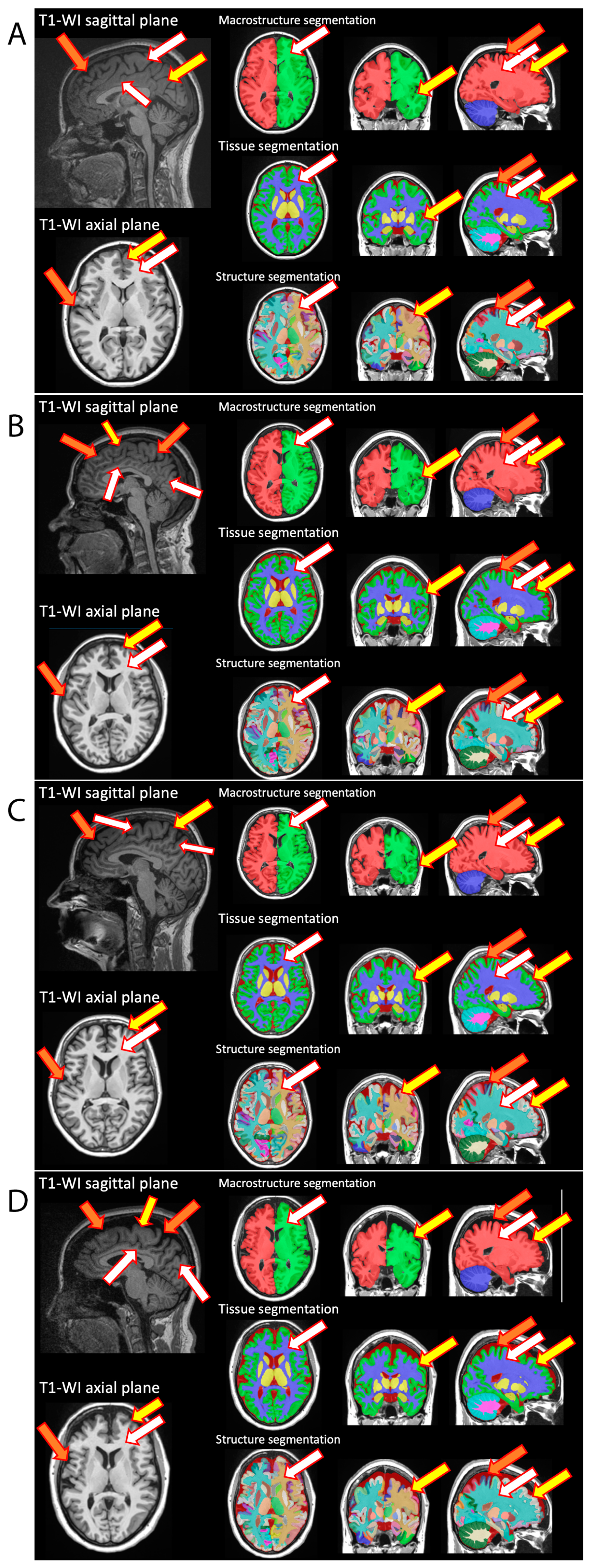

| T2_tra | T2_tirm | DWI | T2_cor | T1_MPRAGE | |
|---|---|---|---|---|---|
| Repetition time/TR | 3970.0 ms | 9000.0 ms | 2800.0 ms | 3500.0 ms | 2300 ms |
| Echo time/TE | 95.00 ms | 96.0 ms | 79.00 ms | 95.00 ms | 2.98 ms |
| FoV | 220 mm | 220 mm | 220 mm | 220 mm | 256 mm |
| Slice thickness | 4.0 mm | 4.0 mm | 3.0 mm | 4.0 mm | 1.2 mm |
| Voxel size × (mm), y (mm) | 0.4 × 0.4 × 4.0 mm | 0.7 × 0.7 × 4.0 mm | 1.7 × 1.7 × 3.0 mm | 0.2 × 0.2 × 4.0 mm | 1.0 × 1.0 × 1.1 mm |
| Study time | 2:05 | 3:56 | 3:37 | 2:01 | 5:12 |
| Patients (n) | Age (years) | Scope of Surgery | Chemotherapy | Hormone Therapy | Radiotherapy | |||||||
|---|---|---|---|---|---|---|---|---|---|---|---|---|
| According to Madden | Sectoral Resection | Subcutaneous Mastectomy with Single-Stage Mammoplasty | FAC | DOC | AC | CAP | CAF | Tamoxifen | ||||
| Patients | 86 (100%) | 43.27 ± 4.38 | 49 (57.0%) | 24 (27.9%) | 13 (15.1%) | 6 (6.9%) | 60 (69.7%) | 27 (31.3%) | 1 (1.16%) | 6 (6.98%) | 51 (59.3) | 57 (66.2%) |
| Healthy volunteers | 28 | 44 ± 5.68 | - | - | - | - | - | - | - | - | - | - |
| Treatment Combination | Number of Patients | Percentage (%) |
|---|---|---|
| Chemotherapy + Hormone Therapy + Radiotherapy | 41 | 47.9 |
| Chemotherapy + Hormone Therapy | 11 | 12.1 |
| Chemotherapy + Radiotherapy | 24 | 28.4 |
| Hormone Therapy + Radiotherapy | 5 | 5.8 |
| Chemotherapy | 5 | 5.8 |
| Hormone Therapy | 0 | 0.0 |
| Radiotherapy | 0 | 0.0 |
| Clinical Presentation | Patients, n | Percentage (%) |
|---|---|---|
| Headaches | 48 | 55.8 |
| Dizziness | 27 | 31.4 |
| Syncopal | 6 | 7 |
| Unsteadiness while walking | 36 | 41.9 |
| Memory decline | 69 | 80.2 |
| Reduced attention span | 66 | 76.7 |
| Difficulty finding words | 38 | 44.2 |
| Region of Interest | Healthy Volunteers, Volume (cm3) | Breast Cancer Survivors | Statistical Analysis | ||
|---|---|---|---|---|---|
| First Visit (cm3) | Second Visit (cm3) | p-Value, Cohen's d (First Visit vs. Healthy Volunteers) | p-Value, Cohen's d (Second Visit vs. Healthy Volunteers) | ||
| Amygdala, left | 1.1 ± 0.1 | 1.0 ± 0.1 | 1.0 ± 0.2 | 0.008, 0.39 | 0.001, 0.56 |
| Amygdala, total | 2.2 ± 0.2 | 2.1 ± 0.3 | 2.0 ± 0.4 | 0.085, 0.31 | 0.016, 0.38 |
| Anterior cingulate gyrus, right | 5.5 ± 1.3 | 4.9 ± 0.8 | 4.8 ± 1.1 | 0.009, 0.34 | 0.010, 0.34 |
| Anterior insula, left | 4.4 ± 0.4 | 4.1 ± 0.5 | 4.0 ± 0.8 | 0.004, 0.51 | 0.002, 0.53 |
| Anterior insula, right | 4.3 ± 0.4 | 4.0 ± 0.4 | 3.9 ± 0.8 | 0.007, 0.61 | 0.004, 0.64 |
| Anterior insula, total volume | 8.7 ± 0.7 | 8.1 ± 0.9 | 7.9 ± 1.6 | 0.003, 0.59 | 0.002, 0.61 |
| Basal forebrain, right | 0.3 ± 0.1 | 0.3 ± 0.0 | 0.3 ± 0.1 | 0.029, 0.65 | 0.030, 0.65 |
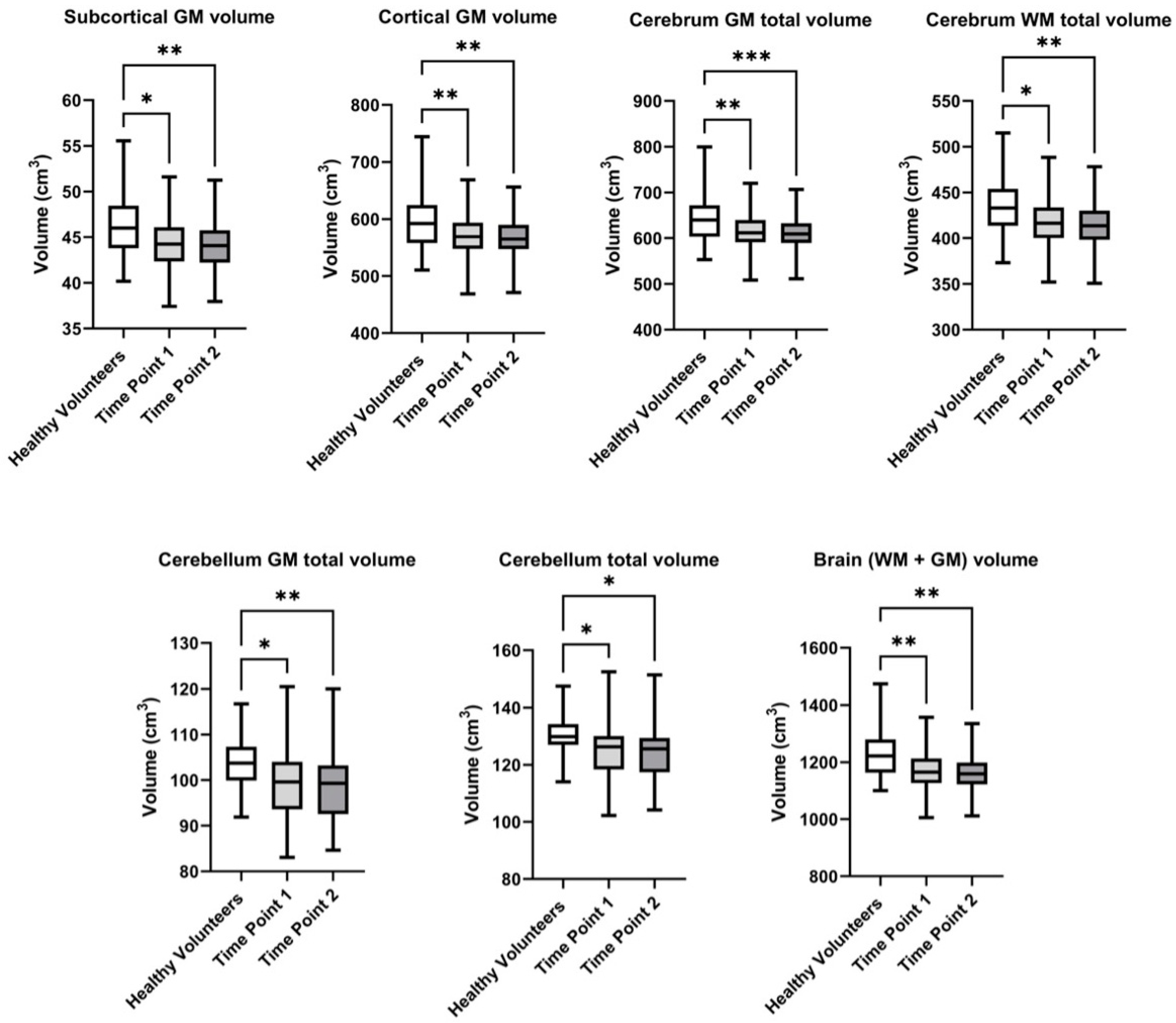
| Brain (WM + GM) | 1221.3 ± 83.0 | 1165.0 ± 73.5 | 1134.2 ± 217.7 | 0.002, 0.47 | 0.004, 0.74 |
| Calcarine cortex, left | 4.5 ± 1.2 | 3.9 ± 0.8 | 3.9 ± 0.9 | 0.011, 0.30 | 0.008, 0.25 |
| Calcarine cortex, total volume | 8.8 ± 2.2 | 8.0 ± 1.4 | 7.8 ± 1.9 | 0.024, 0.25 | 0.024, 0.26 |
| Caudate, left | 3.1 ± 0.4 | 2.9 ± 0.5 | 2.8 ± 0.6 | 0.004, 0.55 | 0.004, 0.73 |
| Caudate, right | 3.2 ± 0.4 | 2.9 ± 0.5 | 2.9 ± 0.7 | 0.003, 0.52 | 0.003, 0.67 |
| Caudate, total volume | 6.3 ± 0.8 | 5.8 ± 0.9 | 5.6 ± 1.3 | 0.003, 0.54 | 0.004, 0.70 |
| Cerebellar GM | 115.5 ± 8.0 | 110.1 ± 8.2 | 107.3 ± 20.8 | 0.002, 0.35 | 0.004, 0.44 |
| Cerebellum GM, left | 51.0 ± 3.4 | 48.9 ± 3.6 | 47.6 ± 9.2 | 0.004, 0.30 | 0.009, 0.39 |
| Cerebellum GM, right | 52.3 ± 3.2 | 49.9 ± 4.0 | 48.6 ± 9.5 | 0.001, 0.40 | 0.002, 0.51 |
| Cerebellum GM, total volume | 103.4 ± 6.4 | 98.8 ± 7.5 | 96.2 ± 18.7 | 0.002, 0.36 | 0.004, 0.46 |
| Cerebellum, left | 64.3 ± 4.1 | 61.7 ± 4.4 | 60.1 ± 11.6 | 0.004, 0.41 | 0.007, 0.44 |
| Cerebellum, right | 65.5 ± 4.2 | 62.7 ± 4.7 | 61.0 ± 11.8 | 0.003, 0.53 | 0.005, 0.55 |
| Cerebellum, total volume | 129.8 ± 8.0 | 124.4 ± 9.1 | 121.1 ± 23.4 | 0.003, 0.48 | 0.004, 0.51 |
| Cerebrum GM, left | 322.3 ± 26.3 | 305.0 ± 19.2 | 297.0 ± 57.1 | 0.002, 0.55 | 0.004, 0.78 |
| Cerebrum GM, right | 322.3 ± 25.8 | 307.0 ± 19.7 | 298.7 ± 57.4 | 0.005, 0.44 | 0.009, 0.65 |
| Cerebrum GM, total volume | 644.7 ± 52.0 | 615.9 ± 39.9 | 612.1 ± 38.7 | 0.003, 0.83 | 0.0007, 0.83 |
| Cerebrum, left | 540.4 ± 39.0 | 513.4 ± 33.4 | 499.9 ± 96.3 | 0.002, 0.51 | 0.003, 0.80 |
| Cerebrum, right | 539.0 ± 38.3 | 515.9 ± 33.8 | 502.1 ± 96.6 | 0.004, 0.39 | 0.008, 0.68 |
| Cerebrum, total volume | 1079.4 ± 77.1 | 1029.3 ± 67.1 | 1002.1 ± 192.9 | 0.003, 0,45 | 0.004, 0.74 |
| Cerebrum WM, left | 218.0 ± 15.2 | 208.3 ± 16.1 | 203.0 ± 39.7 | 0.004, 0.40 | 0.007, 0.74 |
| Cerebrum WM, right | 216.6 ± 14.6 | 208.9 ± 17.4 | 203.5 ± 40.1 | 0.013, 0.24 | 0.021, 0.67 |
| Cerebrum WM, total volume | 434.7 ± 29.8 | 418.2 ± 29.8 | 415.4 ± 29.1 | 0.029, 0.32 | 0.009, 0.71 |
| Cortical GM | 598.6 ± 49.4 | 571.9 ± 38.1 | 568.3 ± 37.03 | 0.004, 0.67 | 0.001, 0.77 |
| Cuneus, left | 5.3 ± 1.0 | 4.9 ± 0.8 | 4.8 ± 1.1 | 0.046, 0.37 | 0.073, 0.65 |
| Cuneus, right | 5.6 ± 1.1 | 5.1 ± 0.8 | 5.0 ± 1.2 | 0.053, 0.55 | 0.049, 0.62 |
| Cuneus, total volume | 10.9 ± 1.9 | 9.9 ± 1.5 | 9.7 ± 2.2 | 0.015, 0.49 | 0.019, 0.72 |
| Frontal lobe, left | 98.1 ± 9.3 | 91.7 ± 7.3 | 89.4 ± 17.5 | 0.001, 0.44 | 0.002, 0.64 |
| Frontal lobe, right | 97.2 ± 10.5 | 92.8 ± 7.3 | 90.5 ± 17.7 | 0.016, 0.32 | 0.021, 0.42 |
| Frontal lobe, total volume | 195.3 ± 19.3 | 184.5 ± 13.9 | 179.9 ± 35.0 | 0.004, 0.40 | 0.007, 0.53 |
| Gray matter (GM) | 760.1 ± 57.2 | 722.2 ± 44.0 | 703.1 ± 134.7 | 0.002, 0.49 | 0.004, 0.71 |
| Gyrus rectus, left | 1.9 ± 0.3 | 1.8 ± 0.3 | 1.8 ± 0.4 | 0.037, 0.03 | 0.153, 0.24 |
| Inf. occipital gyrus, left | 7.7 ± 1.6 | 6.8 ± 1.2 | 6.7 ± 1.6 | 0.028, 0.78 | 0.029, 0.93 |
| Inf. temporal gyrus, left | 13.7 ± 1.6 | 12.7 ± 1.5 | 12.3 ± 2.6 | 0.013, 0.47 | 0.004, 0.43 |
| Inf. temporal gyrus, right | 13.6 ± 2.1 | 12.6 ± 1.4 | 12.3 ± 2.5 | 0.021, 0.59 | 0.024, 0.61 |
| Inf. temporal gyrus, total volume | 27.3 ± 3.1 | 25.3 ± 2.5 | 24.5 ± 4.9 | 0.006, 0.62 | 0.003, 0.61 |
| Insular cortex, left | 15.4 ± 1.4 | 14.7 ± 1.7 | 14.3 ± 3.0 | 0.043, 0.27 | 0.033, 0.28 |
| Intracranial cavity (IC) | 1425.3 ± 104.3 | 1359.0 ± 95.7 | 1324.5 ± 256.7 | 0.003, 0.39 | 0.007, 0.69 |
| Lateral orbital gyrus, left | 2.7 ± 0.6 | 2.5 ± 0.5 | 2.4 ± 0.6 | 0.020, 0.34 | 0.003, 0.34 |
| Lateral orbital gyrus, right | 2.6 ± 0.6 | 2.4 ± 0.5 | 2.3 ± 0.6 | 0.030, 0.31 | 0.013, 021 |
| Lateral orbital gyrus, total volume | 5.4 ± 0.7 | 4.9 ± 0.9 | 4.7 ± 1.1 | 0.008, 0.42 | 0.001, 0.37 |
| Lateral ventricle, left | 9.4 ± 3.2 | 8.4 ± 5.2 | 8.5 ± 5.4 | 0.013, 0.25 | 0.019, 0.13 |
| Lateral ventricle, total volume | 17.7 ± 6.0 | 16.4 ± 9.2 | 16.6 ± 9.6 | 0.047, 0.16 | 0.060, 026 |
| Limbic cortex, right | 21.8 ± 2.6 | 20.4 ± 2.0 | 19.9 ± 4.0 | 0.006, 0.17 | 0.007, 0.39 |
| Lobules VI-VII | 2.8 ± 0.4 | 2.6 ± 0.3 | 2.6 ± 0.5 | 0.023, 0.37 | 0.026, 0.39 |
| Medial orbital gyrus, left | 4.9 ± 0.7 | 4.5 ± 0.6 | 4.5 ± 1.0 | 0.007, 0.16 | 0.021, 0.52 |
| Middle occipital gyrus, left | 6.3 ± 1.5 | 5.5 ± 1.1 | 5.4 ± 1.4 | 0.012, 0.52 | 0.010, 0.83 |
| Middle occipital gyrus, total | 12.1 ± 1.9 | 11.2 ± 1.5 | 10.9 ± 2.3 | 0.074, 0.44 | 0.044, 0.70 |
| Occipital lobe, left | 45.2 ± 5.7 | 41.8 ± 3.7 | 40.9 ± 8.1 | 0.003, 0.50 | 0.004, 0.74 |
| Occipital lobe, total volume | 90.8 ± 10.8 | 85.3 ± 6.8 | 83.1 ± 16.3 | 0.009, 0.47 | 0.013, 0.71 |
| Opercular inf. frontal gyrus, right | 3.9 ± 1.1 | 3.4 ± 0.7 | 3.3 ± 0.8 | 0.032, 0.63 | 0.016, 0.51 |
| Opercular inf. frontal gyrus, total volume | 7.3 ± 1.2 | 6.7 ± 1.1 | 6.5 ± 1.5 | 0.018, 0.75 | 0.018, 0.70 |
| Orbital inf. frontal gyrus, right | 1.6 ± 0.5 | 1.4 ± 0.4 | 1.4 ± 0.4 | 0.070, 0.22 | 0.029, 0.02 |
| Parietal lobe, left | 58.6 ± 5.5 | 55.8 ± 7.5 | 54.6 ± 12.0 | 0.005, 0.28 | 0.012, 1.02 |
| Parietal lobe, right | 59.9 ± 5.0 | 56.9 ± 6.7 | 55.5 ± 11.7 | 0.004, 0.29 | 0.007, 0.93 |
| Parietal lobe, total volume | 118.4 ± 10.2 | 112.7 ± 14.1 | 110.1 ± 23.6 | 0.003, 0.29 | 0.006, 1.00 |
| Planum polare, left | 2.2 ± 0.5 | 2.0 ± 0.3 | 2.0 ± 0.4 | 0.025, 0.58 | 0.028, 0.67 |
| Postcentral gyrus medial segment, right | 1.3 ± 0.3 | 1.1 ± 0.3 | 1.1 ± 0.4 | 0.018, 0.67 | 0.018, 0.87 |
| Postcentral gyrus medial segment, total volume | 2.4 ± 0.5 | 2.1 ± 0.5 | 2.0 ± 0.6 | 0.011, 0.59 | 0.012, 0.81 |
| Postcentral gyrus, right | 12.1 ± 1.7 | 11.0 ± 1.2 | 10.9 ± 2.2 | 0.002, 0.53 | 0.005, 0.88 |
| Postcentral gyrus, total volume | 25.0 ± 3.0 | 23.0 ± 2.1 | 22.6 ± 4.5 | 0.001, 0.78 | 0.003, 1.09 |
| Posterior cingulate gyrus, right | 5.3 ± 0.7 | 4.8 ± 0.8 | 4.8 ± 1.1 | 0.001, 0.03 | 0.003, 0.36 |
| Posterior cingulate gyrus, total volume | 10.3 ± 1.4 | 9.7 ± 1.4 | 9.5 ± 2.1 | 0.030, 0.06 | 0.035, 0.21 |
| Posterior insula, left | 2.4 ± 0.3 | 2.2 ± 0.3 | 2.2 ± 0.5 | 0.008, 0.52 | 0.013, 0.60 |
| Posterior insula, right | 2.5 ± 0.3 | 2.3 ± 0.3 | 2.3 ± 0.5 | 0.054, 0.34 | 0.041, 0.20 |
| Posterior insula, total volume | 4.9 ± 0.5 | 4.5 ± 0.6 | 4.4 ± 0.9 | 0.009, 0.45 | 0.016, 0.43 |
| Precentral gyrus, left | 14.3 ± 1.8 | 13.4 ± 1.2 | 13.1 ± 2.6 | 0.010, 0.31 | 0.020, 0.54 |
| Precentral gyrus medial segment, right | 3.1 ± 0.5 | 2.8 ± 0.5 | 2.8 ± 0.7 | 0.004, 0.41 | 0.007, 0.60 |
| Precentral gyrus medial segment, total volume | 6.1 ± 1.1 | 5.6 ± 0.9 | 5.5 ± 1.3 | 0.029, 0.38 | 0.031, 0.50 |
| Precentral gyrus, right | 14.0 ± 1.9 | 13.5 ± 1.3 | 13.3 ± 2.6 | 0.031, 0.03 | 0.044, 0.28 |
| Precentral gyrus, total volume | 28.3 ± 3.5 | 26.9 ± 2.3 | 26.4 ± 5.2 | 0.010, 0.16 | 0.025, 042 |
| Precuneus, left | 12.1 ± 1.6 | 11.4 ± 1.4 | 11.2 ± 2.3 | 0.031, 0.40 | 0.045, 0.70 |
| Putamen, left | 4.5 ± 0.4 | 4.3 ± 0.6 | 4.2 ± 0.9 | 0.018, 0.54 | 0.020, 0.63 |
| Putamen, right | 4.5 ± 0.4 | 4.3 ± 0.6 | 4.2 ± 0.9 | 0.020, 0.56 | 0.017, 0.66 |
| Putamen, total | 9.1 ± 0.8 | 8.6 ± 1.2 | 8.4 ± 1.9 | 0.019, 0.55 | 0.016, 0.65 |
| Subcortical GM | 46.1 ± 3.3 | 44.4 ± 3.0 | 43.8 ± 5.2 | 0.095, 0.67 | 0.018, 0.77 |
| Sup. frontal gyrus, left | 16.3 ± 2.1 | 15.0 ± 2.4 | 14.6 ± 3.4 | 0.002, 0.40 | 0.001, 1.02 |
| Sup. frontal gyrus medial segment, left | 6.8 ± 1.1 | 6.2 ± 1.0 | 6.1 ± 1.4 | 0.033, 0.55 | 0.026, 0.49 |
| Sup. frontal gyrus, right | 15.9 ± 2.2 | 14.6 ± 1.8 | 14.3 ± 3.0 | 0.009, 0.62 | 0.013, 0.69 |
| Sup. frontal gyrus, total volume | 32.2 ± 3.9 | 29.6 ± 3.8 | 28.9 ± 6.2 | 0.002, 0.54 | 0.003, 0.91 |
| Sup. occipital gyrus, left | 4.9 ± 0.8 | 4.2 ± 0.8 | 4.1 ± 1.0 | 0.001, 0.59 | 0.001, 1.00 |
| Sup. occipital gyrus, total volume | 10.0 ± 1.4 | 8.9 ± 1.3 | 8.7 ± 1.9 | 0.001, 0.61 | 0.001, 1.07 |
| Sup. parietal lobule, left | 12.4 ± 1.5 | 11.9 ± 2.5 | 11.6 ± 3.0 | 0.032, 0.07 | 0.030, 0.36 |
| Sup. parietal lobule, right | 12.7 ± 1.4 | 12.0 ± 1.9 | 11.7 ± 2.7 | 0.011, 0.07 | 0.005, 0.43 |
| Sup. parietal lobule, total volume | 25.1 ± 2.5 | 23.9 ± 4.2 | 23.3 ± 5.7 | 0.014, 0.01 | 0.011, 0.42 |
| Sup. temporal gyrus, left | 7.9 ± 1.0 | 7.1 ± 0.7 | 6.9 ± 1.4 | 0.002, 0.94 | 0.001, 1.00 |
| Sup. temporal gyrus, total volume | 15.3 ± 2.0 | 14.0 ± 1.6 | 13.6 ± 2.8 | 0.020, 0.72 | 0.018, 0.81 |
| Supplementary motor cortex, left | 6.0 ± 1.0 | 5.6 ± 0.7 | 5.5 ± 1.1 | 0.011, 0.19 | 0.018, 0.37 |
| Supplementary motor cortex, total volume | 11.6 ± 1.5 | 10.9 ± 1.4 | 10.7 ± 2.3 | 0.040, 0.24 | 0.049, 0.39 |
| Supramarginal gyrus, total volume | 19.3 ± 3.1 | 18.0 ± 2.5 | 17.6 ± 3.9 | 0.037, 0.53 | 0.041, 0.77 |
| Temporal lobe, left | 60.6 ± 4.9 | 57.5 ± 4.1 | 55.9 ± 10.8 | 0.010, 0.54 | 0.011, 0.63 |
| Temporal lobe, right | 60.0 ± 4.9 | 57.4 ± 4.5 | 55.7 ± 10.8 | 0.018, 0.32 | 0.012, 0.46 |
| Temporal lobe, total volume | 120.6 ± 9.5 | 115.0 ± 8.3 | 111.6 ± 21.6 | 0.011, 0.44 | 0.011, 0.56 |
| Thalamus, left | 8.1 ± 0.8 | 7.6 ± 0.9 | 7.4 ± 1.6 | 0.006, 0.53 | 0.007, 0.71 |
| Thalamus, right | 8.1 ± 0.7 | 7.7 ± 0.9 | 7.5 ± 1.6 | 0.020, 0.41 | 0.032, 0.60 |
| Thalamus, total | 16.2 ± 1.5 | 15.3 ± 1.9 | 15.0 ± 3.2 | 0.010, 0.47 | 0.017, 0.66 |
| Triangular inf. frontal gyrus, left | 4.1 ± 0.9 | 3.7 ± 0.7 | 3.6 ± 0.9 | 0.041, 0.73 | 0.028, 0.33 |
| Ventral DC, left | 5.0 ± 0.4 | 4.7 ± 0.3 | 4.6 ± 0.9 | 0.003, 0.90 | 0.004, 1.11 |
| Ventral DC, right | 4.9 ± 0.4 | 4.6 ± 0.4 | 4.4 ± 0.9 | 0.001, 0.91 | 0.001, 1.23 |
| Ventral DC, total volume | 9.8 ± 0.9 | 9.3 ± 0.7 | 9.0 ± 1.7 | 0.001, 0.95 | 0.001, 1.19 |
| Vermis | 12.1 ± 2.3 | 11.3 ± 1.0 | 11.1 ± 2.2 | 0.025, 0.16 | 0.044, 0.18 |
| White matter (WM) | 461.2 ± 30.8 | 442.9 ± 33.7 | 431.5 ± 84.2 | 0.007, 0.35 | 0.014, 0.72 |
Disclaimer/Publisher’s Note: The statements, opinions and data contained in all publications are solely those of the individual author(s) and contributor(s) and not of MDPI and/or the editor(s). MDPI and/or the editor(s) disclaim responsibility for any injury to people or property resulting from any ideas, methods, instructions or products referred to in the content. |
© 2025 by the authors. Licensee MDPI, Basel, Switzerland. This article is an open access article distributed under the terms and conditions of the Creative Commons Attribution (CC BY) license (https://creativecommons.org/licenses/by/4.0/).
Share and Cite
Nikolaeva, A.; Pospelova, M.; Krasnikova, V.; Makhanova, A.; Tonyan, S.; Efimtsev, A.; Levchuk, A.; Trufanov, G.; Voynov, M.; Sklyarenko, M.; et al. MRI Voxel Morphometry Shows Brain Volume Changes in Breast Cancer Survivors: Implications for Treatment. Pathophysiology 2025, 32, 11. https://doi.org/10.3390/pathophysiology32010011
Nikolaeva A, Pospelova M, Krasnikova V, Makhanova A, Tonyan S, Efimtsev A, Levchuk A, Trufanov G, Voynov M, Sklyarenko M, et al. MRI Voxel Morphometry Shows Brain Volume Changes in Breast Cancer Survivors: Implications for Treatment. Pathophysiology. 2025; 32(1):11. https://doi.org/10.3390/pathophysiology32010011
Chicago/Turabian StyleNikolaeva, Alexandra, Maria Pospelova, Varvara Krasnikova, Albina Makhanova, Samvel Tonyan, Aleksandr Efimtsev, Anatoliy Levchuk, Gennadiy Trufanov, Mark Voynov, Matvey Sklyarenko, and et al. 2025. "MRI Voxel Morphometry Shows Brain Volume Changes in Breast Cancer Survivors: Implications for Treatment" Pathophysiology 32, no. 1: 11. https://doi.org/10.3390/pathophysiology32010011
APA StyleNikolaeva, A., Pospelova, M., Krasnikova, V., Makhanova, A., Tonyan, S., Efimtsev, A., Levchuk, A., Trufanov, G., Voynov, M., Sklyarenko, M., Samochernykh, K., Alekseeva, T., Combs, S. E., & Shevtsov, M. (2025). MRI Voxel Morphometry Shows Brain Volume Changes in Breast Cancer Survivors: Implications for Treatment. Pathophysiology, 32(1), 11. https://doi.org/10.3390/pathophysiology32010011