Review

Implications and Practical Applications of the Chemical Speciation of Iodine in the Biological Context

by
Astrid N. Espino-Vázquez
,
Flor C. Rojas-Castro
and
Liria Mitzuko Fajardo-Yamamoto
*
Research Department, Sanoviv Medical Institute, Tijuana-Ensenada FWY, 39th KM, Unnumbered, Int. 6 Rancho Santini, Rosarito 22710, Mexico
*
Author to whom correspondence should be addressed.
Future Pharmacol. 2022, 2(4), 377-414; https://doi.org/10.3390/futurepharmacol2040026
Submission received: 19 July 2022 / Revised: 20 August 2022 / Accepted: 29 August 2022 / Published: 3 October 2022

Abstract

:
Iodine is a highly reactive element with a single natural and stable isotopic form (127I). In the biosphere, it is one of the 30 essential elements for life, and its chemical speciation defines its availability and biological activities. The most relevant chemical species are iodate (IO3) and iodide (I) as the major sources of iodine, with molecular iodine (I2) and hypoiodous acid (HIO) as the most reactive species, and thyroid hormones (THs) as the representative organic compounds. In human biology, THs are master regulators of metabolism, while inorganic species serve for the iodination of organic molecules and contribute to the innate immune system and the antioxidant cellular defense. Additionally, I, I2, δ-lactone (6-IL), and α-iodohexadecanal (α-IHDA) have shown therapeutic potential in counteracting oxidative stress, cancer, and inflammation. Both inorganic and organic species have applications in the health science industry, from the manufacturing of disinfection and wound care products to supplements, medicines, and contrast media for radiography. Even after nuclear disasters, intake of high doses of iodine prevents the accumulation of radioactive iodine in the body. Conversely, the controlled production of iodine radioisotopes such as 123I, 124I, 125I, and 131I is exploited in nuclear medicine for radiotherapy and diagnostics.

Graphical Abstract

1. Introduction

Life originated in the oceans, where iodine is available in high amounts, and it is believed that, since then, it has had an ancient role in the biology of emergent organisms [1]. This element is essential in the metabolism of countless living beings from the three major domains of life [1]. Yet, depending on the chemical species and their amount, iodine acts as a nutrient showing protective roles or as a cellular stressor. For instance, catalytic inorganic iodine species inhibit microorganisms, attacking structural components such as nucleic acids, proteins, and lipids in a dose-dependent manner [2]. Contrary, in appropriate amounts, eukaryotic cells have taken advantage of the reactivity of some inorganic species to synthesize new compounds, counteract cellular oxidative stress, and modulate cell signaling pathways [3].
Despite the vast information around iodine, we consider that most recent studies have not entirely regarded critical aspects of its chemistry and stability when discussing the novel roles of this element in biological systems. This review compiles the multidisciplinary evidence of iodine, considering the characteristics of its biologically active forms to better understand the functions, modes of action, and applications. For that, we first describe the basic features of the element, such as the history, physicochemical properties, and geological distribution. Then, we list the most relevant inorganic and organic iodine species and state the fundaments of the chemical speciation, especially under biological conditions. With those bases, we further expose and discuss the known roles and activities of the main iodine species, considering evolutive aspects. Lastly, we connect the presented information with practical applications in the industry and health sciences, taking into account the radioisotopes of iodine.

2. History and Chemical Characteristics

In 1811, the French chemist Bernard Courtois discovered iodine when he tried to obtain saltpeter from seaweed and accidentally added too much sulfuric acid to the mixture, producing a purple vapor. Two years later, iodine was recognized as a new element receiving its name from the Ancient Greek term “οειδής” or “ioeides” which means violet [4].
The periodic table represents iodine by the I letter, which has an atomic number of 53, an atomic mass of 126.9045 Da, and belongs to group 17 of halogens [5]. There are 37 known isotopes of the element with masses from 108 to 144 (108I – 144I). Most are artificially produced and lose energy via radiation (radiation decay) in days, hours, or less; except for 127I, the natural and only 100% stable isotope, and 129I, with a half-life of 15.7 million years, which is naturally produced in traces by spontaneous fission and cosmic-ray reactions (see applications of radioisotopes in Section 7.4) [6,7].
Iodine lacks an electron to fulfill the valence shell; hence, it cannot exist in nature as a free element (neutral atom) because of its high reactivity and electronegativity (2.66 on the Pauling scale). Instead, the diatomic form of I2, formed by a nonpolar covalent bond between two iodine atoms, is recognized as the stable state of the element [5,8]. Likewise, iodine can combine with most elements (except noble gases and most synthetic elements), yielding broad inorganic and organic chemical forms (iodine species) with oxidation states of −1, 0, +1, +3, +5, and +7, distributed along the lithosphere, hydrosphere, and atmosphere.

3. Distribution of Iodine

In the Earth’s crust, the richest inorganic source of iodine is oceanic sediments (68.2%) and continental sedimentary rocks (27.7%), followed by igneous and metamorphic rocks (2.7%), seawater (0.81%), and mafic oceanic crust (0.68%) [9]. In the atmosphere, sea spray aerosolization, volcanic gases, and human activities contribute to iodine emissions, but the highest discharge is given by biological conversion to volatile methyl forms, such as methyl iodide (CH3I).
Cycling of iodine involves biotic and abiotic processes through the lithosphere, hydrosphere, and atmosphere. Into seawater, iodine species are cycled during subduction of the oceanic crust (descending of a tectonic plate below another) and via decomposition of marine organisms; whereas the flux from the atmosphere to the lithosphere occurs during rainfall, wet deposition, leaching, and runoff [9,10,11]. Furthermore, an important amount of iodine is accumulated and used by many living organisms, including algae, plants, corals, sponges, anemones, lobworms, shellfishes, arthropods, and bacteria, that cycle organic and inorganic iodine species in the biosphere (Figure 1) [11].
Quantities and cycling of natural iodine are determined by 127I, and ratios between radioisotopes and 127I serve to measure the anthropogenic impact [9]. After nuclear fission accidents, from all discharged contaminants, 131I is one of the most released radioisotopes to the environment during the first days, along with significant amounts of 132I and 133I. In the long term, 129I serves as a marker of nuclear contamination because of its long radiation decay, and it has the longest half-life of all fission products [7,12]. Quantification of 129I also serves for the age dating of groundwater and meteorites [13,14].

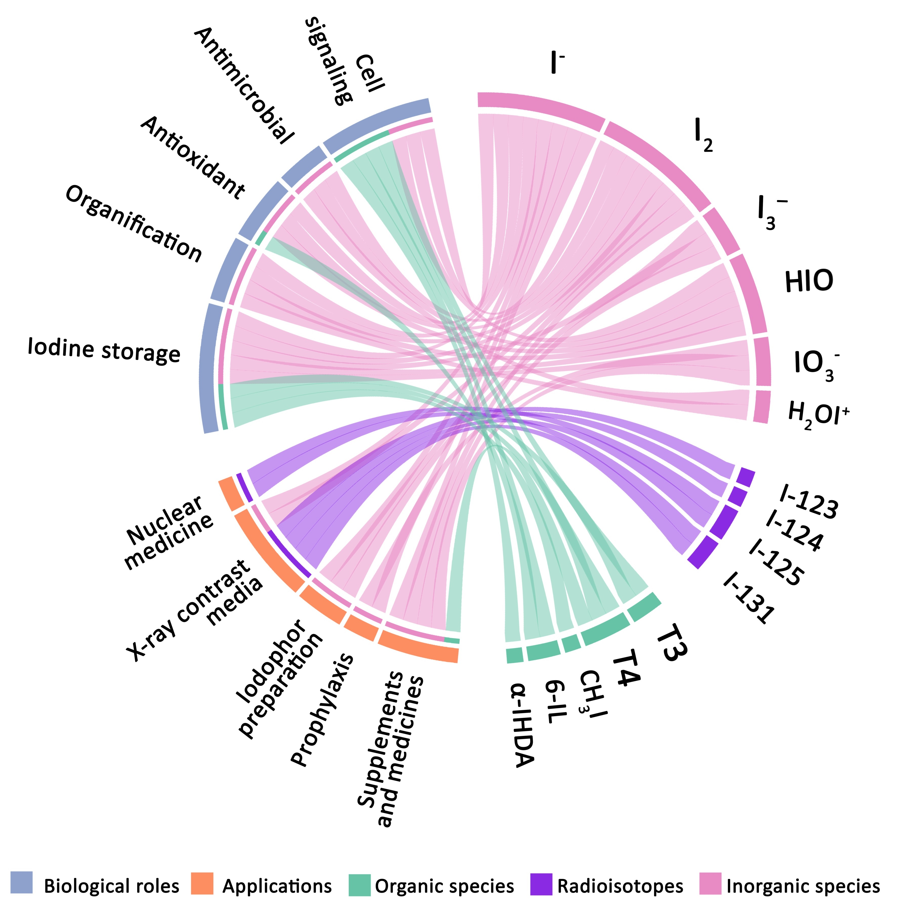
4. Relevant Iodine Species

4.1. Main Inorganic Forms

Dominant iodine species in the solid state are potassium iodate (KIO3) and potassium iodide (KI), whereas IO3 and I ions lead to aqueous systems [15]. I2 and CH3I are the most abundant species in the atmosphere, produced by biotic and abiotic processes [16]. Atmospheric photochemistry promotes the speciation of gaseous iodine species (e.g., HI, IO, HIO, IONO2 INO2, and IO2) [17,18]. Some of them (mainly I2 and HIO), together with other halogen compounds, are responsible for ozone (O3) depletion in the lower troposphere when interacting with hydrogen–oxygen species and nitrogen oxides [19].
Iodide salts are produced by electrostatic attraction between iodine and metals, held together by ionic bonds. Except for lead, silver, and mercuric iodides (PbI2, AgI, and HgI2), all iodide salts have good solubility in water, where they dissociate into charged particles (ions) [4,15]. Conversely, I2 has a much lower solubility than salts like KI (I2 = 0.03 g/100 g H2O at 20 °C; KI = 148 g/100 g H2O at 25 °C), but it is easily dissolved in ethanol (I2 = 271.7 g/kg EtOH at 25 °C) and many organic solvents such as benzene (I2= 164.0 g/kg Bz at 25 °C), hexane (I2 = 13.2 g/kg Hx at 25 °C), or chloroform (I2 = 49.7 g/kg CHCl₃ at 25 °C) [20]. The water solubility of I2 increases when iodides are added; for example, at 20 °C in 10 g/100 mL of sodium iodide (NaI), its solubility increases 32.7-fold (from 0.03 g/100 g H2O to 0.96 g/100 g H2O) [8,21].
Up to 10 different iodine species can be formed when I2 and I combine in water systems (Reactions (1)–(9)), where the chemical speciation is highly influenced by iodide amount (c(I)), pH, and redox potential (Eh) (Figure 2) [22,23,24]. An excellent example is Lugol’s solution (typically made of 5% I2 and 10% KI) used as a disinfectant and for other commercial purposes (see Section 6, Section 7.1 and Section 7.2). Under those conditions, I2, I, triiodide (I3), pentaiodide (I5), and hexaiodide (I62−) coexist. I2 is the most reactive of those, showing several biological activities (see Section 5.1, Section 5.4, Section 5.5 and Section 5.6), I3 is responsible for staining, and I5/I62− barely represents ~8% of the oxidizing potential [24]. However, only I, I2, and I3 are formed and available in significant amounts under physiological conditions [24,25].
I 2 + H 2 O   HIO + I +   H + .
HIO   OI +   H + .
I 2 + I     I 3 .
HIO +   H + H 2 OI + .
I 3 + I 2 I 5   .
2 I 3 I 6   2 .
OI +   I + H 2 O   HI 2 O + OH .
HI 2 O   I 2 O + H + .
3 HIO   IO 3 + 2 I + 3 H + .
Iodine in iodides I3 and I5 have oxidation states of -1, in contrast to an oxidation state of 0 in I2. Higher values exist when iodine combines with oxygen, such as in HIO, iodite (IO2), iodate (IO3), and periodate (IO4 or IO65−) having oxidation states of +1, +3, +5, and +7, respectively [10]. The oxidation state refers to the electronic configuration of an atom in a chemical species and represents the number of electrons gained (-) or lost (+) [10].
Hypoiodous acid and its derivatives (OI, HI2O, I2O2−, and H2OI+) are very reactive species [27,28]; however, despite their potential biological activity, their amount and stability are limited in the physiological pH range (5–8) [24]. For instance, HIO dominates only in basic pH ranges (8–9) under high I dilution (Reactions (1), (2), (4), and (9)), and iodine cation (H2OI+) is only produced at pH <1 (Reactions (4) and (8)) [24,29,30].
Iodate is the most thermodynamically stable of all iodine species and has no oxidative action in the basic and neutral pH range; it only achieves some reactivity at very low pH, such as HIO3 (pKa = 0.77) [24]. The main sources are calcium iodate (Ca(IO3)2), potassium iodate (KIO3), and potassium biiodate (KH(IO3)2), some of them with better water solubility than iodides (e.g., HIO3 = 308 g/100 g H2O at 25 °C vs. KI = 148 g/100 g H2O at 25 °C) [31]. IO3 represents a major source of iodine for living beings and is well absorbed in several organisms. Hence, it needs to be transformed into a different species to exhibit an active biological role, as described in sections below [24].

4.2. Representative Organic Species

Hundreds of iodinated organic compounds are found in nature; nevertheless, for most living organisms, the main iodine sources come from two simple inorganic species, I and IO3 [10]. Intracellularly, marine organisms reduce IO3 into I or I2 and store it or use it to produce organic compounds [32]. Some of the iodinated organic molecules found in algae and seaweed are carbonyl diiodide (COI2), 3-iodopropan-1-ol (C3H7IO), 1-bromo-3-iodopropan-2-ol (C3H6BrIO), 1-bromo-1,1-dichloro-2-iodoethane (C2H2BrCl2I), 2-bromo-2-iodoacetic acid (C2H2BrIO2), and 2-iodoacetamide (C2H4INO) [32,33]. Furthermore, algae, phytoplankton, rice paddies, fungi, and bacteria produce and emit large amounts of volatile metabolites containing carbon–iodine bonds such as CH3I (the most abundant iodine species in the atmosphere), diiodomethane (CH2I2), chloro(iodo)methane (CH2ClI), 1-iodocyclopenta-1,3-diene (C5H5I), iodoethane (C2H5I), or 1-iodopropane (C3H7I) [9,10,11,32].
Land plants can absorb and accumulate iodine in different tissues and organs (mainly roots, leaves, fruits, and seeds) [34,35]. Although, their content is significantly lower (0.18–0.24 mg/kg) than in algae and sea plants (~75–6750 mg/kg) [32]. Some examples of organic species of iodine in crops are salicylic acid (iodosalicylates) and benzoic acid derivatives (iodobenzoates), and proteins (iodoproteins) [36,37].
Tyrosine residues (Tyr) containing iodine atoms covalent-bonded (iodotyrosines) are the protagonists of organic iodine species because of their hormonal role in vertebrate animals (see Section 5.3). A human adult has, on average, 0.02 mg/kg of iodine in the body, and most of it is retained by the thyroid gland (70–80%) to produce THs (organification). The remainder is distributed in other organs (salivary glands, lactating breast tissue, kidney, intestine, placenta, ovary, fallopian tubes, testicles, etc.) with different purposes [38,39]. THs comprise (i) 3-iodotyrosine or monoiodotyrosine (MIT or T1) and 3,5-diiodotyrosine (DIT or T2) as precursors, (ii) 3′,5,3-triiodothyronine (T3) and thyroxine (T4), typically considered as the active hormones, and (iii) 3′,5′,3-triiodothyronine or reverse T3 (rT3), 3′,5′-diiodotyrosine (3′,5′-DIT or 3′,5′-T2), and 3′- monoiodotyrosine (3′-MIT or 3′-T1), the T4 derivatives [40]. Other TH derivatives such as thyronamines (TAMs) and thyroacetic acids (e.g., 3,3′,5-triiodothyroacetic acid (Triac) and 3,3′,5,5′-tetraiodothyroacetic acid (Tetrac)) can be produced by decarboxylation, deamination (removal of the carboxyl group and amine group, respectively), and/or deiodination [41].
Thyroid peroxidase (TPO) is the enzyme responsible for synthesizing THs using thyroglobulin (TG) as a substrate. In fact, TG is the main iodinated compound in the thyroid gland [42,43,44]. TPO is also responsible for the iodination of lipids (iodolipids) as α-IHDA and 6-IL. Noticeably, 6-IL was recently related to antioxidant, anti-inflammatory, and anticarcinogenic activities (see Section 5.4, Section 5.5 and Section 5.6) [45,46]. Other peroxidase enzymes such as lactoperoxidase (LPO) are responsible for the synthesis of iodolactones (e.g., δ-lactone of 6-iodo-4-hydroxy-eicosa-8,11,14-trienoic acid, and ε-lactone of 5-iodo-4-hydroxy-7-docosapentaenoic acid) using docosahexaenoic acid (DHA) as a substrate [32]. TPO and LPO belong to the peroxidase/cyclooxygenase superfamily, a class of oxidoreductase enzymes broadly distributed in the tree of life. Plants, fungi, bacteria, and animals use them to maintain oxidative status and synthesize new compounds or even as part of the innate host defense [47].
Some organisms unable to produce their own iodinated organic compounds depend on symbiotic relationships to supply them. Sea urchins, for example, need THs to metamorphose from larva to adult rudiment but obtain them from algae feeding [48]. Symbiosis also impacts the iodinated compounds profile of microorganisms. The enteric bacterium Escherichia coli cannot use I, I2, MIT, or DIT, but takes T4 and T3 to iodinate proteins [49,50]. Even viruses influence the production of iodinated organic compounds of their hosts. Recently, it was observed that a cyanophage (cyanobacteria virus) encodes for a halogenase (an enzyme that replaces hydrogen atoms with halogen atoms in a molecule) with the potential of iodinating organic molecules of its host [51]. Moreover, the presence of viruses in Emiliania huxleyi algae changes its metabolic profile. After viral infections, algal blooms (excessive growth of marine photosynthetic organisms) discharge a large amount of organic matter containing chloro and iodinated metabolites [52]. More examples of iodinated organic compounds can be consulted in [32].

5. Biological Roles of Iodine

5.1. Antimicrobial Action

Iodine shows a broad antibacterial spectrum against several microorganisms, including resistant forms (e.g., spores, cysts, and biofilms) [53,54,55]. Of all inorganic iodine species with antimicrobial activity, I2 is the most abundant and the leading antimicrobial agent in iodine-based disinfectants [24]. Other iodine species recognized as antimicrobials are HIO and H2OI+; however, as mentioned, they are restricted to extreme pH ranges and by the amount of I [27,28]. Within solvents like alcohol, the solvated forms I2 H2O or I2 ROH are proposed as the antimicrobial species [2].
Contrary to antibiotics or antifungals, microorganisms do not develop antimicrobial resistance to iodine, probably because of its several simultaneous mechanisms of action. In bacteria, iodine inhibits glycolysis, respiration, glucose transport, and DNA and RNA synthesis, and it compromises the integrity of the plasma membrane and cell wall. Antimicrobial iodine species are also responsible for protein (and potentially DNA) denaturalization in bacteria and viruses and damage viral envelopes [27,56,57].
The molecular modes of action described for I2 and HIO are oxidation of nucleotides (adenine, cytosine, and guanine) and aromatic rings of amino acids (mainly Tyr) [57], interfering with the synthesis and folding of proteins via iodination of amino (–NH) (Reaction (13) and (14)) and sulfhydryl (–SH) groups (Reactions (15)–(17)) [28,30,57], or disrupting disulfide bonds (R–S–S–R) between cysteines (Reactions (10)–(12)) [58]. They also have a great affinity to unsaturated fatty acids, attacking the carbon–carbon double bonds (Reaction (18)) [30,59]. Cation H2OI+ is also responsible for attacking the –SH groups of some amino acids (Reactions (19) and (20)) and iodination of tyrosine and histidine residues (Reactions (21)–(23)) [56,57]. The following reactions have been proposed for antimicrobial iodine species (most of them as chemical skeletal equations):
R S S R + 2 I 2 + 2 H 2 O   2 R SOH + 2 HI .
R SOH + I 2 + H 2 O   R SO 2 H + 2 HI .
R SO 2 H + I 2 + H 2 O   R SO 3 H + 2 HI .
R NH 2 + I 2   R NHI .
R NH + HIO   R NI + H 2 O .
R SH + I 2   R S I + I + H + .
R S I + H 2 O   R S OH + I + H + .
R SH + HIO   R SI + H 2 O R SOH + H + + I .
R CH = CH R 1 + I 2   R CHI CHIR 1 .
R S H + H 2 OI +   R S I + H + + H 2 O .
R S I + R S H RS RS + H + + I .
Tyr + H 2 OI +   TyrI + H + + H 2 O .
TyrI + H 2 OI +   TyrII + H + + H 2 O .
His + H 2 OI +   HisI + H + + H 2 O .
2 I + H 2 O 2   LPO I 2 + H 2 O .
Eukaryotes take advantage of the reactivity of some iodine species to counteract infections, among other biological activities (see Section 5.4, Section 5.5 and Section 5.6). In mammals, antimicrobial iodine species can be produced as byproducts of peroxidases. Depending on the pH and I amounts, LPO can produce HIO and I2 by reducing hydrogen peroxide (H2O2) to H2O, using I as the electron donor (Reaction (24)) [60,61]. LPO is secreted in mucosal surfaces or exocrine secretions such as saliva, milk, or tears and is essential for the innate immune defense system [28,47]. The sole action of LPO is bacteriostatic; however, in conjunction with I, it becomes bactericidal and antiviral [62,63,64].

5.2. Nutrition and Human Health

Together with vitamin A, iron, zinc, folate, and vitamin B-12, iodine is considered an essential micronutrient for humans and an important marker of nutritional public health status [65]. An optimal iodine status (sufficiency) can be maintained with an appropriate diet and supplements (see Section 7.2); however, inadequate iodine ingestion drives severe health consequences in the long term, known as iodine deficiency disorders (IDD). The most common are hypothyroidism, endemic goiter (enlargement of the thyroid), and thyroid nodules. Pregnant women and fetuses are the most susceptible to IDD, inducing abortions, perinatal mortality, and congenital anomalies [66,67,68]. Other expected health consequences are cretinism and delayed physical development in infants and children. Cognitive impairment and increased susceptibility to nuclear radiation are seen at all ages under iodine deficiency [67,69].
Thyroid disorders in the population and exposure to iodine are correlated in a U-shaped curve, as depicted in Figure 3A, where the higher risk comes with the chronic suboptimal intake of iodine (iodine deficiency) [67,70]. Excess exposure to iodine is less common but can be dangerous, especially in iodine-deficient populations. Excessive intake is frequently associated with high consumption of iodized salt and diet [71,72], but other sources came from medications such as amiodarone (200–400 mg/day) [73], radiologic procedures (one dose of >350 mg) [74], and povidone/iodine (PVP/I)-based antiseptics (<30 mg/day) [75]. Excess iodine increases the risk of developing transient or permanent thyroid dysfunction (iodine-induced hyperthyroidism or hypothyroidism), thyroid autoimmune diseases, metabolic disorders, metabolic syndrome, hypertension, prediabetes, diabetes mellitus, impaired glucose tolerance, central obesity, and dyslipidemia (Figure 3B–F) [70,76,77,78]. Vulnerable groups such as the elderly, fetuses, neonates, and people already having an IDD are more susceptible to iodine-induced hyperthyroidism or hypothyroidism. In the best scenario, hypothyroidism only lasts 2–10 days (escape from acute Wolff–Chaikoff effect), but might be lethal in fetuses or neonates. Iodine-induced hyperthyroidism or Jod–Basedow syndrome has greater incidence in adults and can be related to cardiac complications [39,78].
People with normal thyroid functioning (euthyroid) tolerate excessive iodine well. In the Japanese population, the regular consumption of iodine-rich foods correlates with a higher life span, as well as fewer infant deaths, cardiac diseases, and breast and prostate cancer incidences, than in the US, whose iodine intake is typically much lower. On average, people consume 1–3 mg/day of iodine in Japan, where one of the primary sources is seaweed-based meals such as nori (Porphyra), wakame (Undaria), and kombu (Laminaria) [80]. Interestingly, these health markers are not attributable to genetics; Japanese emigrants increased their risk of mammary cancer from 21.1 to 49.4 per 100,000 population and from 8.4 to 32.2 of prostate cancer per 100,000 just by changing diet [81,82]. Currently, the incidence of both types of cancer in Japan is increasing, probably because of the occidentalization of diet. Prostate cancer had the highest frequency in 2020 (close to 96,000), and breast cancer new cases were over 50,000 in 2018, compared to 2003 [83]. Nevertheless, thyroid cancer represents the rarest cause of death from cancer for Japanese (600 deaths for men in 2018 and 1200 for women in 2019) [83].
Iodine sufficiency areas have a lower risk of developing IDD; however, they are not exempt from Graves’ disease (with ~80% incidence in iodine-sufficient populations), nodular thyroid disease, and thyroiditis [84]. Respecting thyroid cancer (the most common endocrine cancer), some authors conclude that more than the recommended iodine ingestion could increase papillary and thyroid cancer [38,72], but others observed a lower incidence [85]. A more robust meta-analysis of humans and animals found iodine deficiency as a promotor risk factor for follicular, and possibly anaplastic, thyroid cancers [86].

5.3. Thyroid Hormones

5.3.1. TH Synthesis and Metabolism

From the anthropocentric point of view, the leading role of iodine is TH production in the thyroid gland, also known as organification. THs are iodotyrosines that can be produced by peroxidase enzymes (such as LPO and TPO) or via nonenzymatic reactions of iodine oxidative species (such as I2) with Tyr residues (Reaction (25)) [47,87]. This section focuses on the TH synthesis carried out in vertebrate animals. The occurrence of THs in different organisms and their functions is discussed next.
The regulation of the thyroid function is a circuit known as the thyroid axis, where, in normal conditions, TH synthesis and secretion increase in response to the thyroid-stimulating hormone (TSH) produced by the anterior pituitary gland [88]. TSH is stimulated in turn by the thyrotropin-releasing hormone (TRH), released by the hypothalamus [89]. Excess circulating T4 or T3 inhibits the hypothalamus from secreting more TRH, resulting in a decrement in TSH and, hence, in TH production [90].
As an initial step in TH synthesis, iodine must be absorbed into the body. Iodide is not diffused but is actively imported by transmembranal transporters into the bloodstream [91]. Most intake of iodine (>90%) is absorbed in the digestive tract, mainly in the small intestine by the Na+/I symporter (NIS) [92]. Depending on the iodine status (sufficiency, deficiency, or excess), the thyroid imports <10% up to 80% of the circulating iodine. Unused I is mainly excreted in the urine (~90%), depending on the glomerular filtration rate, with a lesser proportion in feces [39,65].
TH synthesis occurs in the thyroid follicles, composed of a colloid (follicular lumen) enclosed by follicular cells and surrounded in the interfollicular space by blood vessels and parafollicular cells [93]. In the basement membrane of follicular cells, I is imported by the Na+/I symporter (NIS) and moves at the apical membrane into the follicular lumen through different transporters such as the anion exchanger Pendrin, the calcium-dependent Cl channel Anoctamin 1 (ANO1), and possibly others such as cystic fibrosis transmembrane conductance regulator (CFTR), Cl/H+ antiporter (ClC5), Cl/H+ exchanger (ClC-3), and the sodium-dependent multivitamin transporter (SMVT) [94,95,96,97]. Once in the lumen, I needs to be oxidated into a more reactive form by the TPO. The specific iodine species formed in this step has not been determined, but the likely candidates are I2, iodinium (I+), iodine free radical (I), and hypoiodite (IO) [98].
TPO, coupled to the apical membrane of the follicles, is responsible for iodination and coupling of Tyr residues using I, H2O2, and thyroglobulin (TG) as the substrate (a large glycoprotein of ~330 kDa) [42]. H2O2 is produced by a complex formed by the dual oxidase 2 (DUOX2, formerly THOX2) and dual oxidase maturation factor 2 (DUOXA2), also coupled to the apical membrane [99]. The sequence followed in TH production is incorporating one iodine atom into Tyr residues at position 3 of the ring to produce MIT (Reaction (26)) and then at position 5 to form DIT (Reaction (27)). Subsequently, T3 is constructed by an oxidative coupling reaction between one DIT and one MIT (Reaction (28)), still incorporated into the TG. At the same time, T4 is formed by coupling two adjacent DITs (Reaction (29)). At the end of the process, only 37 of the ~70 Tyr residues in the TG are known iodination targets [42,43]. To release THs, iodinated TG is transported back into follicular cells by endocytosis and hydrolyzed by proteolysis [42,90]. Most MITs and DITs produced are kept in the follicles and are eventually degraded by iodotyrosine deiodinase (IYD) enzymes into MIT and Tyr, respectively (Reactions (30) and (31)), releasing I which is reused and recycled [100]. However, T3 and T4 are transported into the bloodstream, mostly bound to plasma proteins. The most common are thyroxine-binding globulin (carrying up to 80% of bound T3 and ~65% of bound T4), transthyretin (transporting ~9% of T3 and ~15% of T4), and albumin (with ~11% of T3 and ~20% of T4). A lower percentage of THs circulate as free hormones (approximately 0.2% of T3 and 0.05% of T4) [40].
Tyr + I 2   MIT + HI .
Tyr   residue + I + H 2 O 2 + H + TPO MIT + 2 H 2   O .
MIT + I + H 2 O 2 + H + TPO   DIT + 2 H 2   O .
MIT + DIT + H 2 O 2 + H + TPO T 3 + 2 H 2   O + TG   dehydroalanine .
2 DIT + I + H 2 O 2 + H + TPO T 4 + 2 H 2   O + TG   dehydroalanine .
DIT + NADPH IYD MIT + NADP + + I .
MIT + NADPH IYD Tyr + NADP + + I .
T 4 + NADPH ID T 3 + NADP + + I .
T 4 + NADPH ID rT 3 + NADP + + I .
In the blood, T4 has the highest concentration (54–174 μg/dL) and most prolonged half-life (~7 days), but T3 (half-life < 12 h) has a higher proportion in organs, especially the kidney, liver, small and large intestine, and central nervous system [40]. Just a fraction of T3 (21.8%) is synthesized and released in the same fashion as T4; most of it is generated by the deiodination of T4 out of the thyroid by iodothyronine deiodinases (ID) [90,101]. Iodine removal serves to activate or inactivate THs. D1 and D2 are the two kinds of ID enzymes considered as activators by converting T4 into T3 (Reaction (32)) [90,100]. D3 transforms T4 into rT3 (Reaction (33)), an inactive TH produced to maintain hormone homeostasis [102].
Deiodination is considered the most important process in the peripheral metabolism of THs, followed by conjugation, formation of analogs (via deamination and decarboxylation), and oxidation [103]. Conjugation of THs with sulfate or glucuronic acid (via microsomal glucuronyl or cytoplasmic phenol transferases) inactivates and increases the solubility of hormones, accelerating their deiodination and clearance by biliary excretion [104].
The gut microbiota also significantly contributes to TH metabolism, favoring the cycling of I and THs [101,102]. On the one hand, enteric bacteria are proposed as TH storage because of their ability to absorb and retain T3 and T4 [49,105]; on the other hand, the gut microbiota releases sulfatase and glucuronidase enzymes favoring the enterohepatic circulation (secretion of conjugated THs in the bile to the small intestine and back to the liver after deconjugation) [106]. Moreover, gut bacteria influence the absorption of nutrients that support the normal thyroid metabolism, such as iron, selenium, and zinc [107]. The importance of the gut microbiome in thyroid function is more evident in autoimmune thyroid diseases, where an altered microbiota composition (dysbiosis) is linked to predisposition to Hashimoto’s thyroiditis and Graves’ disease. Furthermore, a reduction in or lack of Lactobacillaceae and Bifidobacteriaceae families in the gut has been related to autoimmune metaplastic atrophic gastritis [107,108].

5.3.2. Actions of THs

THs are needed in all stages of life, participating in numerous biological functions in vertebrates such as regulation of the brain function, hair growth, functioning of gonads (especially in females), thermogenesis (heat produced in the body), and response to the environment and stimuli. At the molecular level, THs regulate essential cellular processes such as development, differentiation, growth, maturation, and metabolism via different mechanisms, as described below [3,40].
Excluding activation/inactivation by deiodination or conjugation, the biological activities of THs are determined by the hormonal levels inside the cells (controlled by TH transporters), thyroid hormone receptors (TRs), and other elements such as corepressors and coactivators [109]. More than 20 TH transporters have been characterized, but those with the highest affinity are the monocarboxylate transporters MCT8 (transporting T4, T3, rT3, and 3,3′-T2) and MCT10 (transporting only T3 and T4), as well as the organic anion-transporting polypeptides, such as OATP1C1 (with affinity to T4, rT3, and T4 sulfate) and OATP-F (with affinity to T4 and rT3) [110,111].
Once inside, THs activate TRs in the nucleus and mitochondria. The complex of THs/TRs binds directly to DNA regions acting as transcription factors regulating the expression of several genes [112]. In humans, two genes encode for TRs (α and β), yielding, via alternative splicing, several TRα and TRβ isoforms depending on the tissues and developmental stage. For example, TRα1 is mainly expressed in the bones, heart, intestine, skeletal muscle, and central nervous system, while TRβ1 is better expressed in the liver, kidney, and inner ear [109,113,114]. Truncated TR forms are also produced by alternative translations of start codons (AUG) or internal promoters in introns. Their cellular location is linked to post-translational modifications such as palmitoylation, phosphorylation, acetylation, ubiquitination, and sumoylation [109].
Concerning the mode of action, THs typically follow two pathways: genomic (carried out in the nucleus) and nongenomic (faster and initiated in the cytoplasm, plasma membrane, mitochondria, and possibly others) [113]. A more recent classification proposes four types of mechanisms: (1) direct interaction of THs/TRs on the DNA (as the classic genomic mechanism), (2) indirect interaction of THs/TRs on the DNA, (3) TH/TR signaling without DNA binding, and (4) TH signaling independent of TRs [115]. The classical genomic mechanism involves the activation of TRs through a complex with T3 and other proteins in the nucleus. After that, the complex binds to regulatory DNA regions known as thyroid hormone response elements (TREs) to regulate their expression [40]. The indirect DNA binding mechanism (also considered a genomic pathway) implicates the interaction of TH and TRs with chromatin-associated proteins leading to chromatin accessibility and upregulation of gene expression [109].
Nongenomic mechanisms comprise alternative cellular compartments, novel interactors, and potentially less recognized THs. A type 2 mechanism (of the new classification) comprises the interaction of TRs with THs and kinases in the cytoplasm to activate cell signaling responses such as the phosphatidylinositol 3-OH kinase pathway (PI3K) (implicated in growth, proliferation, differentiation, etc.). Another nongenomic pathway (or type 3 mechanism in the new classification) starts with the activation of a structural protein in the plasma membrane, the integrin αvβ3 receptor (with binding domains for T4 and T3), which triggers PI3K and the extracellular signal-regulated kinase 1/2 (ERK1/2) pathways, also a crucial regulator of the cell cycle, growth, and proliferation [113]. Similarly, a truncated TRα1 variant (p30) is found in the lipid rafts of the plasma membrane and activates the nitric oxide–cyclic guanosine monophosphate pathway (NO–cGMP) in response to T3 [109].
The mitochondrion is one of the main TH accumulators in the cell. Here, the oxidative phosphorylation system is highly influenced by different mechanisms involving truncated TRα1 variants (p28 in the inner membrane and p43 in the matrix). In the best-described pathway, T3 interacts with p43 on responsive elements of the mitochondrial DNA, regulating the transcription of respiratory genes (such as classic genomic mechanisms) [113,116]. The pathway that p28 follows is still unknown but shows a higher affinity to T3, and its expression affects mitochondrial physiology [117].
Previously, T3 was considered the biologically active hormone due to its higher affinity to most TRs [40]. However, the activities of T4 and other THs has been proven in different ways. For example, T4 but not T3 has the faculty to remodel the actin cytoskeleton of astrocytes and glial cells in the brain via nongenomic mechanisms [118]. Triac (antagonist to αvβ3) is better than T3 for increasing thermogenesis in brown adipocytes [119], and both Triac and Tetrac are preferable treatments for TH-resistant patients [120]. T1AM, the most potent antagonist of the trace amine-associated receptor 1 (TAAR1), can decrease heart output and body temperature [120,121]. T2 may prevent body weight gain, hepatic fat accumulation, and insulin resistance [120,122]. Moreover, T1AM and T2 modulate the mitochondrial oxidative phosphorylation system [116].

5.3.3. THs out of Vertebrates

Evolutionary, when marine organisms move to environments with less availability of iodine, they have to develop strategies to compensate for iodine deficiency. It has been postulated that our marine ancestors developed iodine-storage organs (endostyle and thyroid) when they moved from oceans to fresh waters and lands [1,3]. Lampreys, the most ancient vertebrates, have all the components of the thyroid system. These organisms transform from filter-feeder larvae (ammocoete stage) to juvenile and adult jawless fishes (many of them are fish parasites) by producing high levels of T3 and T4. The TH levels drastically fall during metamorphosis until the complete conversion into fish parasites, when lampreys move from freshwater to seawater [123]. Animals such as amphioxuses (a basal group of chordates) lack the thyroid but have the endostyle, a filter-feeding organ where iodine accumulation and TH synthesis occur (Figure 4). Instead of T3 or T4, Triac is the active hormone in this model, binding with higher affinity to TRs and regulating the metamorphosis [124,125].
Interestingly, many organisms along the three of life produce one or more components of the thyroid system (Figure 4). Nuclear receptors (NRs) are found in all animals and can bind to several hormones (including THs); hence, they are considered ancestors of TRs [126]. TRs can be found in vertebrates and lineages of nonvertebrate deuterostomes [127]. Numerous nonbilaterians can accumulate iodine into specific cells and tissues [125]. However, only chordates have specialized iodine-concentrating organs, the endostyle in cephalochordates and the thyroid gland in vertebrates [126]. Insects, plants, protozoa, and bacteria produce THs (including analogs or derivatives) and deiodinase enzymes [125], even though the incidence of THs might have no hormonal implications outside of chordates. For instance, moon jellyfish Aurelia aurita synthesizes MIT, DIT, and T4 in significant amounts. Nevertheless, the transition from polyp into jellyfish (strobilation) can be induced by temperature and the addition of I, but is not dependent on THs [128]. Moreover, in land plants, it is suggested that THs, more than hormones, could act as protective agents by altering the metabolisms of herbivores or plagues [129].
Due to iodotyrosines being produced via enzymatic and nonenzymatic reactions, it is no coincidence that THs emerged earlier than other components of the thyroid system. Peroxidases with the potential to iodinate proteins are vastly distributed in prokaryotes and eukaryotes [47], and I2 may react with organic matter in the environment. Regarding the hormonal role of THs, as organic compounds become more reactive when iodine is incorporated, it is believed that basal chordates developed the endostyle (afterward thyroid) to store iodine in the first place, and animals later took advantage of the reactivity of iodotyrosines produced and incorporated them into their cellular machinery as cell signaling molecules [3,130].

5.4. Antioxidant Capacity

Photosynthesis in chloroplasts and the respiratory chain in mitochondria yield multiple reactive oxygen species (ROS), including peroxides (H2O2 is the most common), superoxides (O2), hydroxyl radicals (OH), and O3. In appropriate amounts, ROS are involved in host defense, protection from cellular stress, cell signaling, and homeostasis [131,132]. However, some endogen and exogenous stimuli imbalance the amount of ROS, causing irreversible damage to the cells. Under oxidative stress, cellular components and essential molecules (e.g., DNA, RNA, enzymes, and fatty acids) are degraded [131,133]. Thus, cells must produce protective elements such as enzymes and antioxidant molecules (e.g., carotenes and glutathione) to prevent oxidative damage [134,135,136]. Electron donors such as I can also be considered antioxidants by serving as cofactors for peroxidase enzymes and reducing free radicals [28,137]. In addition, catalytic iodine species can react directly with free radicals by scavenging ROS.
Iodine species having antioxidant capabilities are I, HIO, and I2, with I being the least reactive and needed for enzymatic assistance to serve as an antioxidant (Reactions (34) and (35)). IO3 has no biological activity in the physiological pH range (5–8). Either way, once ingested, it is converted to I to be absorbed in the body [24,25]. While most studies focus on the biological activities of I and I2, HIO and its derivatives could also be relevant in studying antioxidant molecules. In water systems, I2 is converted to HIO via hydrolysis (Reaction (1)) in a fast equilibrium reaction and, in basic pH, becomes the dominant species [24]. Both HIO and I2 are involved in enzymatic redox reactions as byproducts (Reactions (34) and (35)) [28,90]. Additionally, as oxidizing molecules, they have the potential to reduce peroxides and free radicals by themselves (Reactions (36)–(38)) [138,139], as well as avoid the oxidation of –SH groups in enzymes and proteins (Reaction (39)) [28,61].
I + H 2 O 2   V IPO OH + HIO .
  LOOH + 2 H + + 2 I peroxidase LOH +   H 2 O + I 2 .
  H 2 O 2 + 2 HIO   O 2 + 2 I + 2 H 2 O .
  HO 2 + I 2   H + + O 2 + + I 2 .
2 R . + 2 I 2 R . . + I 2 , R . = e . g . ,   OH   radical .
2 R SH I red R SS R + 2 H + .
To date, photosynthetic marine organisms are the best iodine accumulators, where the primary function of iodine is to control oxidative stress produced by ROS [140]. Laminaria digitata (brown algae) is a hyperaccumulator of iodine that serves for studying iodine metabolism. In this model, the protective role of iodine was demonstrated not only as an antioxidant [141] but also as an element involved in the immune response [142] and maintenance of osmotic balance [143]. L. digitata transports I in a hydrogen peroxide-dependent diffusion manner, quite different from the active transportation in thyroid follicular cells [144]. In the cell wall of brown algae, a vanadium iodoperoxidase (V-IPO) reduces H2O2 to produce HIO (Reaction (34)). Successively HIO can be concentrated in the cytoplasm, be transformed into I2, or participate in protecting lipid iodinated compounds (Figure 5) [141,143]. Many other algae are excellent iodine accumulators, from unicellular organisms such as Cyanobacteria, the first photosynthetic organism known, to Macrocystis pyrifera, a giant kelp. In these marine organisms, it is hypothesized that iodine protects against ROS produced in photosynthesis [1,140].
In land plants, the antioxidant role of iodine is less evident; molecules such as ascorbic acid, polyphenols, carotenoids, and enzymes (e.g., catalase (CAT), guaiacol peroxidase (G-POX), ascorbate peroxidase (APX), and superoxide dismutase (SOD)) are best known for counteracting ROS [146,147]. Nonetheless, in lettuce, tomato, water spinach, and soybean crops, some of these antioxidant markers improve after fertilization with KIO3 and KI [148,149,150].
Non-photosynthetic organisms, including bacteria, fungi, and invertebrates, express different oxidoreductase enzymes with the potential to scavenge H2O2 by using I as an electron donor [47]. For example, the marine bacterium Pseudomonas iodooxidans has an extracellular peroxidase system that reduces H2O2 by converting I into I2. Fungus Caldariomyces fumago has a chloroperoxidase (CPO) responsible for the oxidation of I and production of several organic compounds (including iodotyrosines) [32]. Furthermore, as mentioned in Section 5.3.3, moon jellyfish contain THs and other iodinated compounds as a strategy to reduce oxidative stress [128].
Mammals have four classes of peroxidases: TPO, LPO, myeloperoxidases (MPO), and eosinophil peroxidases (EPO), all with the capability of using I [47]. LPO, MPO, and EPO are involved in the immune response (see Section 5.5 and Section 5.6), while TPO is expressed in the thyroid gland, the organ most subjected to oxidative stress. TH synthesis in vertebrates seems paradoxical because organification controls the oxidative stress caused by H2O2, but TH production demands large amounts of H2O2 [40]. In large part, the activity of TPO reduces the H2O2 produced, but other antioxidant enzymes are needed to protect follicular cells from oxidative damage, such as glutathione reductase, thioredoxin reductase, peroxiredoxins, and CATs [151].

5.4.1. Markers of Oxidative Stress in Mammals

In eukaryotic cells, oxidative stress and other connected biological processes have characteristic biochemical signatures that can be used as molecular markers to evaluate the oxidative status. In human and animal models, iodine changes oxidative stress markers depending on the chemical species and dosage.
Experimentally, the antioxidant capacity is typically valued as the total antioxidant capacity (TAC), determined by the moles of free radicals scavenged and excluding the activity of antioxidant enzymes [152]. Other common markers include ROS levels, reactive nitrogen species, lipid peroxidation byproducts (e.g., malondialdehyde (MDA)), protein peroxidation byproducts (e.g., advanced lipoxidation end products (ALEs)), reduced/oxidized glutathione (GSH/GSSG), antioxidant enzymes such as SOD and CAT, and other enzymes such as nitric oxide synthase (NOS) [132,153].
As programmed cell death is triggered when cells suffer from oxidative stress damage, apoptosis markers can be considered when studying antioxidants. Apoptosis is characterized by cellular shrinkage, DNA fragmentation, changes in morphology (like reduction in cellular size without losing membrane integrity), activation of caspases (a family of proteases in charge of degrading the cellular structures), etc. [154].
Both, I2 and I species have demonstrated antioxidant effects in vitro. In an antioxidant assay (ferric reducing/antioxidant power assay), the TAC of I2 was 30 to 60 times higher than in KI, Lugol’s solution (containing I, I2, and I3), and IO3. In this assay, I2 had 10 times more antioxidant power than ascorbic acid, used as a positive control [155]. In a cell-free system, concentrations of 25 μM and 50 μM I2 (but not 10 μM) significantly reduced the conversion of nitrate (NO3) to nitrite (NO2), a reactive nitrogen species [156]. In human sera, the TAC was enhanced by adding 15 μM KI at a comparable level to 50 μM ascorbic acid (vitamin C) [157]. Moreover, KI and sodium selenite (Na₂SeO₃) protected human placenta explants from DNA damage (using 8-hydroxy-2′-deoxyguanosine as a marker) and apoptosis (via Caspase-3 activity) under oxidative stress with menadione (vitamin K3) and antimycin A (an inhibitor of cellular respiration) [158].
Excess I can be an antioxidant or stressor in animals, depending on the model. In bulls, higher doses of KI in the diet (50% and 100%) improve the TAC, hormonal status (testosterone and T4), and semen quality [159]. A similar observation was registered in boar feed with organic iodine (Prost fodder additive), which increased 43.8% of the blood iodine content and showed better antioxidant, hormonal, and fertility parameters [160]. However, an excess of 100% and 500% in rats (KI 0.7/100 g body weight and 3.5/100 g body weight, respectively) increased oxidative stress markers (lipid peroxidation, SOD levels, and heme oxygenase 1 (HO-1) expression), as well as markers of inflammatory response and apoptosis, while also leading to testicular degeneration changes, reductions in sperm count and motility, and deformities [161,162].

5.4.2. Regulation of Antioxidant Pathways

The response to oxidative stress in eukaryotic cells also involves cell signaling pathways derived in the regulation of the gene expression, such as the Nrf2–ARE pathway. ARE (antioxidant responsive element) is a DNA enhancer sequence involved in the regulation of phase II detoxification enzymes (e.g., NAD(P)H:quinone oxidoreductase-1, glutathione S-transferase, and glutamate–cysteine ligase) [163] and related proteins such as SOD and CAT [164]. The transcriptional factor Nrf2 (nuclear factor erythroid 2-related factor 2) binds to ARE to regulate several target genes. In normal conditions, Keap1 (Kelch-like ECH-associated protein 1) sequesters Nrf2 in the cytoplasm by forming a complex (Nrf2/Keap1). However, under oxidative stress, the complex disassociates, and Nrf2 is translocated to the nucleus, where it binds to ARE (possibly with the transcription factor Maf), upregulating several antioxidant genes [163].
The activity of iodine directly impacts the Nrf2–ARE pathway and, as a result, indirectly influences the cellular response to oxidative stress. In human skin cells, it was demonstrated that I2 (250 µM) and KI (750 µM) activate the Nrf2–ARE pathway, inducing the expression of CAT and SOD enzymes [165]. It is hypothesized that iodine favors the release of Nrf2 by targeting the cysteine residues of Keap1 and changing its 3D conformation (Figure 6) [166]. In rats, supplementation with high doses of KI (3.5–5 mg per 100 g body weight) raised the oxidative status in the testis and diminished sperm quality. In affected tissues, a higher expression of oxidative response genes (Nrf2 and HO-1) and related elements (NF-κB and follistatin) was registered, together with apoptotic markers (p53, Bcl-2 proteins, Survivin, cytochrome C, PPAR, Caspase-3, and Caspase-9) [161].
The activity of I2 also results in the synthesis of iodolactones such as α-IHDA and 6-IL from arachidonic acid (AA) [46]. AA is a nonessential fatty acid in the plasma membrane involved in cell signaling pathways. The activation of the AA cascades begins with its release from the plasma membrane via nonenzymatic action of O2 or via the enzymatic activity of cyclooxygenase (COX), lipoxygenase (LOX), or cytochrome P450 [167]. TPO and LPO can also react with AA and I to form iodolactones with antioxidant and other significant biological activities, as seen in next two sections [45,168].

5.5. Immunomodulation

The immune response can be influenced indirectly by iodine through the regulation of oxidative stress, which could be a consequence of infections and inflammation processes, or via direct participation, such as its role in the LPO system, part of the innate immune response [62]. The enzymatic activity of LPO yields HIO and other ions such as I3 and I2 in the presence of I [28,64,169]. LPO is secreted in mucosal surfaces or exocrine secretions such as saliva, milk, or tears and is essential for the innate immune defense system [28,47]. Similarly, the leukocyte and eosinophil peroxidase enzymes (MPO and EPO) from white blood cells have the potential to react with I and produce HIO or I2 to kill pathogens [170].
Human leukocytes also express components of the thyroid system such as NIS and Pendrin transporters, TG, TPO, and IDs with the potential of modulating TH metabolism [171,172]. The addition of NaI (1 mM) to white blood cells significantly impacted the gene expression and synthesis of immunomodulating agents like immunoglobulin G (IgG), without altering the viability [173]. Upregulated genes include proliferation elements (IL-2, IL-24, and CSF2), inflammatory proteins (IFN-γ, IL-6, IL-1β, and IL-13), and especially chemokines (CCL7, CXCL5, and CXCL6). Likewise, Lugol’s solution (I, I2, and I3) decreases interferon-gamma (IFN-γ) levels and is involved in both early innate and adaptive immune responses. The treatment with Lugol also increases the release of IL-6, IL-10, and CXCL8 by the immune cells [171].
In the skin, inorganic iodine species revealed an anti-inflammatory potential. As mentioned in the Section 5.4.2, I and I2 activate the Nrf2–ARE pathway in human skin cells. Moreover, in ex vivo experiments in human skin explants with induced inflammation (using lipopolysaccharides), only the KI treatment reduced proinflammatory cytokines IL-6 and IL-8. In the same study, skin explants were exposed to 24 h UVB irradiation (300 mJ/cm2), and both I2 and KI showed a significant protective effect reflected in the higher cell viability and fewer apoptotic events [165].
Iodolipids produced by inorganic iodine species also showed immunomodulatory effects. Ex vivo culturing of follicular cells revealed that 40 times lower doses of 6-IL (0.05 and 0.5 µM) were needed to induce apoptosis in thyroid cells in a similar way to KI (2–20 µM) [174]. The proliferation of thyroid cell lines seems to be regulated by I and 6-IL by inhibition of the epidermal growth factor (EGF) and inositol 1,4,5-triphosphate (IP3) production (in a dose-dependent manner) but does not impact transforming growth factor beta 1 (TGF beta 1) [175,176,177]. α-IHDA could also be implicit in thyroid function by inhibiting adenyl cyclase, NADPH-oxidase, and TPO in thyroid cells, activating the cyclic adenosine-3′,5′-monophosphate (cAMP) pathway [178], a master regulator of the adoptive and innate immune cell function [179].
As discussed in the next section, organic and inorganic species’ immunomodulatory effects have great pharmacological potential in diseases. For example, in hypothyroid chinchilla with pancreatitis and insulitis (induced by methimazole), supplementation with 0.2 mg/kg I2 prevents lipoxidation in the serum and pancreas and increases the anti-inflammatory response via upregulation of PPARγ receptors. However, a higher dose (2 mg/kg) exacerbates pancreatic damage and induces fibrosis and abnormal amyloid deposits [180].
PPARs are nuclear receptors activated by lipid metabolites (e.g., linolenic acids and prostaglandins) that control the expression of numerous genes involved in inflammation and metabolism. PPARα activates fatty acid oxidation pathways, and PPARγ regulates adipogenesis, lipid metabolism, insulin sensitivity, glucose and lipid homeostasis, and anticarcinogenic pathways [181,182]. Furthermore, 6-IL (produced either with I or I2) can induce apoptosis and inhibit cell migration of cancer cells in a dependent PPARγ activation mechanism (see Figure 6) [182].

5.6. Anticarcinogenic Effect

First observations of iodine as a protective agent were noticed in the Japanese population, whose iodine intake is typically higher and whose lifespan and health status are better than in other countries with lower iodine ingest [65]. The antioxidant capacity of iodine and its impact on several cell signaling pathways could explain these benefits. In this section, the protective role of iodine is addressed in mammal models in the context of cancer and other diseases (excluding the direct intervention of THs).
Higher oxidative status is related to aging and numerous diseases, including thyroid disorders and all types of cancer [98,132]. In these conditions, biological processes such as apoptosis, proliferation, or inflammation are connected, each with characteristic physiological phenotypes and specific molecular markers. In normal conditions, apoptosis is promoted when cells suffer irreparable damage. However, cancer cells are characterized by the accumulation of DNA mutations, uncontrolled proliferation, and disruption in caspase activation pathways (e.g., p53 and Bcl-2 pathways), among other hallmarks [154]. These patterns can be modified with iodine supplementation depending on the target (organ, tissue, and specific type of cell), doses, and chemical species used.

5.6.1. Effects on Cells and Tissues

In the literature, three iodine species are best explored as potential anticarcinogenic agents in mammals: I, I2, and 6-IL. Experiments in vitro showed that excessive I did not affect non-thyroid normal and cancer cell lines (human dermal fibroblasts (PG1), osteosarcoma cells (SaOS), and endometrial carcinoma cells (HeLa)). Contrarily, on immortalized thyroid cells, I induced apoptosis (seen by morphological changes, DNA fragmentation, and protein crosslinking) and increased oxidative stress (higher ROS and MDA levels). In this study, the programmed cell death process was not dependent on protein synthesis, activation of p53, or proteins from the Bcl-2 family [183]. Proteins from the Bcl-2 family include both antiapoptotic (BCL-2, BCL-XL, BCL-w, MCL-1, and BFL-1/A1) and proapoptotic members (BAX, BAK, BIM, BID, BAD, BMF, and PUMA), and their expression can be linked to promotion or prevention of mitochondrial permeabilization [154].
Molecular iodine, in contrast, exhibits differential activity against breast cells and tissues. Breast cancer cell lines with low and elevated invasive potential (MCF-7 and MDA-MB231, respectively) significantly reduced their viability at 100 to 400 µM I2 concentrations, but normal epithelial breast cells were not inhibited at the same doses [184]. In a different study, I2 induced apoptosis in four of five breast cancer cell lines in a time- and dose-dependent manner. In those treatments, mitochondrial membrane potential was dissipated, and expression of Bcl-2 proteins was modified (Bax was upregulated, and Bcl-2 was downregulated) [185]. As caspase activation can be stimulated via two main mechanisms, the extrinsic via activation of death receptors (e.g., TNF or TRAIL) and the intrinsic starting through mitochondrial membrane permeabilization and release of cytochrome C [186], these results suggest that I2 activates the intrinsic apoptosis pathway mediated by Bcl-2 proteins.
As mentioned, inorganic iodine species can react with AA to generate 6-IL with or without the intervention of peroxidases (Figure 6) [187]. In breast cancer cells, low doses of 6-IL (5 µM and 10 µM) and higher doses of I2 (100 µM and 500 µM), but not I, reduce the proliferation rate [176,187]. In the breast cancer cell line MCF-7, the incorporation of iodine from I2, but not from I, to produce 6-IL was demonstrated by autoradiography [187]. Moreover, 6-IL formation (with I2) was related to lower expression of the proliferating cell nuclear antigen (PCNA), vascular endothelial growth factor (VEGF), and urokinase-type plasminogen activator (uPA), while it regulated PPAR type alpha and gamma (PPARα and PPARγ) [188]. Additionally, a higher expression of PPARγ in cervix cancer cells HeLa and SiHa was observed with I2 (200 µM), together with a lower proliferation and aggregation of the cell lines, as well as a lower expression of stemness genes (CD49f, CK17, OCT-4, NANOG, SOX2, and KLF4), related to self-renewal, generation, and pluripotency [189].

5.6.2. Animal Models and Humans

The effect of iodine species in cancer progression has also been studied in animals after tumor induction with drugs or implantation of exogenous abnormal cells (xenografts). In Sprague-Dawley rats with breast-induced tumors (with N-methyl-N-nitrosourea), the absorption of radioisotopic 125I and 125I2 was comparable. However, the incidence in tumors was different when the drinking water was supplemented with I, I2, and T4. In the long term (16 weeks), continuous supplementation with I2 exposed lower MDA levels and lower incidence of cancer (30%) than KI (93.7%), T4 (81.8%), or controls (72.7%). Serum T3 or NIS, LPO, and the p53 gene expression were not affected in I2 treatments [190].
On immunodeficient animals (athymic nude mice) with implanted human breast cancer cells, the incidence and size of tumors were significantly lower under I2 supplementation (25–50% and 0.4–0.5 cm3, respectively) compared with nonsupplemented controls (91.6% and 1.4–2.4 cm3, respectively). In this study, animals who drank water with I2 maintained normal body weight gain, expressed higher levels of PPARγ, and had decreased expression in the proliferating cell nuclear antigen (PCNA) [184], a marker of proliferation in breast cancer diagnosis [191].
Furthermore, I2 has demonstrated adjuvant properties when supplemented with chemotherapy. Mice with neuroblastoma-induced cancer (using the cell line SK-N-BE(2)) had a preventive effect on bladder damage and a similar reduction in tumor size with I2 (8 mg/kg/day) and chemotherapy (cyclophosphamide 20 mg/kg/day)). At the molecular level, all the treatments with I2 downregulated the expression of proto-oncogenes (MYCN and TrkB) and upregulated genes associated with neuronal differentiation (p53, PPARγ, TrkA, and Bax) [192]. Furthermore, doxorubicin chemotherapy in combination with I2 (3–6 mg/day) had the best anticarcinogenic effect and heart protection in female rats with induced mammary cancer (using methyl nitrosourea). Treated rats showed a significantly developed tumor size and lower proliferation rate, together with reduced MDA levels in the heart. Noticeably, I2 suppresses tumor chemoresistance in the long-term by downregulating Bcl2 and survivin genes and upregulating Bax and PPARγ expression [155].
Induced benign prostate tumors in rats had a comparative antioxidant and antiproliferative improvement after supplementation with I2 (2.5 mg/day/rat) and celecoxib (1.25 mg/day/rat), an anti-inflammatory drug [156]. I2 supplementation and celecoxib prevented lipoxidation and production of reactive nitrogen species (NO2). Likewise, I2 repressed the expression of the PCNA gene and inhibited pro-oxidant NOS and proinflammatory Cox2 enzymes. Cox2 generates prostaglandin H2 from AA, especially during inflammatory processes [193]. Overexpression of Cox2 is related to carcinogenesis in the prostate and other organs [194]; hence, its inhibitors potentially improve cancer progression. The anti-inflammatory and antiproliferative mechanisms of iodine related to COX expression could be complex to address, but they can be initiated by simply reducing available AA and preventing prostaglandin production by producing 6-IL.
In humans, there was one randomized pilot study in women diagnosed with early and advanced breast cancer (stages II and III, respectively) treated with I2 (5 mg/day) and I2 combined with a chemotherapeutic cocktail (5-fluorouracil/epirubicin/cyclophosphamide or taxotere/epirubicin). In all treatments containing I2, the chemoresistance and side-effects improved in the patients. Most important, the 5 year survival rate was significantly higher in chemotherapy combined with I2 than alone (82% and 46%, respectively). Transcriptional responses under I2 and I2 chemotherapy included upregulation in pathways related to inflammation, immune response, and survival, while antiapoptotic and chemoresistance pathways were downregulated [195]. Further transcriptomic analysis of the treated tumors revealed a higher antitumor response in I2 supplementation alone or with chemotherapy by activating the Th1 differentiation pathway (involved in eliminating pathogens and cancerous cells). The immune cells profile in the I2 treatments showed cytotoxic activity of natural killer and CD8+ cells and higher infiltration of nonactivated macrophages and B lymphocytes [196].

5.6.3. Additional Considerations

A key feature in the biological activity of iodine is the expression of transporters on the surface of target cells. The main transporters of I are NIS and Pendrin, but there are others such as ANO1, CFTR, and SMVT [94,95]. To date, it is known that I2 is not transported by NIS, Pendrin, or TPO activity but in a protein synthesis-dependent diffusion manner [187,197]. So far, no recent evidence supports the passive diffusion of I2 or any iodine species.
In normal conditions, the thyroid, kidney, salivary glands, lactating mammary glands, and placenta express higher levels of I transporters [97,166]. Contrarily, in cancer, the profile change, and the breast, colon, pancreas, liver, ovary, and skin tumors overexpress I transporters. In the prostate, normal cells better express Pendrin, but NIS and CFTR dominate in the tumoral tissues [97]. The differential expression of iodine transporters could have a practical application in diagnostics. In ovary and nasopharynx cancers, the increment in CFTR expression is correlated to cancer progression and is proposed as a prognostic marker [198,199]. Moreover, higher expression of NIS and Pendrin is seen in women with a history of recurrent abortions (≥2) and could be a marker to predict adverse pregnancy outcomes in women with a subclinical IDD [200].
An important implication of aberrant expression of ion transporters is changing tissular pH balance. Normal cells have an intracellular pH value of ~7.2 and ~7.4 extracellularly. In cancer, these parameters are altered, resulting in a pH gradient from a more acidic pH in the outer space (~6.7–7.1) to an alkaline environment inside the cells (>7.4) [201]. Altered pH is probably the less described hallmark in cancer research and one of the most influential variables in the chemical speciation of iodine and, consequently, a relevant feature when studying its anticarcinogenic activity.
Recent studies have shown promising results with high doses of I2 (such as 5 mg/day) in the treatment of cancer (especially in the breast) [195]; however, this biological activity must imply the presence of I and possibly HIO. This is because, firstly, I2 has poor solubility in water without additional iodide (0.293 g/L) [21] and, secondly, its chemical speciation in water systems renders I and, depending on pH, also HIO (Reaction (1)) [24]. As HIO is formed from I2 in a swift reaction at pH ≥7 [24], the contribution of HIO and probably some of its derivatives (such as OI) inside the tumoral cells could be implicit and must be taken into account [24].
Furthermore, similar anticarcinogenic effects have been observed with 6-IL and I2; thus, it is necessary to consider the formation of organic iodine species (iodoproteins and iodolipids) produced by its catalytic potential. For example, in breast tumors, the composition of the cell membrane is different than in normal cells. In particular, there is a higher content of AA and PPARα [202]. Thus, a feasible explanation of the antitumoral action of I2 might also be given by its conversion to 6-IL. Similarly, the protective effects of I in the thyroid and other tissues expressing peroxidases could be partly mediated by its incorporation into lipids.

6. Iodine in the Industry

Molecular iodine is one the most exploited chemicals in the industry, mainly as a catalyst for organic synthesis (including many heterocyclic compounds used in pharmacy), because of its low cost, efficiency, and high selectivity [203]. I is considered a mild reducing agent, and it is a much less reactive species than I2. IO3 is less reactive and has low toxicology; together with I, it can also be used as a chemical reagent or serve as an ingredient for various products (e.g., supplements and iodophors) [8,204]. Hydrogen iodide (HI), iodine pentoxide (I2O5), iodic acid (HIO3), and sodium periodate (NaIO4) are other frequent chemicals used in the pharmaceutical industry for organic synthesis [205,206]. In this review, the chemical synthesis is not addressed, but it is recommended to consult Küpper et al. (2011) and Wang et al. (2021).
Historically, Lugol’s solution has been one of the most used iodine-based products in medicine and the industry. It was first used in the early 20th century by J.G.A. Lugol as a treatment for tuberculosis and is still considered an essential component of basic healthcare systems [207,208]. Lugol’s solution is used as an antiseptic, disinfectant, supplement, and laboratory reagent, among the most important applications [2,29]. Currently, many iodine-based products are being developed, and the global demand for iodine has increased in the last decades. In 2017, the iodine market value was 833 million USD, and it is forecasted to be ~1.14 billion USD by 2024 [209].
Chile and Japan lead the global production of iodine (having also the biggest reserves worldwide), followed by Turkmenistan, Azerbaijan, Indonesia, and Russia (the US has been excluded from statistics in recent years) [210,211]. In the case of radioisotopes (used in nuclear medicine), it must be produced in nuclear reactors or cyclotrons from tellurium or xenon gases [212,213]. The major industrial applications of iodine include production of X-ray contrast media (XCM) (22%), pharmaceuticals (13%), polarization films (12%), animal feed (8%), iodophors (7%), fluorochemicals (7%), biocides (4%), nylon (4%), and human nutrition supplements (3%) [214]. The section below describes the relevant iodine applications of iodine in biomedicine and some of the representative iodine-based products.

7. Applications in the Health Sciences

7.1. Disinfection, Asepsis, and Wound Care

The first iodine-based tinctures, such as Lugol’s solution, became popular as disinfectants because of their nonselective antimicrobial action against a broad range of microorganisms. However, the use of these kinds of products in drinking water disinfection is not recommended because of the risk of undesirable byproducts. More than 600 peptides are found in water [215], and reactions of chloramine (NH2Cl), H2OI+, and/or HIO with aromatic peptides (e.g., dipeptide Tyr–Gly) form highly toxic iodinated compounds of concern for people and the environment (Reaction (40)) [216]. Reactions of HIO, I2, OI species, and chloramines also affect pyridine nucleotides and glutathione (GSH), a potent and essential cellular antioxidant molecule (Reaction (41)). HIO (resulting from I2 and OH) is a more selective downgrader of reduced dihydro-nicotinamide mononucleotide (NMNH) and nicotinamide adenine dinucleotide phosphate (NADH) than HOCl, and it can directly oxidize GSH or decrease its production by reducing the availability of NADPH (needed in GSH cycling) [217].
Tyr + H 2 OI + TyrI + H 2 O .
GSH + HIO I 2 + OH GSI + H 2 O I GSOH + H + + I I .
The use of iodine-based products in surgical procedures is extended. Nonetheless, classic Lugol’s solution has some drawbacks, such as staining of the skin and materials, local pain, and irritation. In addition, a proportion of I2, the main component responsible for the antimicrobial activity, is lost by volatilization. These problems were solved in the early 1950s by the development of iodophors. The advantages of iodophoric preparations are reduced staining, increased I2 solubility, and slow release to prolong the antimicrobial action [25,218]. Iodophors are made with solubilizing agents such as natural or synthetic polymers or surfactants, and they can be anionic, cationic, or nonionic [24,219]. Natural polymers are mostly starch and cellulose derivatives (e.g., amylose, amylopectin, Cadexomer, carboxymethyl cellulose, and methyl hydroxypropyl cellulose). Chitosan and lecithin are used in more complex iodophoric formulations (e.g., carboxymethyl chitosan and hydroxylated lecithin). Regarding synthetic polymers, PVP/I is the most used, followed by polyvinyl alcohol with iodine (PVAI). Other excellent polymeric carriers include poly(4-vinyl pyridine), poly(3-vinyl-10-methylphenothiazine), poly(2-ethyl-2-oxazoline), and poly(tetramethylene ether) glycol [219,220].
PVP/I was developed in 1956 and is suitable for wound healing because it is well tolerated on inflamed tissue, acting in a pH range of 2.5–7.0. It is also extensively applied as a disinfectant in the industry. For instance, PVP/I is routinely used as a milk and teat disinfectant and is, in fact, an important contributor to the iodine content in dairy products [221]. In the past, PVP/I was associated with bacteremia [222,223,224], but it was later demonstrated that contamination came from stock solutions. When working solutions were prepared according to the manufacturer’s instructions, in all cases, they effectively inactivated all bacteria isolated form nosocomial infections [225,226].
Most antimicrobial effects of iodine were studied using PVP/I formulations. PVP/I has shown biocidal effects on different species of bacteria (e.g., Escherichia coli, Staphylococcus aureus, Pseudomonas aeruginosa, and Chlamydia trachomatis), fungi (e.g., Candida spp. and Trichophyton spp.), protozoa (e.g., Trichomonas and Acanthamoeba spp.), and viruses (e.g., herpes simplex virus type I, adenovirus, influenza A viruses, rotavirus, poliovirus, and human immunodeficiency virus) [57,225,227,228,229,230,231,232,233]. Enveloped viruses are susceptible to iodine, and PVP-I’s efficacy (0.2–5%) was more recently proven in reducing viral loads of SARS-CoV-2 down to undetectable levels. Currently, skin, ophthalmic, nasal, and oral PVP-I-based antiseptic products are recommended to prevent transmissions during medical consultation [234,235,236].
Iodophors have also been exploited in novel wound healing and anti-inflammatory products because of their biocompatibility. PVAI and PVP/I-based hydrogels are good examples; they are constituted by flexible matrices that maintain asepsis and moisture. Moreover, they also absorb exudates, allow wound oxygenation, and have low toxicity and skin irritation [54,237]. Together with other components, hydrogels can serve as scaffolds for tissue regeneration, regulate medication released, or display unique properties to respond to stimuli, such as color change by temperature (thermochromism) or pH/thermal sensitivity [238,239].

7.2. Supplementation

When iodine ingestion is insufficient, there is a need to consume supplements to maintain normal thyroid function (euthyroidism). Iodine intake by groups of age is recommended as follows: 90 µg/day for infants and children 0–59 months, 120 µg/day for school children (6 to 12 years), 150 µg/day for adults, and 200 µg/day for pregnant and breastfeeding women [240]. Upper limits are much more difficult to establish because they are influenced by previous long-term iodine status [84] and even the kind of supplementation [166]. Some studies reported normal thyroid function at high doses of iodine from the diet (mainly from seaweed, 1.2 mg/day) [241], supplements (1–3 mg/day) [242], Lugol’s solution, and PVP-based water disinfectants (4–8 mg/L water) [243]. However, the recommended safe iodine intake for euthyroid people by the WHO is up to 1 mg/day, except for pregnant and lactating women that have no extra benefit receiving >500 µg/day and children of <2 years that should not exceed >180 µg/day [244].
The most common and suitable indicator to evaluate the iodine status in a population is urinary iodine, given that iodine is mostly excreted in urine (~90%). In this sense, the best method is urinary iodine excretion (UIE), which is determined from the whole urine collected for 24 h. Urinary iodine concentration (UIC) from one urine sample (preferably collected in the morning) is enough and more practical to define the general status according to WHO [2013].For most people (including lactating women), >100 µg/L UIC is considered adequate, but <100 µg/L UIC reflects insufficient intake (<20 µg/L UIC suggest severe iodine deficiency), except for pregnant women whose optimal UIC is considered 150–249 µg/L, with <100 µg/L considered insufficient [245]. Nonetheless, urinary concentrations might vary in short periods (days–hours). Thus, to determine iodine status and thyroid function of individuals, it is recommended to screen the levels of TSH and free T4 as a minimum [66,246]. Other parameters are evaluated to monitor thyroid function, such as goiter rate (%), levels of T3, rT3, TG, antibodies for TG (TgAb) or TPO (TPOAb), and thyroid-stimulating immunoglobulin (TSI) [242].
Optimal consumption of iodine can be obtained with an appropriate diet, consuming seafood (e.g., seaweed, fish, crabs, and oyster), dairy products (e.g., milk, yogurt, and cheese), tubers (e.g., turnip and saffron), beef kidney, green beans, eggs, yeast, some condiments (e.g., chili powder and mint), and iodized salt [39,247]. However, some geographical areas might have iodine-deficient water and soils; consequently, the iodine content in food is lower. For example, iodine content in plants can be up to 1 mg/kg or down to 10 μg/kg (dry weight), depending on the soil [66]. Geographic iodine-deficient areas include mountainous and inland regions of South America (e.g., the Andes and inland Brazil), Midwestern United States, Europe (e.g., Alps, Pyrenees, England, Wales, Greece, and the Netherlands), Asia (China, India, Bangladesh, Himalayan hillsides, and Indonesia), Africa (e.g., Atlas Mountains, Nigeria, Cameroon, the Central African Republic Democratic Republic of Congo, Uganda, and Ethiopia), Southern Australia, and Highlands of New Guinea [65].
The additional intake of iodine from supplements is recommended to prevent IDD in iodine-deficient areas (especially among women of reproductive age). Currently, the best strategy to face IDD in the world has been the universal iodization of table salt (with KI, NaI, or KIO3), implemented in 1994 by the International Council for the Control of Iodine Deficiency Disorders (ICCIDD). The advantages of this product lie in the global consumption of salt, cheap cost production, long shelf-life, accessibility for remote communities, and imperceptible taste modification [248]. IDD persists even in developed countries due to the unequal consumption and distribution of iodized salt among low- and high-income households [249,250]. Moreover, it has been demonstrated that iodine can be partially or entirely lost during cooking depending on cookware materials and ingredients such as ascorbic acid, glucose, and some condiments, as well as because of impurities and storage conditions, mainly the kind of package and humidity [251,252].
In the market, there is a wide variety of supplements that can also be taken with one or more of the following components in their formulations: KI, NaI, KIO3, I2, iodized oil, natural products based on seaweed (from species Laminaria spp., Fucus vesiculosus, Alaria esculenta, Ascophyllum nodosum, Ecklonia maxima, Eisenia bicyclis, Hizikia fusiforme, Palmaria palmata, Porphyra tenera, Postelsia palmaeformis, and Sargassum sp.) or plants (e.g., Commiphora mukul and Iris versicolor) [39,253,254]. Among all supplements, natural formulations have the most variability or declare the highest iodine contents and should be avoided for pregnant women or those planning a pregnancy [254,255]. Natural or synthetic thyroid hormones are used not as supplements but under prescription to treat hypothyroidism. Currently, levothyroxine (or l-thyroxine) is the most recommended by endocrine societies (and produced worldwide) for thyroid hormone replacement therapy [256].
Probiotic consumption might improve iodine status, given that the gut microbiome participates in nutrient absorption and cycling of THs. Autoimmune thyroid dysfunctions and thyroid carcinoma are linked to dysbiosis (imbalance in the microbiota). Hypothyroidism and hyperthyroidism are correlated with the reduction in intestinal bacteria from Lactobacillaceae and Bifidobacteriaceae families [257].
Vitamin D and other nutrients such as iron, copper, selenium, and zinc are often altered in thyroid disorders. They can be considered in supplementation and the avoidance of goitrogens [65,257]. Goitrogens decrease iodine absorption, primarily by inhibiting cellular iodine transporters, interfering with TH synthesis, or reducing the conversion of T3 from T4 [65,258,259]. They comprise cyanide (from smoking), nitrates, fluoride, some contaminants (e.g., perchlorate and disulfides), foods rich in glucosides (e.g., cassava, lima beans, linseed, sorghum, and sweet potato), glucosinolates (e.g., broccoli, kale, cauliflower, cabbage, and turnips), and flavonoids (e.g., onions, lettuce, tomatoes, and grapes). Even nutrient deficiencies of iron, selenium, and vitamin A are considered goitrogens [259,260].

7.3. Contrast Agents

X-rays allow us to see structures of high density because the X-ray absorption coefficient (µ) is directly proportional to the atomic mass of the compound. Bones and some organs in the body can be directly distinguished by X-ray, but other less dense structures need contrast agents to visualize them [261]. X-ray contrast media (XCM) are substances that allow visualization of those biological structures, and, depending on the difference in µ, they can be classified as positive or negative. Negative XCM are gases of lower absorption coefficient (e.g., O2 and CO2), and positive XCM contain elements of high atomic number, such as iodine or barium, that stand out in X-ray imaging [261,262].
Positive XCM can be classified as targeted (or specific) and extracellular (or unspecific), depending on whether the contrasting reagent is metabolized in the body [15,261]. Extracellular XCM are water-soluble agents mainly used to visualize the circulatory system. The pharmacokinetics is very similar among extracellular XCM, taking 3–10 min for distribution and typically 1–2 h in elimination (via glomerular filtration). Under renal impairment, the excretion takes up to 10 h with a higher risk of nephrotoxicity [15,261].
On the contrary, targeted XCM (also biliary contrast agents) have good lipophilicity and different distribution and elimination times. The disadvantages of XCM with low hydrophilicity are less tolerability, hepatic or renal toxicity, and higher allergic reactions [261,263]. For those reasons, research on targeted XCM is now developing into noniodine compounds, although promising advances have been accomplished with liposomes and polymers as carriers [263].
The simplest contrast agent is NaI, the first iodinated XCM in the market (in 1918), with an excellent µ value but not as good tolerability at high doses. Currently, most XCM are based on molecules of one or more triiodobenzene rings. Triiodobenzene derivatives are the extracellular XCM of choice because of their stability, tolerability, low toxicity, and wide range of compounds that can be produced. The synthesis is achieved by substituting the hydrogens at positions 1, 3, and 5 of the benzene ring with I atoms. Some good examples are iohexol, iopamidol, ioversol, ioxilan, iopromide, ioxaglic acid, iotrolan, iodixanol, and diatrizoate meglumine [264]. Examples of approved targeted agents are iodipamide meglumine to visualize the bile duct and the gallbladder and lipiodol for imaging hepatocellular carcinoma [265,266,267].

7.4. Applications in Nuclear Medicine

All kinds of radiation can be dangerous for life, but the extent of damage depends on the decay mode and half-life of the radioactive atom. Alpha (α) and beta (β) radiation are weaker but significantly dangerous when particles are inhaled, swallowed, or absorbed in the body. Gamma (γ) and X-rays are much more powerful and penetrating, causing immediate severe cellular damage from external sources [268]. In a controlled fashion, nuclear medicine takes advantage of radioisotopes that can be detected in small amounts and represent a lesser danger for life, to diagnose and treat several diseases.
Diagnostics in nuclear medicine differ from radiology in the study of the functionality of tissues and organs, not only examining anatomic structures. For this purpose, radiopharmaceuticals are injected (most of the time), inhaled, or orally administered to follow their circulation, accumulation, or metabolization in the body [261,269]. For imaging, developed technologies are scintigraphy, single-photon emission computed tomography (SPECT), positron emission tomography (PET), or combinations with computed tomography (CT) such as SPECT/CT and PET/CT [269,270,271].
Radiotherapy also uses radioisotopes to target specific abnormal cells or tissues and destroy them via internal radiation (e.g., hyperthyroidism caused by Graves’ disease toxic nodular goiter) [272]. The most used radioisotope for both diagnostics and therapy is technetium (99Tc), followed by iodine, because of their high intensity and short half-life [269,272]. The main iodine radioisotopes of iodine exploited in medicine and for research are 123I, 124I, 125I, and 131I [273]; their physicochemical properties are described in Table 1.
The simplest agent containing an iodine radioisotope is NaI, which can be orally ingested to evaluate thyroid and parathyroid functioning (using 123I or 124I) or to treat thyroid disorders and thyroid cancer (with 131I) [264,272]. Additionally, radioactive iodides can serve to label molecules by nucleophilic substitution (usually benzene rings derivatives), switching stable iodine (127I) with the radioisotope [269]. Some pharmaceuticals having aromatic rings are meta-iodobenzylguanidine (MIBG), a norepinephrine analog (with 123I, 124I, or 131I) for diagnosis of pheochromocytoma, neuroblastoma, and heart diseases; ioflupane 123I (a cocaine analog) for detection of Parkinson’s disease and dementia; iothalamate 125I to evaluate glomerular filtration; ioetamine 123I to detect ischemic brain diseases, and iodohippurate 123I to diagnose kidney dysfunction [264,275].
Radiotherapy and imaging can combine (theranostic) by using a single agent for visualization and treatment of abnormal cells. For example, by taking advantage of the high accumulation of I in the thyroid, Na131I is administrated to image and treat thyroid cancer and hyperthyroidism. Furthermore, 123I and or 131I combined with meta-iodobenzylguanidine (MIBG), a neurotransmitter analog, is used to treat neuroendocrine tumors [276,277]. Furthermore, theranostics might involve gene and cell-based therapy to deliver pharmaceuticals to specific cells. For example, the increment in NIS expression (by gene transfer) in non-thyroid tissues, such as in the cervix and prostate tumors, facilitates the entry of radioisotopes of iodine for inducing abnormal cell death [278,279]. Other therapeutic approaches include labeled antibodies such as 131I tositumomab, approved for non-Hodgkin’s lymphoma treatment, labeled nanoparticles coated with metals (e.g., gold or copper sulfide) used in SPECT/CT, or even devices such as 125I seeds (within titanium capsules) used in inoperable lung tumors and nonpalpable mammary lesions, among others [272,280,281,282]. 124I has not been used as a routine agent in diagnostics mainly because of its complex decay mode and longer half-life. However, it can be imaged by PET with more sensibility and 3D acquisition in a quantitative manner. Some interesting radiopharmaceuticals developed with it are A14-iodoinsulin 124I, FIAU iodouracil 124I, and AntiCEA minibodies 124I, which exhibited promising results for the treatment and diagnosis of tumors [271].

7.5. Prophylaxis

Radioactive contamination after a nuclear explosion or an accident can bring severe consequences for people and the environment in the short and long term, as occurred with the atomic bombings of Hiroshima and Nagasaki in 1945 or the Chernobyl (former Soviet Union) and Fukushima (Japan) nuclear accidents in 1986 and 2011, respectively. Beyond the immediate damage of radiation, iodine radioisotopes are of particular concern because of their accumulation in the body and potential incorporation into the food chain following regular iodine cycling [283,284,285]. High radiation levels can induce hypothyroidism or acute thyroiditis. Lower exposure to radiation increases the risk of thyroid cancer and benign thyroid nodules in the long term, especially in infants, children, and adolescents [69].
To prevent the absorption of radioactive isotopes (prophylaxis), high doses of stable iodine (127I) can be promptly distributed in the population [242]. Prophylaxis is not used to restore iodine status; instead, it takes advantage of the Wolff–Chaikoff effect, a transient inhibition of TH production induced by excess iodine. In most cases, the thyroid function is restored in 1–2 weeks but vulnerable groups (e.g., fetuses, neonates, and patients with autoimmune thyroiditis and Graves’ disease or treated with antithyroid drugs) may not “escape” from this effect [286].
Prophylaxis is recommended when exposure to vulnerable groups (neonates, infants, and children) is ≥10 mGy (1 Gy is equal to 1 J of radiation absorbed per kg) [287]. Older adults (>40 years) have a minimal thyroid cancer risk and more side-effects with excessive iodine supplementation; therefore, prophylaxis is recommended at ≥100 mGy. According to WHO, a single dose of I2, KI, or KIO3 is sufficient for all groups of age, as indicated in Table 2. Repetition doses are only prescribed for pregnant women from iodine-deficient areas and vulnerable people with prolonged exposure (for example, infants inhaling radioactive material) [287].

8. Conclusions

The chemical speciation of iodine in aqueous systems is complex and influenced by pH, redox potential, dilution, and the occurrence of chemical compounds. In this universe, four inorganic species have leading roles in biological systems: I, I2, IO3, and HIO. Iodate represents a major source of iodine and is well absorbed in several organisms. However, it only achieves some reactivity at extremely low pH; hence, it needs to be transformed into a different species to exhibit a more active role. HIO and its derivatives are reactive iodine species with excellent antimicrobial and antioxidant properties; however, their amount and stability are limited or negligible under pH 7. In contrast, I2 is also a reactive species that can be predominant in conditions compatible with life. I2 has poor solubility in water; hence, it needs to be combined with I to increase its availability. Lastly, I has good water solubility, and it is probably the most versatile of all. I is not as reactive as I2 or HIO but can be used as an electron donor in several enzymatic reactions, yielding numerous inorganic and organic species. I and I2 are constantly cycled in the environment and can be linked to virtually all known biological processes involving iodine. Thus, both can be considered the main inorganic species in the biological context.
Iodine plays a fundamental role as an essential nutrient needed in all stages of life. Its deficiency has severe consequences for human health, from congenital anomalies in the prenatal and neonatal stages to severe metabolic disorders. On the contrary, iodine in higher doses can significantly improve health status by increasing the antioxidant capacity and innate immune response. Certain iodine species also show anticarcinogenic and immunomodulatory activities in some types of cancer. Moreover, after nuclear disasters, one single very high dose has the potential to protect the population from iodine radioisotopes released into the environment.
In general terms, the biological activities of iodine can be divided into (1) iodination of organic compounds (e.g., TH synthesis), (2) degradation of molecules (including neutralization of ROS and antimicrobial activities), and (3) intervention in cell signaling (from hormonal roles to cell signaling regulation). Each has a direct application in medicine, biology, biotechnology, and many other fields.
Even though hormone synthesis has the protagonist role in the performance of iodine, there is sufficient evidence to support that inorganic species are as important not only in supplementation but as active elements in the evolution of life as we know it.

Author Contributions

Conceptualization, A.N.E.-V., L.M.F.-Y. and F.C.R.-C.; investigation, A.N.E.-V.; writing—original draft preparation, A.N.E.-V.; writing—review and editing, A.N.E.-V., L.M.F.-Y. and F.C.R.-C..; visualization, A.N.E.-V. and L.M.F.-Y.; supervision, L.M.F.-Y. All authors have read and agreed to the published version of the manuscript.

Funding

This research received no external funding.

Institutional Review Board Statement

Not applicable.

Informed Consent Statement

Not applicable.

Data Availability Statement

Not applicable.

Acknowledgments

First, the authors want to thank Myron W. Wentz, founder of Sanoviv Medical Institute, for his interest in generating scientific knowledge to benefit humanity. The authors also thank the Research and Medical Departments at Sanoviv, especially the Medical Director, A. Isaac Meza Pereyra, for the support. Lastly, the authors want to recognize Alexander Ramos-Díaz’s technical support in generating the graphical abstract and the artistic intervention of L. Jazmín García-Román in Figure 1, Figure 3, Figure 4 and Figure 5.

Conflicts of Interest

The authors declare no conflict of interest.

Abbreviations

AA Arachidonic Acid, ALE Advanced lipoxidation end products, ANO1 Calcium-dependent Cl− channel Anoctamin 1, APX Ascorbate peroxidase, ARE Antioxidant Response Element, Bcl-2 B-cell lymphoma 2, CAT Catalase, CCL Chemokine ligand, CFTR Cystic Fibrosis Transmembrane Conductance Regulator, CH3I Methyl iodide, ClC5 Cl/H+ antiporter, ClC-3 Cl/H+ exchanger, COX Cyclooxygenase, Cox2 Cyclooxygenase type 2, CPO Chloroperoxidase, CT Computed Tomography, CXC Chemokine, DHA Docosahexaenoic acid, DIT 3,5-Diiodotyrosine, EGF Epidermal Growth factor, Eh Redox potential, EPO Eosinophil peroxidase, ERK1/2 Extracellular signal-Regulated Kinase 1/2, G-POX Guaiacol peroxidase, GSH Reduced glutathione, GSSG Oxidized glutathione, H2O2 Hydrogen peroxide, H2OI+ Iodine cation, HDL-C High-density lipoprotein cholesterol, HI Hydrogen iodide, HIO Hypoiodous acid, HIO3 Iodic acid, HO-1 Heme oxygenase 1, HOCl Hypochlorous acid, I Iodine element, I Iodide, I2 molecular iodine, I2O5 Iodine pentoxide, I3 Triiodide, I5 Pentaiodide, I62− Hexaiodide, ID Iodothyronine deiodinase, IDD Iodine Deficiency Disorders, IFN-γ Interferon-gamma, IGT Impaired glucose tolerance, IL Interleukin, IO3 Iodate ion, IP3 Inositol 1,4,5-triphosphate, IYD Iodotyrosine deiodinases, Keap1 Kelch-like ECH-associated protein 1, KI Potassium iodide, KIO3 Potassium iodate, LDL-C Low-density lipoprotein cholesterol, LOX Lipoxygenase, LPO Lactoperoxidase, MCT Monocarboxylate transporter, MDA Malondialdehyde, MetS Metabolic syndrome, MIBG Meta-iodobenzylguanidine, MIT Monoiodotyrosine, MPO Myeloperoxidase, Na₂SeO₃ Sodium selenite, NAD Nicotinamide adenine dinucleotide, NADH Nicotinamide adenine dinucleotide, NADPH Reduced nicotinamide adenine dinucleotide phosphate, NaI sodium iodide, NaIO4 Sodium periodate, NF-κB Nuclear factor kappa-light-chain-enhancer of activated B, NH2C Chloramine, NIS Na+/I symporter, NMNH Reduced dihydronicotinamide mononucleotide, NO–cGMP Nitric oxide–cyclic guanosine monophosphate pathway, NOS nitric oxide synthase, NR Nuclear Receptors, Nrf2 Nuclear factor erythroid 2-related factor 2, O2 Superoxides, O3 Ozone, OATP Organic Anion-Transporting Polypeptide, OH Hydroxyl radical, OI Hypoiodite ion, PCNA Proliferating Cell Nuclear Antigen, PCNA Proliferating Cell Nuclear Antigen, PDD Hydrogen peroxide-dependent diffusion, PECT Positron Emission Tomography, PI3K Phosphatidylinositol 3-OH Kinase pathway, PPARα Peroxisome Proliferator-Activated Receptor type alpha, PPARγ Peroxisome Proliferator-Activated Receptor type gamma, PVAI Polyvinyl Alcohol Iodine, PVP-I Povidone-Iodine, RIA Radioimmunoassay, rT3 Reverse T3 or 3′,5′,3 triiodothyronine, SMVT Sodium-dependent Multivitamin Transporter, SOD Superoxide dismutase, SPECT Single-Photon Emission Computed Tomography, T3 3′,5,3-Triiodothyronine, T4 Thyroxine, TAAR Trace amine-associated receptor, TAC Total Antioxidant Capacity, TAMs Thyronamines, Tetrac 3,3′,5,5′-Tetraiodothyroacetic acid, TG Thyroglobulin, TGF Transforming Growth Factor, THs Thyroid Hormones, TPO Thyroid Peroxidase, TR Thyroid hormone receptors, TREs Response Elements, TRH Thyrotropin-Releasing Hormone, Triac 3,3′,5-triiodothyroacetic acid, TRs Thyroid hormone Receptors, TSH Thyroid-Stimulating Hormone, Tyr Tyrosine, UIC Urinary Iodine Concentration, VEGF Vascular Endothelial Growth Factor, V-IPO Vanadium Iodoperoxidase, XCM X-ray Contrast Media, 6-IL δ-lactone, α-IHDA α-Iodohexadecanal, β Beta particle emitter, β+ positron emitter, ε Electron capture.

References

  1. Venturi, S.; Donati, F.M.; Venturi, A.; Venturi, M. Environmental Iodine Deficiency: A Challenge to the Evolution of Terrestrial Life? Thyroid 2000, 10, 727–729. [Google Scholar] [CrossRef] [PubMed]
  2. Gottardi, W. Iodine as Disinfectant. In Iodine Chemistry and Applications; Kaiho, T., Ed.; John Wiley & Sons, Inc.: Hoboken, NJ, USA, 2014; Volume 9781118466, pp. 375–410. [Google Scholar]
  3. Crockford, S.J. Evolutionary Roots of Iodine and Thyroid Hormones in Cellcell Signaling. Integr. Comp. Biol. 2009, 49, 155–166. [Google Scholar] [CrossRef] [PubMed] [Green Version]
  4. Küpper, F.C.; Feiters, M.C.; Olofsson, B.; Kaiho, T.; Yanagida, S.; Zimmermann, M.B.; Carpenter, L.J.; Luther, G.W.; Lu, Z.; Jonsson, M.; et al. Commemorating Two Centuries of Iodine Research: An Interdisciplinary Overview of Current Research. Angew. Chemie Int. Ed. 2011, 50, 11598–11620. [Google Scholar] [CrossRef]
  5. National Center for Biotechnology Information PubChem Element Summary for AtomicNumber 53, Iodine. Available online: https://pubchem.ncbi.nlm.nih.gov/element/Iodine (accessed on 24 April 2022).
  6. Meija, J.; Coplen, T.B.; Berglund, M.; Brand, W.A.; De Bièvre, P.; Gröning, M.; Holden, N.E.; Irrgeher, J.; Loss, R.D.; Walczyk, T.; et al. Isotopic Compositions of the Elements 2013 (IUPAC Technical Report). Pure Appl. Chem. 2016, 88, 293–306. [Google Scholar] [CrossRef] [Green Version]
  7. Edwards, R.R. Iodine-129: Its Occurrence in Nature and Its Utility as a Tracer. Science 1962, 137, 851–853. [Google Scholar] [CrossRef]
  8. Kaiho, T. Inorganic Iodides. In Iodine Chemistry and Applications; John Wiley & Sons, Inc.: Hoboken, NJ, USA, 2014; Volume 9781118466, pp. 55–73. [Google Scholar]
  9. Muramatsu, Y.; Yoshida, S.; Fehn, U.; Amachi, S.; Ohmomo, Y. Studies with Natural and Anthropogenic Iodine Isotopes: Iodine Distribution and Cycling in the Global Environment. J. Environ. Radioact. 2004, 74, 221–232. [Google Scholar] [CrossRef]
  10. Moreda-Piñeiro, A.; Romarís-Hortas, V.; Bermejo-Barrera, P. A Review on Iodine Speciation for Environmental, Biological and Nutrition Fields. J. Anal. At. Spectrom. 2011, 26, 2107. [Google Scholar] [CrossRef]
  11. Amachi, S. Microbial Contribution to Global Iodine Cycling: Volatilization, Accumulation, Reduction, Oxidation, and Sorption of Iodine. Microbes Environ. 2008, 23, 269–276. [Google Scholar] [CrossRef] [Green Version]
  12. Kocher, D.C. A Dynamic Model of the Global Iodine Cycle and Estimation of Dose to the World Population from Releases of Iodine-129 to the Environment. Environ. Int. 1981, 5, 15–31. [Google Scholar] [CrossRef]
  13. Elmore, D.; Gove, H.E.; Ferraro, R.; Kilius, L.R.; Lee, H.W.; Chang, K.H.; Beukens, R.P.; Litherland, A.E.; Russo, C.J.; Purser, K.H.; et al. Determination of 129I Using Tandem Accelerator Mass Spectrometry. Nature 1980, 286, 138–140. [Google Scholar] [CrossRef]
  14. Brown, C.F.; Geiszler, K.N.; Lindberg, M.J. Analysis of 129I in Groundwater Samples: Direct and Quantitative Results below the Drinking Water Standard. Appl. Geochemistry 2007, 22, 648–655. [Google Scholar] [CrossRef]
  15. Crivello, J.V. Diaryliodonium Salt Photoacid Generators. In Iodine Chemistry and Applications; John Wiley & Sons, Inc.: Hoboken, NJ, USA, 2014; pp. 457–478. [Google Scholar]
  16. Carpenter, L.J. Atmospheric Chemistry of Iodine. In Iodine Chemistry and Applications; John Wiley & Sons, Inc.: Hoboken, NJ, USA, 2014; pp. 591–601. [Google Scholar]
  17. Cox, R.A.; Bloss, W.J.; Jones, R.L.; Rowley, D.M. OIO and the Atmospheric Cycle of Iodine. Geophys. Res. Lett. 1999, 26, 1857–1860. [Google Scholar] [CrossRef]
  18. Chameides, W.L.; Davis, D.D. Iodine: Its Possible Role in Tropospheric Photochemistry. J. Geophys. Res. Ocean. 1980, 85, 7383–7398. [Google Scholar] [CrossRef]
  19. Raso, A.R.W.; Custard, K.D.; May, N.W.; Tanner, D.; Newburn, M.K.; Walker, L.; Moore, R.J.; Huey, L.G.; Alexander, L.; Shepson, P.B.; et al. Active Molecular Iodine Photochemistry in the Arctic. Proc. Natl. Acad. Sci. USA 2017, 114, 10053–10058. [Google Scholar] [CrossRef] [Green Version]
  20. Seidell, A. Solubilities of Inorganic and Organic Compounds. A Compilation of Quantitative Solubility Data from the Periodical Literature. J. Am. Med. Assoc. 1928, 91, 1131. [Google Scholar]
  21. Lewis, R.A. Hawley’s Condensed Chemical Dictionary, 15th ed.; John Wiley & Sons, Inc.: Hoboken, NJ, USA, 2007. [Google Scholar]
  22. Yuita, K. Dynamics of Iodine, Bromine, and Chlorine in Soil: II. Chemical Forms of Iodine in Soil Solutions. Soil Sci. Plant Nutr. 1992, 38, 281–287. [Google Scholar] [CrossRef]
  23. Li, J.; Wang, Y.; Guo, W.; Xie, X.; Zhang, L. Factors Controlling Spatial Variation of Iodine Species in Groundwater of the Datong Basin, Northern China. Procedia Earth Planet. Sci. 2013, 7, 483–486. [Google Scholar] [CrossRef] [Green Version]
  24. Gottardi, W. Iodine and Disinfection: Theoretical Study on Mode of Action, Efficiency, Stability, and Analytical Aspects in the Aqueous System. Arch. Pharm. 1999, 332, 151–157. [Google Scholar] [CrossRef]
  25. Gottardi, W. Potentiometrische Bestimmung Der Gleichgewichtskonzentrationen an Freiem Und Komplex Gebundenem Iod in W~iflrigen Liisungen von Polyvinylpyrrolidon-Iod (PVP-Iod) Polyvinylpyrrolidon-Iod (PVP-Iod). Fresenius’ Zeitschrift für Anal. Chemie 1983, 314, 582–585. [Google Scholar] [CrossRef]
  26. Bowen, H.J.M. Environmental Chemistry of the Elements; Academic Press: London, UK, 1979. [Google Scholar]
  27. Rackur, H. New Aspects of Mechanism of Action of Povidone-Iodine. J. Hosp. Infect. 1985, 6, 13–23. [Google Scholar] [CrossRef]
  28. Bafort, F.; Parisi, O.; Perraudin, J.-P.; Jijakli, M.H. Mode of Action of Lactoperoxidase as Related to Its Antimicrobial Activity: A Review. Enzyme Res. 2014, 2014, 517164. [Google Scholar] [CrossRef]
  29. Cooper, R.A. Iodine Revisited. Int. Wound J. 2007, 4, 124–137. [Google Scholar] [CrossRef]
  30. Punyani, S.; Narayana, P.; Singh, H.; Vasudevan, P. Iodine Based Water Disinfection: A Review. J. Sci. Ind. Res. 2006, 65, 116–120. [Google Scholar]
  31. Information National Center for Biotechnology PubChem Compound Summary for CID 23665710, Potassium Iodate. Available online: https://pubchem.ncbi.nlm.nih.gov/compound/Potassium-iodate (accessed on 24 April 2022).
  32. Dembitsky, V.M. Biogenic Iodine and Iodine-Containing Metabolites. Nat. Prod. Commun. 2006, 1, 1934578X0600100. [Google Scholar] [CrossRef] [Green Version]
  33. Edmonds, J.S.; Morita, M. The Determination of Iodine Species in Environmental and Biological Samples (Technical Report). Pure Appl. Chem. 1998, 70, 1567–1584. [Google Scholar] [CrossRef]
  34. Cakmak, I.; Prom-u-thai, C.; Guilherme, L.R.G.; Rashid, A.; Hora, K.H.; Yazici, A.; Savasli, E.; Kalayci, M.; Tutus, Y.; Phuphong, P.; et al. Iodine Biofortification of Wheat, Rice and Maize through Fertilizer Strategy. Plant Soil 2017, 418, 319–335. [Google Scholar] [CrossRef]
  35. Kiferle, C.; Gonzali, S.; Holwerda, H.T.; Ibaceta, R.R.; Perata, P. Tomato Fruits: A Good Target for Iodine Biofortification. Front. Plant Sci. 2013, 4, 205. [Google Scholar] [CrossRef] [Green Version]
  36. Smoleń, S.; Kowalska, I.; Halka, M.; Ledwożyw-Smoleń, I.; Grzanka, M.; Skoczylas, Ł.; Czernicka, M.; Pitala, J. Selected Aspects of Iodate and Iodosalicylate Metabolism in Lettuce Including the Activity of Vanadium Dependent Haloperoxidases as Affected by Exogenous Vanadium. Agronomy 2019, 10, 1. [Google Scholar] [CrossRef] [Green Version]
  37. Kiferle, C.; Martinelli, M.; Salzano, A.M.; Gonzali, S.; Beltrami, S.; Salvadori, P.A.; Hora, K.; Holwerda, H.T.; Scaloni, A.; Perata, P. Evidences for a Nutritional Role of Iodine in Plants. Front. Plant Sci. 2021, 12, 1–18. [Google Scholar] [CrossRef] [PubMed]
  38. Koukkou, E.G.; Roupas, N.D.; Markou, K.B. Effect of Excess Iodine Intake on Thyroid on Human Health. Minerva Med. 2017, 108, 136–146. [Google Scholar] [CrossRef] [PubMed]
  39. Zicker, S.; Schoenherr, B. Focus on Nutrition: The Role of Iodine in Nutrition and Metabolism. Compend. Contin. Educ. Vet. 2012, 34, E1-4. [Google Scholar] [PubMed]
  40. Hulbert, A.J. Thyroid Hormones and Their Effects: A New Perspective. Biol. Rev. 2000, 75, 519–631. [Google Scholar] [CrossRef] [PubMed]
  41. Köhrle, J. Thyroid Hormones and Derivatives: Endogenous Thyroid Hormones and Their Targets. In Thyroid Hormone Nuclear Receptor; Springer: Berlin, Germany, 2018; Volume 1897, pp. 85–104. [Google Scholar]
  42. Citterio, C.E.; Targovnik, H.M.; Arvan, P. The Role of Thyroglobulin in Thyroid Hormonogenesis. Nat. Rev. Endocrinol. 2019, 15, 323–338. [Google Scholar] [CrossRef]
  43. Dedieu, A.; Gaillard, J.-C.; Pourcher, T.; Darrouzet, E.; Armengaud, J. Revisiting Iodination Sites in Thyroglobulin with an Organ-Oriented Shotgun Strategy. J. Biol. Chem. 2011, 286, 259–269. [Google Scholar] [CrossRef] [Green Version]
  44. Fong, P. Thyroid Iodide Efflux: A Team Effort? J. Physiol. 2011, 589, 5929–5939. [Google Scholar] [CrossRef]
  45. Pereira, A.; Braekman, J.C.; Dumont, J.E.; Boeynaems, J.M. Identification of a Major Iodolipid from the Horse Thyroid Gland as 2-Iodohexadecanal. J. Biol. Chem. 1990, 265, 17018–17025. [Google Scholar] [CrossRef]
  46. Aceves, C.; Anguiano, B.; Delgado, G. Is Iodine a Gatekeeper of the Integrity of the Mammary Gland? J. Mammary Gland Biol. Neoplasia 2005, 10, 189–196. [Google Scholar] [CrossRef] [PubMed]
  47. Zamocky, M.; Jakopitsch, C.; Furtmüller, P.G.; Dunand, C.; Obinger, C. The Peroxidase-Cyclooxygenase Superfamily: Reconstructed Evolution of Critical Enzymes of the Innate Immune System. Proteins Struct. Funct. Bioinforma. 2008, 72, 589–605. [Google Scholar] [CrossRef]
  48. Chino, Y.; Saito, M.; Yamasu, K.; Suyemitsu, T.; Ishihara, K. Formation of the Adult Rudiment of Sea Urchins Is Influenced by Thyroid Hormones. Dev. Biol. 1994, 161, 1–11. [Google Scholar] [CrossRef]
  49. Salvatore, G.; Covelli, I.; Roche, J. La Fixation Des Hormones Thyroidiennes Par Escherichia Coli et Son Mécanisme. Gen. Comp. Endocrinol. 1963, 3, 15–25. [Google Scholar] [CrossRef]
  50. Gräsbeck, R.; Lamberg, B.-A.; Björkstén, F. The Formation of Thyroxine Metabolites by Escherichia Coli. Acta Endocrinol. 1960, XXXIV, 113–120. [Google Scholar] [CrossRef] [PubMed]
  51. Gkotsi, D.S.; Ludewig, H.; Sharma, S.V.; Connolly, J.A.; Dhaliwal, J.; Wang, Y.; Unsworth, W.P.; Taylor, R.J.K.; McLachlan, M.M.W.; Shanahan, S.; et al. A Marine Viral Halogenase That Iodinates Diverse Substrates. Nat. Chem. 2019, 11, 1091–1097. [Google Scholar] [CrossRef] [PubMed]
  52. Kuhlisch, C.; Schleyer, G.; Shahaf, N.; Vincent, F.; Schatz, D.; Vardi, A. Viral Infection of Algal Blooms Leaves a Unique Metabolic Footprint on the Dissolved Organic Matter in the Ocean. Sci. Adv. 2021, 7, 1–14. [Google Scholar] [CrossRef]
  53. Capriotti, K.; Pelletier, K.; Barone, S.; Capriotti, J. Efficacy of Dilute Povidone-Iodine against Multi- Drug Resistant Bacterial Biofilms, Fungal Biofilms and Fungal Spores. J. Clin. Res. Dermatology 2018, 5, 1–5. [Google Scholar]
  54. Hoekstra, M.J.; Westgate, S.J.; Mueller, S. Povidone-Iodine Ointment Demonstrates in Vitro Efficacy against Biofilm Formation. Int. Wound J. 2017, 14, 172–179. [Google Scholar] [CrossRef]
  55. Chang, S.L. The Use of Active Iodine as a Water Disinfectant. J. Am. Pharm. Assoc. 1958, 47, 417–423. [Google Scholar] [CrossRef]
  56. HSU, Y.-C.; NOMURA, S.; KRUSÉ, C.W. Some Bactericidal and Virucidal Properties of Iodine Not Affecting Infectious RNA and DNA. Am. J. Epidemiol. 1965, 82, 317–328. [Google Scholar] [CrossRef]
  57. Schreier, H.; Erdos, G.; Reimer, K.; König, B.; König, W.; Fleischer, W. Molecular Effects of Povidone-Iodine on Relevant Microorganisms: An Electron-Microscopic and Biochemical Study. Dermatology 1997, 195, 111–116. [Google Scholar] [CrossRef] [PubMed]
  58. Simonsen, D.G. The Oxidation of Cysteine with Iodine: Formation of a Sulfinic Acid. J. Biol. Chem. 1933, 101, 35–42. [Google Scholar] [CrossRef]
  59. Apostolov, K. The Effects of Iodine on the Biological Activities of Myxoviruses. J. Hyg. 1980, 84, 381–388. [Google Scholar] [CrossRef] [Green Version]
  60. Kohler, H.; Jenzer, H. Interaction of Lactoperoxidase with Hydrogen Peroxide. Formation of Enzyme Intermediates and Generation of Free Radicals. Free Radic. Biol. Med. 1989, 6, 323–339. [Google Scholar] [CrossRef]
  61. Winkler, R. Iodine—A Potential Antioxidant and the Role of Iodine/Iodide in Health and Disease. Nat. Sci. 2015, 07, 548–557. [Google Scholar] [CrossRef] [Green Version]
  62. Thomas, E.L.; Aune, T.M. Lactoperoxidase, Peroxide, Thiocyanate Antimicrobial System: Correlation of Sulfhydryl Oxidation with Antimicrobial Action. Infect. Immun. 1978, 20, 456–463. [Google Scholar] [CrossRef] [PubMed]
  63. Thomas, E.L.; Aune, T.M. Cofactor Role of Iodide in Peroxidase Antimicrobial Action against Escherichia Coli. Antimicrob. Agents Chemother. 1978, 13, 1000–1005. [Google Scholar] [CrossRef] [Green Version]
  64. Fischer, A.J.; Lennemann, N.J.; Krishnamurthy, S.; Pócza, P.; Durairaj, L.; Launspach, J.L.; Rhein, B.A.; Wohlford-Lenane, C.; Lorentzen, D.; Bánfi, B.; et al. Enhancement of Respiratory Mucosal Antiviral Defenses by the Oxidation of Iodide. Am. J. Respir. Cell Mol. Biol. 2011, 45, 874–881. [Google Scholar] [CrossRef] [PubMed] [Green Version]
  65. Rohner, F.; Zimmermann, M.; Jooste, P.; Pandav, C.; Caldwell, K.; Raghavan, R.; Raiten, D.J. Biomarkers of Nutrition for Development—Iodine Review. J. Nutr. 2014, 144, 1322S–1342S. [Google Scholar] [CrossRef] [Green Version]
  66. Zimmermann, M.B.; Jooste, P.L.; Pandav, C.S. Iodine-Deficiency Disorders. Lancet 2008, 372, 1251–1262. [Google Scholar] [CrossRef]
  67. Laurberg, P.; Pedersen, I.; Carlé, A.; Andersen, S.; Knudsen, N.; Ovesen, L. The U-Shaped Curve of Iodine Intake and Thyroid Disorders. In Comprehensive Handbook on Iodine: Nutritional, Endocrine and Pathological Aspects; Preedy, V.R., Burrow, G.N., Ross Watson, R., Eds.; Elsevier: Amsterdam, The Netherlands, 2009; pp. 449–555. [Google Scholar]
  68. McCarrison, R. Observations on Endemic Cretinism In The Chitral And Gilgit Valleys.1. Lancet 1908, 172, 1275–1280. [Google Scholar] [CrossRef] [Green Version]
  69. Dreger, S.; Pfinder, M.; Christianson, L.; Lhachimi, S.K.; Zeeb, H. The Effects of Iodine Blocking Following Nuclear Accidents on Thyroid Cancer, Hypothyroidism, and Benign Thyroid Nodules: Design of a Systematic Review. Syst. Rev. 2015, 4, 126. [Google Scholar] [CrossRef] [Green Version]
  70. Jin, M.; Zhang, Z.; Li, Y.; Teng, D.; Shi, X.; Ba, J.; Chen, B.; Du, J.; He, L.; Lai, X.; et al. U-Shaped Associations Between Urinary Iodine Concentration and the Prevalence of Metabolic Disorders: A Cross-Sectional Study. Thyroid 2020, 30, 1053–1065. [Google Scholar] [CrossRef]
  71. Pretell, E.A.; Delange, F.; Hostalek, U.; Corigliano, S.; Barreda, L.; Higa, A.M.; Altschuler, N.; Barragán, D.; Cevallos, J.L.; Gonzales, O.; et al. Iodine Nutrition Improves in Latin America. Thyroid 2004, 14, 590–599. [Google Scholar] [CrossRef] [PubMed]
  72. Michikawa, T.; Inoue, M.; Shimazu, T.; Sawada, N.; Iwasaki, M.; Sasazuki, S.; Yamaji, T.; Tsugane, S. Seaweed Consumption and the Risk of Thyroid Cancer in Women: The Japan Public Health Center-Based Prospective Study. Eur. J. Cancer Prev. 2012, 21, 254–260. [Google Scholar] [CrossRef]
  73. Bogazzi, F.; Tomisti, L.; Bartalena, L.; Aghini-Lombardi, F.; Martino, E. Amiodarone and the Thyroid: A 2012 Update. J. Endocrinol. Investig. 2012, 35, 340–348. [Google Scholar]
  74. Rhee, C.M. Association Between Iodinated Contrast Media Exposure and Incident Hyperthyroidism and Hypothyroidism. Arch. Intern. Med. 2012, 172, 153. [Google Scholar] [CrossRef] [PubMed]
  75. Nobukuni, K.; Hayakawa, N.; Namba, R.; Ihara, Y.; Sato, K.; Takada, H.; Hayabara, T.; Kawahara, S. The Influence of Long-Term Treatment with Povidone-Iodine on Thyroid Function. Dermatology 1997, 195, 69–72. [Google Scholar] [CrossRef]
  76. Fiore, E.; Latrofa, F.; Vitti, P. Iodine, Thyroid Autoimmunity and Cancer. Eur. Thyroid J. 2015, 4, 26. [Google Scholar] [CrossRef] [Green Version]
  77. Teti, C.; Panciroli, M.; Nazzari, E.; Pesce, G.; Mariotti, S.; Olivieri, A.; Bagnasco, M. Iodoprophylaxis and Thyroid Autoimmunity: An Update. Immunol. Res. 2021, 69, 129–138. [Google Scholar] [CrossRef]
  78. Leung, A.M.; Braverman, L.E. Consequences of Excess Iodine. Nat. Rev. Endocrinol. 2014, 10, 136–142. [Google Scholar] [CrossRef] [Green Version]
  79. Laurberg, P.; Andersen, S.; Pedersen, I.B.; Carlé, A. How Do We Optimize Iodine Intake to Minimize the Occurrence of Thyroid Disorders in Europe? Hot Thyroidol. 2007. [Google Scholar]
  80. Zava, T.T.; Zava, D.T. Assessment of Japanese Iodine Intake Based on Seaweed Consumption in Japan: A Literature-Based Analysis. Thyroid Res. 2011, 4, 1–7. [Google Scholar] [CrossRef] [Green Version]
  81. Buell, P. Changing Incidence of Breast Cancer in Japanese-American Women. JNCI J. Natl. Cancer Inst. 1973, 51, 1479–1483. [Google Scholar] [CrossRef]
  82. Shimizu, H.; Ross, R.; Bernstein, L.; Yatani, R.; Henderson, B.; Mack, T. Cancers of the Prostate and Breast among Japanese and White Immigrants in Los Angeles County. Br. J. Cancer 1991, 63, 963–966. [Google Scholar] [CrossRef] [PubMed] [Green Version]
  83. Statista Research Department Statistics in State of Health. Available online: https://www.statista.com (accessed on 15 April 2022).
  84. Sharma, A.; Stan, M.N. Thyrotoxicosis: Diagnosis and Management. Mayo Clin. Proc. 2019, 94, 1048–1064. [Google Scholar] [CrossRef] [Green Version]
  85. Horn-Ross, P.L.; Morris, J.S.; Lee, M.; West, D.W.; Whittemore, A.S.; McDougall, I.R.; Nowels, K.; Stewart, S.L.; Spate, V.L.; Shiau, A.C.; et al. Iodine and Thyroid Cancer Risk among Women in a Multiethnic Population: The Bay Area Thyroid Cancer Study. Cancer Epidemiol Biomarkers Prev. 2001, 10, 979–985. [Google Scholar]
  86. Zimmermann, M.B.; Galetti, V. Iodine Intake as a Risk Factor for Thyroid Cancer: A Comprehensive Review of Animal and Human Studies. Thyroid Res. 2015, 8, 8. [Google Scholar] [CrossRef] [PubMed] [Green Version]
  87. Huwiler, M.; Bürgi, U.; Kohler, H. Mechanism of Enzymatic and Non-Enzymatic Tyrosine Iodination. Inhibition by Excess Hydrogen Peroxide and/or Iodide. Eur. J. Biochem. 1985, 147, 469–476. [Google Scholar] [CrossRef] [PubMed]
  88. Song, Y.; Massart, C.; Chico-Galdo, V.; Jin, L.; De Maertelaer, V.; Decoster, C.; Dumont, J.E.; Van Sande, J. Species Specific Thyroid Signal Transduction: Conserved Physiology, Divergent Mechanisms. Mol. Cell. Endocrinol. 2010, 319, 56–62. [Google Scholar] [CrossRef]
  89. Ren, B.; Zhu, Y. A New Perspective on Thyroid Hormones: Crosstalk with Reproductive Hormones in Females. Int. J. Mol. Sci. 2022, 23, 2708. [Google Scholar] [CrossRef]
  90. Milanesi, A.; Brent, G.A. Iodine and Thyroid Hormone Synthesis, Metabolism, and Action. In Molecular, Genetic, and Nutritional Aspects of Major and Trace Minerals; Elsevier: Amsterdam, The Netherlands, 2017; Volume 1896, pp. 143–150. [Google Scholar]
  91. Filetti, S.; Bidart, J.; Arturi, F.; Caillou, B.; Russo, D.; Schlumberger, M. Sodium/Iodide Symporter: A Key Transport System in Thyroid Cancer Cell Metabolism. Eur. J. Endocrinol. 1999, 443–457. [Google Scholar] [CrossRef] [Green Version]
  92. Nicola, J.P.; Basquin, C.; Portulano, C.; Reyna-Neyra, A.; Paroder, M.; Carrasco, N. The Na+/I- Symporter Mediates Active Iodide Uptake in the Intestine. Am. J. Physiol.-Cell Physiol. 2009, 296, 654–662. [Google Scholar] [CrossRef] [PubMed] [Green Version]
  93. Khan, Y.S.; Farhana, A. Histology, Thyroid Gland. StatPearls 2019. [Google Scholar]
  94. Darrouzet, E.; Lindenthal, S.; Marcellin, D.; Pellequer, J.L.; Pourcher, T. The Sodium/Iodide Symporter: State of the Art of Its Molecular Characterization. Biochim. Biophys. Acta - Biomembr. 2014, 1838, 244–253. [Google Scholar] [CrossRef] [PubMed] [Green Version]
  95. Silveira, J.C.; Kopp, P.A. Pendrin and Anoctamin as Mediators of Apical Iodide Efflux in Thyroid Cells. Curr. Opin. Endocrinol. Diabetes Obes. 2015, 22, 374–380. [Google Scholar] [CrossRef] [PubMed]
  96. Yu, M.; Wei, Y.; Wang, P.; Deng, Z.; Mao, J.; Zhu, L.; Chen, L.; Peng, S.; Wang, L. Excess Iodide-Induced Reactive Oxygen Species Elicit Iodide Efflux via β-Tubulin-Associated ClC-3 in Thyrocytes. Biochem. J. 2022, 479, 629–640. [Google Scholar] [CrossRef]
  97. De La Vieja, A.; Santisteban, P. Role of Iodide Metabolism in Physiology and Cancer. Endocr. Relat. Cancer 2018, 25, R225–R245. [Google Scholar] [CrossRef] [Green Version]
  98. Smyth, P.P.A. Role of Iodine in Antioxidant Defence in Thyroid and Breast Disease. BioFactors 2003, 19, 121–130. [Google Scholar] [CrossRef] [PubMed]
  99. Vermot, A.; Petit-Härtlein, I.; Smith, S.M.E.; Fieschi, F. NADPH Oxidases (NOX): An Overview from Discovery, Molecular Mechanisms to Physiology and Pathology. Antioxidants 2021, 10, 890. [Google Scholar] [CrossRef]
  100. Kopp, P.A. Reduce, Recycle, Reuse—Iodotyrosine Deiodinase in Thyroid Iodide Metabolism. N. Engl. J. Med. 2008, 358, 1856–1859. [Google Scholar] [CrossRef]
  101. Nicoloff, J.T.; Low, J.C.; Dussault, J.H.; Fisher, D.A. Simultaneous Measurement of Thyroxine and Triiodothyronine Peripheral Turnover Kinetics in Man. J. Clin. Invest. 1972, 51, 473–483. [Google Scholar] [CrossRef] [PubMed]
  102. Gomes-Lima, C.; Burman, K.D. Reverse T 3 or Perverse T 3? Still Puzzling after 40 Years. Cleve. Clin. J. Med. 2018, 85, 450–455. [Google Scholar] [CrossRef] [Green Version]
  103. Kelly, G.S. Peripheral Metabolism of Thyroid Hormones: A Review. Altern. Med. Rev. 2000, 5, 306–333. [Google Scholar]
  104. DISTEFANO, J.J. Excretion, Metabolism and Enterohepatic Circulation Pathways and Their Role in Overall Thyroid Hormone Regulation in the Rat. Am. Zool. 1988, 28, 373–387. [Google Scholar] [CrossRef]
  105. DiStefano, J.J.; De Luze, A.; Nguyen, T.T. Binding and Degradation of 3,5,3′-Triiodothyronine and Thyroxine by Rat Intestinal Bacteria. Am. J. Physiol. -Endocrinol. Metab. 1993, 264, 6. [Google Scholar] [CrossRef]
  106. Rutgers, M.; Heusdens, F.A.; Bonthuis, F.; de Herder, W.W.; Hazenberg, M.P.; Visser, T.J. Enterohepatic Circulation of Triiodothyronine (T3) in Rats: Importance of the Microflora for the Liberation and Reabsorption of T3 from Biliary T3 Conjugates. Endocrinology 1989, 125, 2822–2830. [Google Scholar] [CrossRef]
  107. Fröhlich, E.; Wahl, R. Microbiota and Thyroid Interaction in Health and Disease. Trends Endocrinol. Metab. 2019, 30, 479–490. [Google Scholar] [CrossRef] [PubMed] [Green Version]
  108. Bargiel, P.; Szczuko, M.; Stachowska, L.; Prowans, P.; Czapla, N.; Markowska, M.; Petriczko, J.; Kledzik, J.; Jędrzejczyk-Kledzik, A.; Palma, J.; et al. Microbiome Metabolites and Thyroid Dysfunction. J. Clin. Med. 2021, 10, 3609. [Google Scholar] [CrossRef] [PubMed]
  109. Anyetei-Anum, C.S.; Roggero, V.R.; Allison, L.A. Thyroid Hormone Receptor Localization in Target Tissues. J. Endocrinol. 2018, 237, R19–R34. [Google Scholar] [CrossRef] [Green Version]
  110. Visser, W.E.; Friesema, E.C.H.; Visser, T.J. Minireview: Thyroid Hormone Transporters: The Knowns and the Unknowns. Mol. Endocrinol. 2011, 25, 1–14. [Google Scholar] [CrossRef] [PubMed]
  111. Pizzagalli, F.; Hagenbuch, B.; Stieger, B.; Klenk, U.; Folkers, G.; Meier, P.J. Identification of a Novel Human Organic Anion Transporting Polypeptide as a High Affinity Thyroxine Transporter. Mol. Endocrinol. 2002, 16, 2283–2296. [Google Scholar] [CrossRef] [Green Version]
  112. Bakker, O. Thyroid Hormone Receptors. In Encyclopedia of Endocrine Diseases; Elsevier: Amsterdam, The Netherlands, 2004; pp. 490–495. [Google Scholar]
  113. Cheng, S.-Y.; Leonard, J.L.; Davis, P.J. Molecular Aspects of Thyroid Hormone Actions. Endocr. Rev. 2010, 31, 139–170. [Google Scholar] [CrossRef] [Green Version]
  114. De Vito, P.; Incerpi, S.; Pedersen, J.Z.; Luly, P.; Davis, F.B.; Davis, P.J. Thyroid Hormones as Modulators of Immune Activities at the Cellular Level. Thyroid 2011, 21, 879–890. [Google Scholar] [CrossRef] [PubMed]
  115. Flamant, F.; Cheng, S.-Y.; Hollenberg, A.N.; Moeller, L.C.; Samarut, J.; Wondisford, F.E.; Yen, P.M.; Refetoff, S. Thyroid Hormone Signaling Pathways: Time for a More Precise Nomenclature. Endocrinology 2017, 158, 2052–2057. [Google Scholar] [CrossRef]
  116. Cioffi, F.; Giacco, A.; Goglia, F.; Silvestri, E. Bioenergetic Aspects of Mitochondrial Actions of Thyroid Hormones. Cells 2022, 11, 997. [Google Scholar] [CrossRef] [PubMed]
  117. Pessemesse, L.; Lepourry, L.; Bouton, K.; Levin, J.; Cabello, G.; Wrutniak-Cabello, C.; Casas, F. P28, a Truncated Form of TRα1 Regulates Mitochondrial Physiology. FEBS Lett. 2014, 588, 4037–4043. [Google Scholar] [CrossRef] [Green Version]
  118. Leonard, J.L.; Farwell, A.P. Thyroid Hormone-Regulated Actin Polymerization in Brain. Thyroid 2009, 7, 147–151. [Google Scholar] [CrossRef] [PubMed]
  119. Medina-Gomez, G.; Hernández, A.; Calvo, R.M.; Martin, E.; Obregón, M.J. Potent Thermogenic Action of Triiodothyroacetic Acid in Brown Adipocytes. Cell. Mol. Life Sci. C 2003, 60, 1957–1967. [Google Scholar] [CrossRef] [PubMed]
  120. Senese, R.; Cioffi, F.; de Lange, P.; Goglia, F.; Lanni, A. Thyroid: Biological Actions of ‘Nonclassical’ Thyroid Hormones. J. Endocrinol. 2014, 221, R1–R12. [Google Scholar] [CrossRef] [PubMed] [Green Version]
  121. Chiellini, G.; Frascarelli, S.; Ghelardoni, S.; Carnicelli, V.; Tobias, S.C.; DeBarber, A.; Brogioni, S.; Ronca-Testoni, S.; Cerbai, E.; Grandy, D.K.; et al. Cardiac Effects of 3-Iodothyronamine: A New Aminergic System Modulating Cardiac Function. FASEB J. 2007, 21, 1597–1608. [Google Scholar] [CrossRef] [PubMed]
  122. Giammanco, M.; Di Liegro, C.M.; Schiera, G.; Di Liegro, I. Genomic and Non-Genomic Mechanisms of Action of Thyroid Hormones and Their Catabolite 3,5-Diiodo-L-Thyronine in Mammals. Int. J. Mol. Sci. 2020, 21, 4140. [Google Scholar] [CrossRef] [PubMed]
  123. Manzon, R.G.; Manzon, L.A. Lamprey Metamorphosis: Thyroid Hormone Signaling in a Basal Vertebrate. Mol. Cell. Endocrinol. 2017, 459, 28–42. [Google Scholar] [CrossRef]
  124. Klootwijk, W.; Friesema, E.C.H.; Visser, T.J. A Nonselenoprotein from Amphioxus Deiodinates Triac But Not T3: Is Triac the Primordial Bioactive Thyroid Hormone? Endocrinology 2011, 152, 3259–3267. [Google Scholar] [CrossRef] [PubMed] [Green Version]
  125. Taylor, E.; Heyland, A. Evolution of Thyroid Hormone Signaling in Animals: Non-Genomic and Genomic Modes of Action. Mol. Cell. Endocrinol. 2017, 459, 14–20. [Google Scholar] [CrossRef]
  126. Bertrand, S.; Brunet, F.G.; Escriva, H.; Parmentier, G.; Laudet, V.; Robinson-Rechavi, M. Evolutionary Genomics of Nuclear Receptors: From Twenty-Five Ancestral Genes to Derived Endocrine Systems. Mol. Biol. Evol. 2004, 21, 1923–1937. [Google Scholar] [CrossRef] [PubMed]
  127. Wu, W.; Niles, E.G.; LoVerde, P.T. Thyroid Hormone Receptor Orthologues from Invertebrate Species with Emphasis on Schistosoma Mansoni. BMC Evol. Biol. 2007, 7, 1–16. [Google Scholar] [CrossRef] [PubMed] [Green Version]
  128. Berking, S.; Czech, N.; Gerharz, M.; Herrmann, K.; Hoffmann, U.; Raifer, H.; Sekul, G.; Siefker, B.; Sommerei, A.; Vedder, F. A Newly Discovered Oxidant Defence System and Its Involvement in the Development of Aurelia Aurita (Scyphozoa, Cnidaria): Reactive Oxygen Species and Elemental Iodine Control Medusa Formation. Int. J. Dev. Biol. 2005, 49, 969–976. [Google Scholar] [CrossRef] [Green Version]
  129. Eales, J.G. Iodine Metabolism and Thyroid-Related Functions in Organisms Lacking Thyroid Follicles: Are Thyroid Hormones Also Vitamins? Exp. Biol. Med. 1997, 214, 302–317. [Google Scholar] [CrossRef]
  130. Mourouzis, I.; Lavecchia, A.M.; Xinaris, C. Thyroid Hormone Signalling: From the Dawn of Life to the Bedside. J. Mol. Evol. 2020, 88, 88–103. [Google Scholar] [CrossRef]
  131. Schieber, M.; Chandel, N.S. ROS Function in Redox Signaling and Oxidative Stress. Curr. Biol. 2014, 24, R453–R462. [Google Scholar] [CrossRef] [PubMed]
  132. Kochman, J.; Jakubczyk, K.; Bargiel, P.; Janda-Milczarek, K. The Influence of Oxidative Stress on Thyroid Diseases. Antioxidants 2021, 10, 1442. [Google Scholar] [CrossRef]
  133. Lushchak, V.I. Free Radicals, Reactive Oxygen Species, Oxidative Stress and Its Classification. Chem. Biol. Interact. 2014, 224, 164–175. [Google Scholar] [CrossRef] [PubMed]
  134. Nagababu, E.; Mohanty, J.G.; Friedman, J.S.; Rifkind, J.M. Role of Peroxiredoxin-2 in Protecting RBCs from Hydrogen Peroxide-Induced Oxidative Stress. Free Radic. Res. 2013, 47, 164–171. [Google Scholar] [CrossRef] [Green Version]
  135. Jung, C.; Rong, Y.; Doctrow, S.; Baudry, M.; Malfroy, B.; Xu, Z. Synthetic Superoxide Dismutase/Catalase Mimetics Reduce Oxidative Stress and Prolong Survival in a Mouse Amyotrophic Lateral Sclerosis Model. Neurosci. Lett. 2001, 304, 157–160. [Google Scholar] [CrossRef]
  136. Polefka, T.G.; Meyer, T.A.; Agin, P.P.; Bianchini, R.J. Cutaneous Oxidative Stress. J. Cosmet. Dermatol. 2012, 11, 55–64. [Google Scholar] [CrossRef] [PubMed]
  137. Küpper, F.C.; Schweigert, N.; Gall, E.A.; Legendre, J.; Vilter, H.; Kloareg, B. Iodine Uptake in Laminariales Involves Extracellular, Haloperoxidase-Mediated Oxidation of Iodide. Planta 1998, 207, 163–171. [Google Scholar] [CrossRef]
  138. Mohammad, A.; Liebhafsky, H.A. The Kinetics of the Reduction of Hydrogen Peroxide by the Halides. J. Am. Chem. Soc. 1934, 56, 1680–1685. [Google Scholar] [CrossRef]
  139. Stanbury, D.M. Reduction Potentials Involving Inorganic Free Radicals in Aqueous Solution. In Advances in Inorganic Chemistry; Elsevier: Amsterdam, The Netherlands, 1989; Volume 33, pp. 69–138. [Google Scholar]
  140. Küpper, F.C.; Carrano, C.J. Key Aspects of the Iodine Metabolism in Brown Algae: A Brief Critical Review. Metallomics 2019, 11, 756–764. [Google Scholar] [CrossRef]
  141. Küpper, F.C.; Carpenter, L.J.; Leblanc, C.; Toyama, C.; Uchida, Y.; Maskrey, B.H.; Robinson, J.; Verhaeghe, E.F.; Malin, G.; Luther, G.W.; et al. In Vivo Speciation Studies and Antioxidant Properties of Bromine in Laminaria Digitata Reinforce the Significance of Iodine Accumulation for Kelps. J. Exp. Bot. 2013, 64, 2653–2664. [Google Scholar] [CrossRef] [PubMed] [Green Version]
  142. Cosse, A.; Potin, P.; Leblanc, C. Patterns of Gene Expression Induced by Oligoguluronates Reveal Conserved and Environment-specific Molecular Defense Responses in the Brown Alga Laminaria Digitata. New Phytol. 2009, 182, 239–250. [Google Scholar] [CrossRef]
  143. Nitschke, U.; Stengel, D.B. Iodine Contributes to Osmotic Acclimatisation in the Kelp Laminaria Digitata (Phaeophyceae). Planta 2014, 239, 521–530. [Google Scholar] [CrossRef]
  144. Miller, A.E.M.M.; Heyland, A. Iodine Accumulation in Sea Urchin Larvae Is Dependent on Peroxide. J. Exp. Biol. 2012, 216, 915–926. [Google Scholar] [CrossRef] [PubMed] [Green Version]
  145. Medrano-Macías, J.; Leija-Martínez, P.; González-Morales, S.; Juárez-Maldonado, A.; Benavides-Mendoza, A. Use of Iodine to Biofortify and Promote Growth and Stress Tolerance in Crops. Front. Plant Sci. 2016, 7, 1146. [Google Scholar] [CrossRef] [Green Version]
  146. Blokhina, O.; Virolainen, E.; Fagerstedt, K. V Antioxidants, Oxidative Damage and Oxygen Deprivation Stress: A Review. Ann. Bot. 2003, 91 Spec No, 179–194. [Google Scholar] [CrossRef] [Green Version]
  147. Khanna-Chopra, R.; Semwal, V.K. Superoxide Dismutase and Ascorbate Peroxidase Are Constitutively More Thermotolerant than Other Antioxidant Enzymes in Chenopodium Album. Physiol. Mol. Biol. Plants 2011, 17, 339–346. [Google Scholar] [CrossRef] [Green Version]
  148. Gupta, N.; Shukla Bajpai, M.; Singh Majumdar, R.; Mishra, P.K. Response of Iodine on Antioxidant Levels of Glycine Max L. Grown under Cd Stress 2+. Adv. Biol. Res. (Rennes). 2015, 9, 40–48. [Google Scholar]
  149. Halka, M.; Smoleń, S.; Ledwożyw-Smoleń, I.; Sady, W. Comparison of Effects of Potassium Iodide and Iodosalicylates on the Antioxidant Potential and Iodine Accumulation in Young Tomato Plants. J. Plant Growth Regul. 2020, 39, 282–295. [Google Scholar] [CrossRef] [Green Version]
  150. Weng, H.-X.; Yan, A.-L.; Hong, C.-L.; Xie, L.-L.; Qin, Y.-C.; Cheng, C.Q. Uptake of Different Species of Iodine by Water Spinach and Its Effect to Growth. Biol. Trace Elem. Res. 2008, 124, 184–194. [Google Scholar] [CrossRef] [PubMed]
  151. Ameziane El Hassani, R.; Buffet, C.; Leboulleux, S.; Dupuy, C. Oxidative Stress in Thyroid Carcinomas: Biological and Clinical Significance. Endocr. Relat. Cancer 2019, 26, R131–R143. [Google Scholar] [CrossRef] [Green Version]
  152. Bartosz, G. Total Antioxidant Capacity. Adv. Clin. Chem. 2003, 37, 219–292. [Google Scholar] [PubMed]
  153. Weidinger, A.; Kozlov, A.V. Biological Activities of Reactive Oxygen and Nitrogen Species: Oxidative Stress versus Signal Transduction. Biomolecules 2015, 5, 472–484. [Google Scholar] [CrossRef]
  154. Letai, A. Apoptosis and Cancer. Annu. Rev. Cancer Biol. 2017, 1, 275–294. [Google Scholar] [CrossRef]
  155. Alfaro, Y.; Delgado, G.; Cárabez, A.; Anguiano, B.; Aceves, C. Iodine and Doxorubicin, a Good Combination for Mammary Cancer Treatment: Antineoplastic Adjuvancy, Chemoresistance Inhibition, and Cardioprotection. Mol. Cancer 2013, 12, 45. [Google Scholar] [CrossRef] [Green Version]
  156. Quintero-García, M.; Delgado-González, E.; Sánchez-Tusie, A.; Vázquez, M.; Aceves, C.; Anguiano, B. Iodine Prevents the Increase of Testosterone-Induced Oxidative Stress in a Model of Rat Prostatic Hyperplasia. Free Radic. Biol. Med. 2018, 115, 298–308. [Google Scholar] [CrossRef] [PubMed]
  157. Winkler, R.; Griebenow, S.; Wonisch, W. Effect of Iodide on Total Antioxidant Status of Human Serum. Cell Biochem. Funct. 2000, 18, 143–146. [Google Scholar] [CrossRef]
  158. Habibi, N.; Labrinidis, A.; Leemaqz, S.Y.L.; Jankovic-Karasoulos, T.; McCullough, D.; Grieger, J.A.; Gilbert, S.; Ricciardelli, C.; Zhou, S.J.; Perkins, A.V.; et al. Effect of Selenium and Iodine on Oxidative Stress in the First Trimester Human Placenta Explants. Nutrients 2021, 13, 800. [Google Scholar] [CrossRef] [PubMed]
  159. Srivastava, R.; Tiwari, S.; Banakar, P.S.; Bhakat, M.; Mani, V.; Mohanty, T.K.; Mondal, G. Iodine Supplementation Improved Antioxidant Status, Hormonal Status, Sexual Behavior, and Semen Production Performance of Bos Indicus Bulls Under Tropical Climatic Condition. Biol. Trace Elem. Res. 2022, 1–14. [Google Scholar] [CrossRef] [PubMed]
  160. Fomichev, Y.P.; Nikanova, L.A. Increasing of the Reproductive Properties of Boar Semen While Using Organic Iodine in Feeding. Russ. Agric. Sci. 2017, 43, 419–422. [Google Scholar] [CrossRef]
  161. Chakraborty, A.; Singh, V.; Singh, K.; Rajender, S. Excess Iodine Impairs Spermatogenesis by Inducing Oxidative Stress and Perturbing the Blood Testis Barrier. Reprod. Toxicol. 2020, 96, 128–140. [Google Scholar] [CrossRef]
  162. Chakraborty, A.; Mandal, J.; Mondal, C.; Sinha, S.; Chandra, A.K. Effect of Excess Iodine on Oxidative Stress Markers, Steroidogenic-Enzyme Activities, Testicular Morphology, and Functions in Adult Male Rats. Biol. Trace Elem. Res. 2016, 172, 380–394. [Google Scholar] [CrossRef]
  163. Lee, J.-M.; Johnson, J.A. An Important Role of Nrf2-ARE Pathway in the Cellular Defense Mechanism. BMB Rep. 2004, 37, 139–143. [Google Scholar] [CrossRef]
  164. Kim, H.J.; Vaziri, N.D. Contribution of Impaired Nrf2-Keap1 Pathway to Oxidative Stress and Inflammation in Chronic Renal Failure. Am. J. Physiol.-Ren. Physiol. 2010, 298, 662–671. [Google Scholar] [CrossRef] [Green Version]
  165. Ben-Yehuda Greenwald, M.; Frušić-Zlotkin, M.; Soroka, Y.; Ben-Sasson, S.; Bianco-Peled, H.; Kohen, R. A Novel Role of Topical Iodine in Skin: Activation of the Nrf2 Pathway. Free Radic. Biol. Med. 2017, 104, 238–248. [Google Scholar] [CrossRef] [PubMed]
  166. Aceves, C.; Mendieta, I.; Anguiano, B.; Delgado-González, E. Molecular Iodine Has Extrathyroidal Effects as an Antioxidant, Differentiator, and Immunomodulator. Int. J. Mol. Sci. 2021, 22, 1228. [Google Scholar] [CrossRef] [PubMed]
  167. Brash, A.R. Arachidonic Acid as a Bioactive Molecule. J. Clin. Invest. 2001, 107, 1339–1345. [Google Scholar] [CrossRef] [PubMed] [Green Version]
  168. Dugrillon, A.; Uedelhoven, W.; Pisarev, M.; Bechtner, G.; Gärtner, R. Identification of δ-Iodolactone in Iodide Treated Human Goiter and Its Inhibitory Effect on Proliferation of Human Thyroid Follicles. Horm. Metab. Res. 1994, 26, 465–469. [Google Scholar] [CrossRef]
  169. Schlorke, D.; Flemmig, J.; Birkemeyer, C.; Arnhold, J. Formation of Cyanogen Iodide by Lactoperoxidase. J. Inorg. Biochem. 2016, 154, 35–41. [Google Scholar] [CrossRef]
  170. Williams, R. Killing Controversy. J. Exp. Med. 2006, 203, 2404. [Google Scholar] [CrossRef] [Green Version]
  171. Bilal, M.Y.; Dambaeva, S.; Kwak-Kim, J.; Gilman-Sachs, A.; Beaman, K.D. A Role for Iodide and Thyroglobulin in Modulating the Function of Human Immune Cells. Front. Immunol. 2017, 8, 1573. [Google Scholar] [CrossRef] [PubMed] [Green Version]
  172. Stevenson, T.J.; Onishi, K.G.; Bradley, S.P.; Prendergast, B.J. Cell-Autonomous Iodothyronine Deiodinase Expression Mediates Seasonal Plasticity in Immune Function. Brain. Behav. Immun. 2014, 36, 61–70. [Google Scholar] [CrossRef] [Green Version]
  173. Weetman, A.P.; McGregor, A.M.; Campbell, H.; Lazarus, J.H.; Ibbertson, H.K.; Hall, R. Iodide Enhances IgG Synthesis by Human Peripheral Blood Lymphocytes in Vitro. Eur. J. Endocrinol. 1983, 103, 210–215. [Google Scholar] [CrossRef]
  174. Langer, R.; Burzler, C.; Bechtner, G.; Gärtner, R. Influence of Iodide and Iodolactones on Thyroid Apoptosis. Evidence That Apoptosis Induced by Iodide Is Mediated by Iodolactones in Intact Porcine Thyroid Follicles. Exp. Clin. Endocrinol. Diabetes 2003, 111, 325–329. [Google Scholar] [CrossRef]
  175. Gärtner, R.; Dugrillon, A.; Bechtner, G. Evidence That Iodolactones Are the Mediators of Growth Inhibition by Iodine on the Thyroid. Acta Med. Austriaca 1996, 23, 47–51. [Google Scholar]
  176. Gärtner, R.; Rank, P.; Ander, B. The Role of Iodine and δ-Iodolactone in Growth and Apoptosis of Malignant Thyroid Epithelial Cells and Breast Cancer Cells. Hormones 2010, 9, 60–66. [Google Scholar] [CrossRef] [PubMed]
  177. Gärtner, R.; Schopohl, D.; Schaefer, S.; Dugrillon, A.; Erdmann, A.; Toda, S.; Bechtner, G. Regulation of Transforming Growth Factor Beta 1 Messenger Ribonucleic Acid Expression in Porcine Thyroid Follicles in Vitro by Growth Factors, Iodine, or Delta-Iodolactone. Thyroid 1997, 7, 633–640. [Google Scholar] [CrossRef]
  178. Dugrillon, A. Iodolactones and Iodoaldehydes—Mediators of Iodine in Thyroid Autoregulation. Exp. Clin. Endocrinol. Diabetes 2009, 104, 41–45. [Google Scholar] [CrossRef] [PubMed]
  179. Sassone-Corsi, P. The Cyclic AMP Pathway. Cold Spring Harb. Perspect. Biol. 2012, 4, a011148. [Google Scholar] [CrossRef]
  180. Rodríguez-Castelán, J.; Delgado-González, E.; Rodríguez-Benítez, E.; Castelán, F.; Cuevas-Romero, E.; Aceves, C. SAT-561 Protective Effect of Moderated Dose of Iodine in Pancreatic Alterations during Hypothyroidism. J. Endocr. Soc. 2019, 3, SAT-561. [Google Scholar] [CrossRef]
  181. Ahmadian, M.; Suh, J.M.; Hah, N.; Liddle, C.; Atkins, A.R.; Downes, M.; Evans, R.M. PPARγ Signaling and Metabolism: The Good, the Bad and the Future. Nat. Med. 2013, 19, 557–566. [Google Scholar] [CrossRef] [Green Version]
  182. Nava-Villalba, M.; Nuñez-Anita, R.E.; Bontempo, A.; Aceves, C. Activation of Peroxisome Proliferator-Activated Receptor Gamma Is Crucial for Antitumoral Effects of 6-Iodolactone. Mol. Cancer 2015, 14, 1–11. [Google Scholar] [CrossRef] [Green Version]
  183. Vitale, M.; Di Matola, T.; D’Ascoli, F.; Salzano, S.; Bogazzi, F.; Fenzi, G.; Martino, E.; Rossi, G. Iodide Excess Induces Apoptosis in Thyroid Cells through a P53-Independent Mechanism Involving Oxidative Stress1. Endocrinology 2000, 141, 598–605. [Google Scholar] [CrossRef] [PubMed]
  184. Mendieta, I.; Nuñez-Anita, R.E.; Nava-Villalba, M.; Zambrano-Estrada, X.; Delgado-González, E.; Anguiano, B.; Aceves, C. Molecular Iodine Exerts Antineoplastic Effects by Diminishing Proliferation and Invasive Potential and Activating the Immune Response in Mammary Cancer Xenografts. BMC Cancer 2019, 19, 261. [Google Scholar] [CrossRef]
  185. Shrivastava, A.; Tiwari, M.; Sinha, R.A.; Kumar, A.; Balapure, A.K.; Bajpai, V.K.; Sharma, R.; Mitra, K.; Tandon, A.; Godbole, M.M. Molecular Iodine Induces Caspase-Independent Apoptosis in Human Breast Carcinoma Cells Involving the Mitochondria-Mediated Pathway. J. Biol. Chem. 2006, 281, 19762–19771. [Google Scholar] [CrossRef] [PubMed] [Green Version]
  186. Fulda, S.; Debatin, K.-M. Extrinsic versus Intrinsic Apoptosis Pathways in Anticancer Chemotherapy. Oncogene 2006, 25, 4798–4811. [Google Scholar] [CrossRef] [Green Version]
  187. Arroyo-Helguera, O.; Anguiano, B.; Delgado, G.; Aceves, C. Uptake and Antiproliterative Effect of Molecular Iodine in the MCF-7 Breast Cancer Cell Line. Endocr. Relat. Cancer 2006, 13, 1147–1158. [Google Scholar] [CrossRef] [PubMed] [Green Version]
  188. Aceves, C.; García-Solís, P.; Arroyo-Helguera, O.; Vega-Riveroll, L.; Delgado, G.; Anguiano, B. Antineoplastic Effect of Iodine in Mammary Cancer: Participation of 6-Iodolactone (6-IL) and Peroxisome Proliferator-Activated Receptors (PPAR). Mol. Cancer 2009, 8, 33. [Google Scholar] [CrossRef] [Green Version]
  189. Bigoni-Ordóñez, G.D.; Ortiz-Sánchez, E.; Rosendo-Chalma, P.; Valencia-González, H.A.; Aceves, C.; García-Carrancá, A. Molecular Iodine Inhibits the Expression of Stemness Markers on Cancer Stem-like Cells of Established Cell Lines Derived from Cervical Cancer. BMC Cancer 2018, 18, 1–12. [Google Scholar] [CrossRef]
  190. García-Solís, P.; Alfaro, Y.; Anguiano, B.; Delgado, G.; Guzman, R.C.; Nandi, S.; Díaz-Muñoz, M.; Vázquez-Martínez, O.; Aceves, C. Inhibition of N-Methyl-N-Nitrosourea-Induced Mammary Carcinogenesis by Molecular Iodine (I2) but Not by Iodide (I−) Treatment. Mol. Cell. Endocrinol. 2005, 236, 49–57. [Google Scholar] [CrossRef] [PubMed]
  191. Juríková, M.; Danihel, Ľ.; Polák, Š.; Varga, I. Ki67, PCNA, and MCM Proteins: Markers of Proliferation in the Diagnosis of Breast Cancer. Acta Histochem. 2016, 118, 544–552. [Google Scholar] [CrossRef]
  192. Álvarez-León, W.; Mendieta, I.; Delgado-González, E.; Anguiano, B.; Aceves, C. Molecular Iodine/Cyclophosphamide Synergism on Chemoresistant Neuroblastoma Models. Int. J. Mol. Sci. 2021, 22, 8936. [Google Scholar]
  193. FitzGerald, G.A. COX-2 and beyond: Approaches to Prostaglandin Inhibition in Human Disease. Nat. Rev. Drug Discov. 2003, 2, 879–890. [Google Scholar] [CrossRef]
  194. Hussain, T.; Gupta, S.; Mukhtar, H. Cyclooxygenase-2 and Prostate Carcinogenesis. Cancer Lett. 2003, 191, 125–135. [Google Scholar] [CrossRef]
  195. Moreno-Vega, A.; Vega-Riveroll, L.; Ayala, T.; Peralta, G.; Torres-Martel, J.M.; Rojas, J.; Mondragón, P.; Domínguez, A.; De Obaldía, R.; Avecilla-Guerrero, C.; et al. Adjuvant Effect of Molecular Iodine in Conventional Chemotherapy for Breast Cancer. Randomized Pilot Study. Nutrients 2019, 11, 1623. [Google Scholar] [CrossRef] [PubMed]
  196. Cuenca-Micó, O.; Delgado-González, E.; Anguiano, B.; Vaca-Paniagua, F.; Medina-Rivera, A.; Rodríguez-Dorantes, M.; Aceves, C. Effects of Molecular Iodine/Chemotherapy in the Immune Component of Breast Cancer Tumoral Microenvironment. Biomolecules 2021, 11, 1501. [Google Scholar] [CrossRef] [PubMed]
  197. Aceves, C.; Anguiano, B.; Delgado, G. The Extrathyronine Actions of Iodine as Antioxidant, Apoptotic, and Differentiation Factor in Various Tissues. Thyroid 2013, 23, 938–946. [Google Scholar] [CrossRef] [Green Version]
  198. Xu, J.; Yong, M.; Li, J.; Dong, X.; Yu, T.; Fu, X.; Hu, L. High Level of CFTR Expression Is Associated with Tumor Aggression and Knockdown of CFTR Suppresses Proliferation of Ovarian Cancer in Vitro and in Vivo. Oncol. Rep. 2015, 33, 2227–2234. [Google Scholar] [CrossRef] [Green Version]
  199. Tu, Z.; Chen, Q.; Zhang, J.T.; Jiang, X.; Xia, Y.; Chan, H.C. CFTR Is a Potential Marker for Nasopharyngeal Carcinoma Prognosis and Metastasis. Oncotarget 2016, 7, 76955. [Google Scholar] [CrossRef] [Green Version]
  200. Bilal, M.Y.; Dambaeva, S.; Brownstein, D.; Kwak-Kim, J.; Gilman-Sachs, A.; Beaman, K.D. Iodide Transporters in the Endometrium: A Potential Diagnostic Marker for Women with Recurrent Pregnancy Failures. Med. Princ. Pract. 2020, 29, 412–421. [Google Scholar] [CrossRef] [PubMed]
  201. Webb, B.A.; Chimenti, M.; Jacobson, M.P.; Barber, D.L. Dysregulated PH: A Perfect Storm for Cancer Progression. Nat. Rev. Cancer 2011, 11, 671–677. [Google Scholar] [CrossRef]
  202. Chang, N.W.; Wu, C.T.; Chen, D.R.; Yeh, C.Y.; Lin, C. High Levels of Arachidonic Acid and Peroxisome Proliferator-Activated Receptor-Alpha in Breast Cancer Tissues Are Associated with Promoting Cancer Cell Proliferation. J. Nutr. Biochem. 2013, 24, 274–281. [Google Scholar] [CrossRef]
  203. Wang, X.; Yan, F.; Wang, Q. Molecular Iodine: Catalysis in Heterocyclic Synthesis. Synth. Commun. 2021, 51, 1763–1781. [Google Scholar]
  204. Bürgi, H.; Schaffner, T.; Seiler, J.P. The Toxicology of Iodate: A Review of the Literature. Thyroid 2001, 11, 449–456. [Google Scholar] [CrossRef] [Green Version]
  205. Tekale, S.U.; Kauthale, S.S.; Dake, S.A.; Sarda, S.R.; Pawar, R.P. Molecular Iodine: An Efficient and Versatile Reagent for Organic Synthesis. Curr. Org. Chem. 2012, 16, 1485–1501. [Google Scholar] [CrossRef]
  206. Dohi, T.; Kita, Y. Oxidizing Agents. In Iodine Chemistry and Applications; Kaiho, T., Ed.; John Wiley & Sons, Inc.: Hoboken, NJ, USA, 2014; pp. 277–301. [Google Scholar]
  207. World Health Organization. WHO Model List of Essential Medicines—22nd List; World Health Organization: Geneva, Switzerland, 2021; p. 66. [Google Scholar]
  208. Lugol, J.G.A. Mémoire Sur l’Emploi de l’Iode Dans Les Maladies Scrofuleuses. Glasgow Med. J. 1832, 5, 83–92. [Google Scholar]
  209. Fernández, L. Global Market Value of Iodine 2017 & 2024. Available online: https://www.statista.com/statistics/1001959/market-value-iodine-worldwide/ (accessed on 2 March 2022).
  210. Garside, M. Iodine Global Reserves by Countries 2020. Available online: https://www.statista.com/statistics/264946/global-iodine-reserves-by-countries/ (accessed on 2 March 2022).
  211. Garside, M. Major Countries in Iodine Production 2010–2020. Available online: https://www.statista.com/statistics/264945/major-countries-in-iodine-production/ (accessed on 2 March 2022).
  212. Rayyes, A.; Hamid, A. Technical Meeting of Project Counterparts on Cyclotron Production of I-123. In Proceedings of the Cyclotron production of Iodine-123, Sao Paulo, Brazil, 8–10 August 2001; International Atomic Energy Agency: Vienna, Brazil, 2002; pp. 81–89. [Google Scholar]
  213. Chattopadhyay, S.; Saha Das, S. Recovery of 131I from Alkaline Solution of N-Irradiated Tellurium Target Using a Tiny Dowex-1 Column. Appl. Radiat. Isot. 2010, 68, 1967–1969. [Google Scholar] [CrossRef] [PubMed]
  214. Statista Research Department Global Demand for Iodine by Application 2016. Available online: https://www.statista.com/statistics/862097/global-iodine-demand-share-by-application/ (accessed on 2 March 2022).
  215. Tang, Y.; Xu, Y.; Li, F.; Jmaiff, L.; Hrudey, S.E.; Li, X.-F. Nontargeted Identification of Peptides and Disinfection Byproducts in Water. J. Environ. Sci. 2016, 42, 259–266. [Google Scholar] [CrossRef]
  216. Gao, Y.; Qiu, J.; Ji, Y.; Wawryk, N.J.P.; An, T.; Li, X.-F. Formation Mechanism of Iodinated Aromatic Disinfection Byproducts: Acid Catalysis with H2OI+. Environ. Sci. Technol. 2022, 56, 1791–1800. [Google Scholar] [CrossRef]
  217. Prütz, W.A.; Kissner, R.; Koppenol, W.H.; Rüegger, H. On the Irreversible Destruction of Reduced Nicotinamide Nucleotides by Hypohalous Acids. Arch. Biochem. Biophys. 2000, 380, 181–191. [Google Scholar] [CrossRef] [PubMed]
  218. Reddy, J.M.; Knox, K.; Robin, M.B. Crystal Structure of HI 3 ·2C 6 H 5 CONH 2: A Model of the Starch—Iodine Complex. J. Chem. Phys. 1964, 40, 1082–1089. [Google Scholar] [CrossRef]
  219. Makhayeva, D.N.; Irmukhametova, G.S.; Khutoryanskiy, V.V. Polymeric Iodophors: Preparation, Properties, and Biomedical Applications. Rev. J. Chem. 2020, 10, 40–57. [Google Scholar] [CrossRef] [PubMed]
  220. Sukawa, H.; Yoda, Y.; Sugimoto, H.; Yoshida, S.; Yamamoto, T.; Kuroda, S.; Sanechika, K.; Nishinuma, M. Absorption of Iodine by Polymers and Electrochemical Response of Polymer Film in Aqueous Solution of Iodine. Polym. J. 1989, 21, 403–408. [Google Scholar] [CrossRef] [Green Version]
  221. French, E.A.; Mukai, M.; Zurakowski, M.; Rauch, B.; Gioia, G.; Hillebrandt, J.R.; Henderson, M.; Schukken, Y.H.; Hemling, T.C. Iodide Residues in Milk Vary between Iodine-Based Teat Disinfectants. J. Food Sci. 2016, 81, T1864–T1870. [Google Scholar] [CrossRef]
  222. Panlilio, A.L.; Beck-Sague, C.M.; Siegel, J.D.; Anderson, R.L.; Yetts, S.Y.; Clark, N.C.; Duer, P.N.; Thomassen, K.A.; Vess, R.W.; Hill, B.C.; et al. Infections and Pseudoinfections Due to Povidone-Iodine Solution Contaminated with Pseudomonas Cepacia. Clin. Infect. Dis. 1992, 14, 1078–1083. [Google Scholar] [CrossRef]
  223. Berkelman, R.L.; Lewin, S.; Allen, J.R.; Anderson, R.L.; Budnick, L.D.; Shapiro, S.; Friedman, S.M.; Nicholas, P.; Holzman, R.S.; Haley, R.W. Pseudobacteremia Attributed to Contamination of Povidone-Iodine with Pseudomonas Cepacia. Ann. Intern. Med. 1981, 95, 32–36. [Google Scholar] [CrossRef]
  224. Craven, D.E.; Moody, B.; Connolly, M.G.; Kollisch, N.R.; Stottmeier, K.D.; McCabe, W.R. Pseudobacteremia Caused by Povidone-Iodine Solution Contaminated with Pseudomonas Cepacia. N. Engl. J. Med. 2010, 305, 621–623. [Google Scholar] [CrossRef] [PubMed]
  225. Berkelman, R.L.; Holland, B.W.; Anderson, R.L. Increased Bactericidal Activity of Dilute Preparations of Povidone-Iodine Solutions. J. Clin. Microbiol. 1982, 15, 635–639. [Google Scholar] [CrossRef] [Green Version]
  226. Weber, D.J.; Rutala, W.A.; Sickbert-Bennett, E.E. Outbreaks Associated with Contaminated Antiseptics and Disinfectants. Antimicrob. Agents Chemother. 2007, 51, 4217–4224. [Google Scholar] [CrossRef] [PubMed] [Green Version]
  227. Rösner, H.; Möller, W.; Groebner, S.; Torremante, P. Antiproliferative/Cytotoxic Effects of Molecular Iodine, Povidone-Iodine and Lugol’s Solution in Different Human Carcinoma Cell Lines. Oncol. Lett. 2016, 12, 2159–2162. [Google Scholar] [CrossRef] [Green Version]
  228. Piérard, G.E.; Piérard-Franchimont, C.; Arrese, J.E. Povidone-Iodine Wash Solutions in the Prevention of Superficial Fungal Infections; Predictive Evaluation Using the Corneofungimetry Bioassay. Eur. J. Clin. Pharmacol. 1997, 53, 101–104. [Google Scholar] [CrossRef] [PubMed]
  229. Yu, H.; Tak-Yin, M. The Efficacy of Povidone-Iodine Pessaries in a Short, Low-Dose Treatment Regime on Candidal, Trichomonal and Non-Specific Vaginitis. Postgrad. Med. J. 1993, 69 (Suppl. S3), S58–S61. [Google Scholar] [PubMed]
  230. Gatti, S.; Cevini, C.; Bruno, A.; Penso, G.; Rama, P.; Scaglia, M. In Vitro Effectiveness of Povidone-Iodine on Acanthamoeba Isolates from Human Cornea. Antimicrob. Agents Chemother. 1998, 42, 2232–2234. [Google Scholar] [CrossRef] [PubMed] [Green Version]
  231. Kawana, R.; Kitamura, T.; Nakagomi, O.; Matsumoto, I.; Arita, M.; Yoshihara, N.; Yanagi, K.; Yamada, A.; Morita, O.; Yoshida, Y.; et al. Inactivation of Human Viruses by Povidone-Iodine in Comparison with Other Antiseptics. Dermatology 1997, 195, 29–35. [Google Scholar] [CrossRef]
  232. Asanaka, M. In Vitro Study on Inactivation of Various Viruses Including Human Immunodeficiency Virus (HIV) by PVP-I. Proc. First Asian/Pacific Congr. Antisepsis. 1988. Available online: https://cir.nii.ac.jp/crid/1570009750369814784?lang=en (accessed on 15 March 2022).
  233. Sriwilaijaroen, N.; Wilairat, P.; Hiramatsu, H.; Takahashi, T.; Suzuki, T.; Ito, M.; Ito, Y.; Tashiro, M.; Suzuki, Y. Mechanisms of the Action of Povidone-Iodine against Human and Avian Influenza A Viruses: Its Effects on Hemagglutination and Sialidase Activities. Virol. J. 2009, 6, 124. [Google Scholar] [CrossRef] [Green Version]
  234. Anderson, D.E.; Sivalingam, V.; Kang, A.E.Z.; Ananthanarayanan, A.; Arumugam, H.; Jenkins, T.M.; Hadjiat, Y.; Eggers, M. Povidone-Iodine Demonstrates Rapid In Vitro Virucidal Activity Against SARS-CoV-2, The Virus Causing COVID-19 Disease. Infect. Dis. Ther. 2020, 9, 669–675. [Google Scholar] [CrossRef] [PubMed]
  235. Liang, B.; Yuan, X.; Wei, G.; Wang, W.; Zhang, M.; Peng, H.; Javer, A.; Mendenhall, M.; Julander, J.; Huang, S.; et al. In-Vivo Toxicity Studies and In-Vitro Inactivation of SARS-CoV-2 by Povidone-Iodine In-Situ Gel Forming Formulations. bioRxiv 2020. bioRxiv:2020.05.18.103184. [Google Scholar]
  236. Garcia-Sanchez, A.; Peña-Cardelles, J.F.; Ruiz, S.; Robles, F.; Ordonez-Fernandez, E.; Salgado-Peralvo, A.O.; Balloch, J.; Simon, J.C. Efficacy of Pre-Procedural Mouthwashes against SARS-CoV-2: A Systematic Review of Randomized Controlled Trials. J. Clin. Med. 2022, 11, 1692. [Google Scholar] [CrossRef] [PubMed]
  237. Muppalaneni, S.; Omidian, H. Polyvinyl Alcohol in Medicine and Pharmacy: A Perspective. J. Dev. Drugs 2013, 2, 1–5. [Google Scholar] [CrossRef] [Green Version]
  238. Miao, Z.; Sun, Y.; Tao, Z.; Chen, Y.; Ma, Y.; Zhu, D.; Huang, X.; Zha, Z. Thermochromic Polyvinyl Alcohol-Iodine Hydrogels with Safe Threshold Temperature for Infectious Wound Healing. Adv. Healthc. Mater. 2021, 10, 2100722. [Google Scholar] [CrossRef]
  239. Montaser, A.S.; Rehan, M.; El-Naggar, M.E. PH-Thermosensitive Hydrogel Based on Polyvinyl Alcohol/Sodium Alginate/N-Isopropyl Acrylamide Composite for Treating Re-Infected Wounds. Int. J. Biol. Macromol. 2019, 124, 1016–1024. [Google Scholar] [CrossRef]
  240. World Health Organization/United Nations Children’s Fund (UNICEF)/International Council for Control of Iodine Deficiency Disorders. Review of Findings from 7-Country Study in Africa on Levels of Salt Idodization in Relation to Iodine Deficiency Disorders, Including Iodine-Induced Hyperthyroidism; WHO, UNICEF, ICCIDD: Geneva, Switzerland, 1996. [Google Scholar]
  241. Nagataki, S. The Average of Dietary Iodine Intake Due to the Ingestion of Seaweeds Is 1.2 Mg/Day in Japan. Thyroid 2008, 18, 667–668. [Google Scholar] [CrossRef]
  242. Farebrother, J.; Zimmermann, M.B.; Andersson, M. Excess Iodine Intake: Sources, Assessment, and Effects on Thyroid Function. Ann. N. Y. Acad. Sci. 2019, 1446, 44–65. [Google Scholar] [CrossRef]
  243. Backer, H.; Hollowell, J. Use of Iodine for Water Disinfection: Iodine Toxicity and Maximum Recommended Dose. Environ. Health Perspect. 2000, 108, 679–684. [Google Scholar] [CrossRef]
  244. World Health Organization. Vitamin and Mineral Requirements in Human Nutrition, 2nd ed; World Health Organization: Geneva, Switzerland, 2005. [Google Scholar]
  245. World Health Organization. Urinary Iodine Concentrations for Determining Iodine Status in Populations; World Health Organization: Geneva, Switzerland, 2013. [Google Scholar]
  246. Ristic-Medic, D.; Piskackova, Z.; Hooper, L.; Ruprich, J.; Casgrain, A.; Ashton, K.; Pavlovic, M.; Glibetic, M. Methods of Assessment of Iodine Status in Humans: A Systematic Review. Am. J. Clin. Nutr. 2009, 89, 2052S–2069S. [Google Scholar] [CrossRef] [PubMed] [Green Version]
  247. Milagres, R.C.; de Souza, E.C.G.; Peluzio, M.; Franceschini, S.; Duarte, M.S.L. Food Iodine Content Table Compiled from International Databases. Rev. Nutr. 2020, 33, 1–12. [Google Scholar] [CrossRef]
  248. Venkatesh Mannar, M.G.; Dunn, J.T.; World Health Organization. Salt Iodization for the Elimination of Iodine Deficiency; International Council for Control of Iodine Deficiency Disorders: Brussels; Belgium, 1995. [Google Scholar]
  249. World Health Organization Guideline. In Fortification of Food-Grade Salt with Iodine for the Prevention and Control of Iodine Deficiency Disorders; World Health Organization: Geneva, Switzerland, 2014.
  250. UNICEF Worldwide, 89 per Cent of People Consume Iodized Salt. Available online: https://data.unicef.org/topic/nutrition/iodine/ (accessed on 15 March 2022).
  251. Chavasit, V.; Malaivongse, P.; Judprasong, K. Study on Stability of Iodine in Iodated Salt by Use of Different Cooking Model Conditions. J. Food Compos. Anal. 2002, 15, 265–276. [Google Scholar] [CrossRef]
  252. Diosady, L.L.; Alberti, J.O.; Mannar, M.G.V.; FitzGerald, S. Stability of Iodine in Iodized Salt Used for Correction of Iodine-Deficiency Disorders. II. Food Nutr. Bull. 1998, 19, 240–250. [Google Scholar] [CrossRef]
  253. Stansbury, J.; Saunders, P.; Winston, D. Promoting Healthy Thyroid Function with Iodine, Bladderwrack, Guggul and Iris. J. Restor. Med. 2012, 1, 83–90. [Google Scholar] [CrossRef] [Green Version]
  254. Zimmermann, M.; Delange, F. Iodine Supplementation of Pregnant Women in Europe: A Review and Recommendations. Eur. J. Clin. Nutr. 2004, 58, 979–984. [Google Scholar]
  255. Leung, A.M.; Pearce, E.N.; Braverman, L.E. Iodine Content of Prenatal Multivitamins in the United States. N. Engl. J. Med. 2009, 360, 939–940. [Google Scholar] [CrossRef] [PubMed] [Green Version]
  256. Hennessey, J.V. The Emergence of Levothyroxine as a Treatment for Hypothyroidism. Endocr. 2016 551 2016, 55, 6–18. [Google Scholar]
  257. Knezevic, J.; Starchl, C.; Tmava Berisha, A.; Amrein, K. Thyroid-Gut-Axis: How Does the Microbiota Influence Thyroid Function? Nutrients 2020, 12, 1769. [Google Scholar]
  258. Agrawal, V.; Ghaznavi, S.A.; Paschke, R. Environmental Goitrogens. In Encyclopedia of Endocrine Diseases; Elsevier: Amsterdam, The Netherlands, 2018; pp. 506–511. [Google Scholar]
  259. López-Moreno, M.; Garcés-Rimón, M.; Miguel, M. Antinutrients: Lectins, Goitrogens, Phytates and Oxalates, Friends or Foe? J. Funct. Foods 2022, 89. [Google Scholar]
  260. Gaitan, E. 9 Goitrogens. Baillieres. Clin. Endocrinol. Metab. 1988, 2, 683–702. [Google Scholar] [CrossRef]
  261. Speck, U. X-ray Contrast Media: Overview, Use and Pharmaceutical Aspects; Springer: Berlin/Heidelberg, Germany, 2018. [Google Scholar]
  262. Bourin, M.; Jolliet, P.; Ballereau, F. An Overview of the Clinical Pharmacokinetics of X-ray Contrast Media. Clin. Pharmacokinet. 1997, 32, 180–193. [Google Scholar] [CrossRef]
  263. Krause, W. Iodinated X-ray Contrast Agents. In Iodine Chemistry and Applications; John Wiley & Sons, Inc.: Hoboken, NJ, USA, 2014; Volume 9781118466, pp. 353–374. [Google Scholar]
  264. Zhibin, Y. FDA Approved Radiopharmaceuticals. Foreign Med. Sci. Sect. Radiat. Med. Nucl. Med. 2000, 24, 161–163. [Google Scholar]
  265. Bartolozzi, C.; Lencioni, R.; Caramella, D.; Palla, A.; Bassi, A.M.; Di Candio, G. Small Hepatocellular Carcinoma. Acta Radiol. 1996, 37, 69–74. [Google Scholar] [CrossRef] [PubMed]
  266. Wallers, K.J.; McDermott, P.; James, W.B. Intravenous Cholangiography by Bolus Injection of Meglumine Iotroxamate and Meglumine Iodoxamate: A Comparative Trial of Two New Contrast Media. Clin. Radiol. 1981, 32, 457–459. [Google Scholar] [CrossRef]
  267. Miszczuk, M.A.; Chapiro, J.; Geschwind, J.-F.H.; Thakur, V.; Nezami, N.; Laage-Gaupp, F.; Kulon, M.; van Breugel, J.M.M.; Fereydooni, A.; Lin, M.; et al. Lipiodol as an Imaging Biomarker of Tumor Response After Conventional Transarterial Chemoembolization: Prospective Clinical Validation in Patients with Primary and Secondary Liver Cancer. Transl. Oncol. 2020, 13, 100742. [Google Scholar] [CrossRef]
  268. Karmaker, N.; Maraz, K.M.; Islam, F.; Haque, M.M.; Razzak, M.; Mollah, M.Z.I.; Faruque, M.R.I.; Ruhul, A. Khan Fundamental Characteristics and Application of Radiation. GSC Adv. Res. Rev. 2021, 7, 064–072. [Google Scholar]
  269. Shirakami, Y. Radioactive Iodine. In Iodine Chemistry and Applications; John Wiley & Sons, Inc.: Hoboken, NJ, USA, 2014; Volume 9781118466, pp. 603–624. [Google Scholar]
  270. Zerdoud, S.; Giraudet, A.-L.; Leboulleux, S.; Leenhardt, L.; Bardet, S.; Clerc, J.; Toubert, M.-E.; Al Ghuzlan, A.; Lamy, P.-J.; Bournaud, C.; et al. Radioactive Iodine Therapy, Molecular Imaging and Serum Biomarkers for Differentiated Thyroid Cancer: 2017 Guidelines of the French Societies of Nuclear Medicine, Endocrinology, Pathology, Biology, Endocrine Surgery and Head and Neck Surgery. Ann. Endocrinol. 2017, 78, 162–175. [Google Scholar] [CrossRef]
  271. Braghirolli, A.M.S.; Waissmann, W.; Da Silva, J.B.; Dos Santos, G.R. Production of Iodine-124 and Its Applications in Nuclear Medicine. Appl. Radiat. Isot. 2014, 90, 138–148. [Google Scholar] [CrossRef] [PubMed]
  272. Piel, M.; Rösch, F. Radiopharmaceutical Chemistry; Springer: Berlin/Heidelberg, Germany, 2012; Volume 71. [Google Scholar]
  273. Crișan, G.; Moldovean-Cioroianu, N.S.; Timaru, D.-G.; Andrieș, G.; Căinap, C.; Chiș, V. Radiopharmaceuticals for PET and SPECT Imaging: A Literature Review over the Last Decade. Int. J. Mol. Sci. 2022, 23, 5023. [Google Scholar] [CrossRef]
  274. Kondev, F.G.; Wang, M.; Huang, W.J.; Naimi, S.; Audi, G. The NUBASE2020 Evaluation of Nuclear Physics Properties. Chinese Phys. C 2021, 45, 030001. [Google Scholar] [CrossRef]
  275. Rasmussen, T.; de Nijs, R.; Olsen, L.K.; Kamper, A.L.; Bang, L.E.; Frimodt-Møller, M.; Kelbæk, H.; Sørensen, S.S.; Kjær, A.; Feldt-Rasmussen, B.; et al. Renal 123I-MIBG Uptake before and after Live-Donor Kidney Transplantation. Diagnostics 2020, 10, 802. [Google Scholar] [CrossRef]
  276. Qaim, S.M.; Scholten, B.; Neumaier, B. New Developments in the Production of Theranostic Pairs of Radionuclides. J. Radioanal. Nucl. Chem. 2018, 318, 1493–1509. [Google Scholar] [CrossRef] [Green Version]
  277. Barca, C.; Griessinger, C.; Faust, A.; Depke, D.; Essler, M.; Windhorst, A.; Devoogdt, N.; Brindle, K.; Schäfers, M.; Zinnhardt, B.; et al. Expanding Theranostic Radiopharmaceuticals for Tumor Diagnosis and Therapy. Pharmaceuticals 2021, 15, 13. [Google Scholar] [CrossRef]
  278. Spitzweg, C.; Dietz, A.; O’Connor, M.; Bergert, E.; Tindall, D.; Young, C.; Morris, J. In Vivo Sodium Iodide Symporter Gene Therapy of Prostate Cancer. Gene Ther. 2001, 8, 1524–1531. [Google Scholar] [CrossRef] [PubMed] [Green Version]
  279. Boland, A.; Ricard, M.; Opolon, P.; Bidart, J.M.; Yeh, P.; Filetti, S.; Schlumberger, M.; Perricaudet, M. Adenovirus-Mediated Transfer of the Thyroid Sodium/Iodide Symporter Gene into Tumors for a Targeted Radiotherapy. Cancer Res. 2000, 60, 3484–3492. [Google Scholar]
  280. Chen, E.; Wang, J.; Zhang, H.; Zhang, Y.; Jia, C.; Min, X.; Liang, Y. Analysis of the Efficacy and Safety of Iodine-125 Seeds Implantation in the Treatment of Patients with Inoperable Early-Stage Non-Small Cell Lung Cancer. J. Contemp. Brachytherapy 2021, 13, 347–357. [Google Scholar] [CrossRef]
  281. Pavlicek, W.; Walton, H.A.; Karstaedt, P.J.; Gray, R.J. Radiation Safety with Use of I-125 Seeds for Localization of Nonpalpable Breast Lesions. Acad. Radiol. 2006, 13, 909–915. [Google Scholar] [CrossRef] [PubMed]
  282. Mansouri, M.; Shahbazi-Gahrouei, D. A Review on Theranostic Applications of Iodine Nanoparticles: Recent Findings and Perspectives. Nanomedicine J. 2021, 8, 234–240. [Google Scholar]
  283. Zimmermann, M.B. Iodine Deficiency Disorders and Their Correction Using Iodized Salt and/or Iodine Supplements. In Iodine Chemistry and Applications; John Wiley & Sons, Inc.: Hoboken, NJ, USA, 2014; Volume 9781118466, pp. 421–431. [Google Scholar]
  284. Yoshida, S.; Ojino, M.; Ozaki, T.; Hatanaka, T.; Nomura, K.; Ishii, M.; Koriyama, K.; Akashi, M. Guidelines for Iodine Prophylaxis as a Protective Measure: Information for Physicians. Japan Med. Assoc. J. JMAJ 2014, 57, 113–123. [Google Scholar]
  285. Küpper, F.C.; Kroneck, P.M.H. Iodine Bioinorganic Chemistry. In Iodine Chemistry and Applications; John Wiley & Sons, Inc.: Hoboken, NJ, USA, 2014; pp. 555–589. [Google Scholar]
  286. Markou, K.; Georgopoulos, N.; Kyriazopoulou, V.; Vagenakis, A.G. Iodine-Induced Hypothyroidism. Thyroid 2001, 11, 501–510. [Google Scholar] [CrossRef] [PubMed]
  287. World Health Organization. Protection of the Human Environment. Guidelines for Iodine Prophylaxis Following Nuclear Accidents; Update 1999; World Health Organization: Geneva, Switzerland, 1999; pp. 1–45. [Google Scholar]
Figure 1. Iodine cycling in the environment. Adapted from [10], Royal Society of Chemistry, 2011. Abbreviations: B, brine; M, magma; hw, hydrothermal water; hv, energy of a photon (where h is Planck’s constant = 6.62607015 × 10⁻³⁴ J·s, and v is the frequency of light = c·λ−1).
Futurepharmacol 02 00026 g001
Figure 2. Iodine speciation in aqueous systems. Pourbaix diagram of iodine depicting species stability by pH and Eh [26]. Abbreviations: pH, potential of hydrogen; Eh, redox potential (measured in volts).
Futurepharmacol 02 00026 g002
Figure 3. Association of diseases and iodine intake. (A) Exposure to iodine and the risk of developing IDD ([67] data from [79], Academic Press, 2009). Relationship between UIC (µg/L) and the prevalence (%) of (B) metabolic disorders and MetS, (C) prediabetes, diabetes, and IGT, (D) hypertension and central obesity, (E) hypertriglyceridemia and hypercholesterolemia, and (F) HDL-C and LDL-C (data from [70], Mary Ann Liebert, Inc., publishers). Abbreviations: HDL-C, high-density lipoprotein cholesterol; IGT, impaired glucose tolerance; IDD, iodine deficiency disorders; LDL-C, low-density lipoprotein cholesterol; MetS, metabolic syndrome; UIE, urinary iodine excretion; UIC, urinary iodine concentration.
Futurepharmacol 02 00026 g003
Figure 4. Components of the thyroid system in prokaryotes and eukaryotes Reprinted from Molecular and Cellular Endocrinology, 459, Taylor and Heyland, Evolution of thyroid hormone signaling in animals: Non-genomic and genomic modes of action, Pages 14-20, Copyright (2022), with permission from Elsevier. The presence of a component in a clade does not denote the occurrence in all its species. Abbreviations: I, iodine; ID, iodothyronine deiodinase; NR, nuclear receptors; TG, thyroglobulin; TH, thyroid hormones; TR, thyroid hormone receptors.
Futurepharmacol 02 00026 g004
Figure 5. Iodine metabolism in brown algae. In the seawater Laminaria digitata, iodine uptake comprises the active transportation of I through H2O2 reduction in the cell wall by a V-IPO enzyme (adapted from [145], Frontiers Media, 2016). Intracellularly, I can be transformed into different inorganic species (such as HIO and I2) and organic forms (mostly methylated forms). Abbreviations: HIO, hypoiodous acid; I, iodide ion; I2, molecular iodine; PDD, hydrogen peroxide-dependent diffusion; V-IPO, vanadium iodoperoxidase.
Futurepharmacol 02 00026 g005
Figure 6. Molecular events influenced by iodine in mammal cells. To face the oxidative stress caused by ROS, some of the proposed molecular mechanisms of iodine are as follows: (1) using I as a cofactor in PO reactions (yielding I2, HIO, α-IHDA, or 6-IL); (2) as a direct scavenger of ROS produced by mitochondria (OH radical and O2) by I2 (yielding HIO and HI); (3) activation of the Nrf2–ARE pathway; (4) inhibition of pro-oxidant and proinflammatory enzymes NOS and Cox2 by 6-IL (adapted from [166], MDPI, 2016). Abbreviations: I, iodide; I2, molecular iodine; 6-IL, δ-lactone; AA, arachidonic acid; ARE, antioxidant response element; CAT, catalase; Cox2, cyclooxygenase type 2; Keap1, Kelch-like ECH-associated protein 1; NOS, nitric oxide synthase; Nrf2, nuclear factor erythroid 2-related factor 2; O2, superoxides; OH, hydroxyl radical; PO, peroxidases; PPARγ, peroxisome proliferator-activated receptor type gamma; PSDD, protein synthesis-dependent diffusion; SOD, superoxide dismutase.
Futurepharmacol 02 00026 g006
Table 1. Iodine isotopes of relevance in industry and biology.
IsotopeAbundanceAtomic MassHalf-LifeDecay Mode (%)keVProductionApplication
123ISynthetic122.905613.2232 hβ+ (100%)159CyclotronDiagnosis (SPECT) and therapy
124ISynthetic123.90624.176 dβ+ (100%)603CyclotronResearch and diagnosis (PET)
125ISynthetic124.904659.392 dε (100%)27.5Nuclear reactor and cyclotronTherapy and radioimmunoassay
127INatural (1)126.9045StableNaturalDiagnosis (X-rays and CT) and therapy
131ISynthetic130.90618.0249 dβ (100)364.5Nuclear reactorDiagnosis, therapy, and RIA
CT, computed tomography; PECT, positron emission computed tomography; RIA, radioimmunoassay; SPECT, single-photon emission computed tomography. Abbreviations: d, days; ε, electron capture; β, beta− decay; β+, beta positive decay (includes both ε and positron emission e++ = ε + e+)) physical properties data from [274], IOP Publishing 2021.
Table 2. Recommended iodine dosage as prophylaxis by age group [287].
Age GroupI2
(mg)
KI
(mg)
KIO3 (mg)Fraction of a
Tablet (100 mg)
Adults and adolescents, including lactating women (>12 years)1001301701
Children (3–12 years)5065851/2
Infants (1 month–3 years)2532421/4
Neonates (birth–1 month)12.516211/8
Reprinted from Guidelines for iodine prophylaxis following nuclear accidents: update 1999, World Health Organization. Protection of the Human Environment, page 20, Copyright ID 390332 (2022), https://apps.who.int/iris/handle/10665/66143.
Publisher’s Note: MDPI stays neutral with regard to jurisdictional claims in published maps and institutional affiliations.

