Article

Root Dilaceration and Dentigerous Cyst—A Cause–Effect Relationship?—“Case-Series Study”

by
Aurelia Magdalena Enache
1,
Dana Feștilă
2,*,
Florin Boboc
3,*,
Evelyn Beatrix Nagy
4,*,
Mircea Ghergie
2 and
Mihai Dorobanțu
5
1
Department of Orthodontics and Dentofacial Orthopedics, Faculty of Dental Medicine, “Carol Davilla” University of Medicine and Pharmacy, 020021 Bucharest, Romania
2
Department of Orthodontics and Dentofacial Orthopedics, Faculty of Dental Medicine, University of Medicine and Pharmacy “Iuliu Haţieganu”, 400349 Cluj-Napoca, Romania
3
Clinica de Ortodontie dr A M Enache SRL, 030167 Bucharest, Romania
4
B-Line Dent, 400500 Cluj-Napoca, Romania
5
Clinica NAOS, 011972 Bucharest, Romania
*
Authors to whom correspondence should be addressed.
Appl. Sci. 2022, 12(19), 9854; https://doi.org/10.3390/app12199854
Submission received: 29 July 2022 / Revised: 14 September 2022 / Accepted: 27 September 2022 / Published: 30 September 2022
(This article belongs to the Special Issue Oral Pathology and Medicine: Diagnosis and Therapy)

Abstract

:
Root dilaceration (DL) has a significant impact on orthodontic treatment. However, its etiology is still debatable. One of the etiologic factors of DL is the dentigerous cyst (DC); therefore, the aim of our case-series clinical study was the association between DL and DCs. This retrospective study is comprised of five clinical cases of DL associated with the presence of DCs of developmental origin. In all of these cases, other dental anomalies (impaction, transposition, supernumerary teeth, and congenitally missing teeth) were found, and accordingly, the association between DL and DCs might be of genetic origin in a dental anomaly pattern.

1. Introduction

Root dilaceration (DL) is a dental anomaly that has been defined in various ways over time, depending on the criteria set by different authors. The term dilaceration (DL) was first used by Tomes in 1848, and according to the glossary of dental terms, DL is defined as the deformity of a tooth due to a disturbance between the mineralized and unmineralized portions of the developing tooth germ [1,2]. Its most concise definition describes DL as an angulation, deviation, or sharp bend of either the crown or root axis, differentiating it from smooth curvatures of the root, also known as flexion [3,4].
The criteria for acknowledging DL vary in the literature. According to Hamasha et al., a tooth is considered to have a DL toward the mesial or distal direction if there is a 90-degree angle or more along the axis of the tooth or root [5]. Chohayeb et al. consider that DL is present when there is a deviation of 20 degrees or more of the apical part of the root to the normal axis of the tooth or root [6]. Root angulation (“sickle” incisor) is distinguished from DL as it denotes a curvature of the root resulting from a gradual change in the direction of root development without any evidence of abrupt displacement of the tooth germ during odontogenesis [4,7,8]. Becker has described this condition as “classic” dilaceration [9].
DL can be seen in both permanent and deciduous dentitions, but the incidence in the latter one is significantly lower [10,11,12]. Some researchers reported that the prevalence of DL is higher in posterior teeth and the maxilla, with fewer occurrences among anterior teeth and in the mandible [5]. Bilaterally occurring DL in the same jaw might be seen in many patients, but bilateral DL in both the maxilla and mandible is rarely found [13,14,15]. Most publications concerning DL are case reports, and only a few have reported the prevalence of DL [5,6,10,13,14,16,17,18,19,20,21,22,23,24,25,26,27,28,29,30,31,32,33,34,35]. Although any tooth may be affected by DL, there is no consistency between the most and least affected teeth reported with this anomaly [11].
DL might occur anywhere along the length of the tooth, such as within the crown, at the cemento-enamel junction, along the long axis of the root, or just at the root apex, depending on the extent of root that was formed at the time of injury [3]. There is no sex predilection regarding the DL of the teeth [36].
Without the consensus of the researchers, the etiology of DL is not fully understood. There are two main explanations for the causes of DL. The most widely accepted cause is trauma, an acute mechanical injury to the primary predecessor tooth, which leads to the DL of the underlying developing succedaneous permanent tooth. The calcified part of the permanent tooth germ is shifted in such a manner that the rest, the noncalcified part of the tooth germ, forms an angle to it [18,19,20,37,38,39,40]). Despite the high prevalence of traumatic injuries in primary dentition (ranging from 11–30%), the incidence of dilacerated permanent teeth is minimal because most of the traumatic injuries do not damage the permanent tooth [37,41]. Hence, traumatic injuries to the primary dentition are unlikely to account for all cases of DL, the high-risk injuries being those that reduce the intrabony space for permanent tooth development (i.e., intrusion, ankylosed primary tooth after trauma). An idiopathic developmental disturbance is proposed as another possible cause in cases without clear evidence of traumatic injury [7,18,21].
Dentigerous cyst DC (follicular cyst) is the second most prevalent epithelial-lined developmental odontogenic cyst, associated with partially erupted, developing, or impacted teeth [42,43,44]. It is formed due to an alteration in the reduced epithelium, by the accumulation of fluid between the reduced epithelium and the crown of an unerupted tooth. With a relative incidence among all three types of jaw cysts, it most commonly involves the mandibular third molar, followed by the maxillary canine and the maxillary third molar [45,46]. There is also a dentigerous cyst of inflammatory origin, accompanying an immature tooth, caused by the inflammation from a non-vital deciduous tooth in the mixed dentition, involving especially mandibular premolars [47].
Generally, DCs are unicystic and solitary, rarely bilateral or multiple in different areas of the jaws [43,48]. Diagnosed unexpectedly (most frequently being asymptomatic) by an X-ray exam, DC appears radiographically as a unilocular radiolucent area around the crown of an impacted tooth surrounded by a well-defined sclerotic area [49,50]. The new 3D imaging, CBCT, brings the great advantage of accurate evaluation of the size, shape, and structure of DC for a differential diagnosis. The most important objective of the treatment of DC is saving the impacted teeth, which depends on the depth and position of the impacted tooth [51,52,53].
As stated previously, DC can cause root dilacerations, but to our knowledge, there is no study about this association as the literature is limited to case reports [54,55,56]. Accordingly, our study aims to assess the association between dilaceration and dentigerous cyst as an etiologic factor through a clinical case series, as this association is scarcely discussed in the literature.

2. Materials and Methods
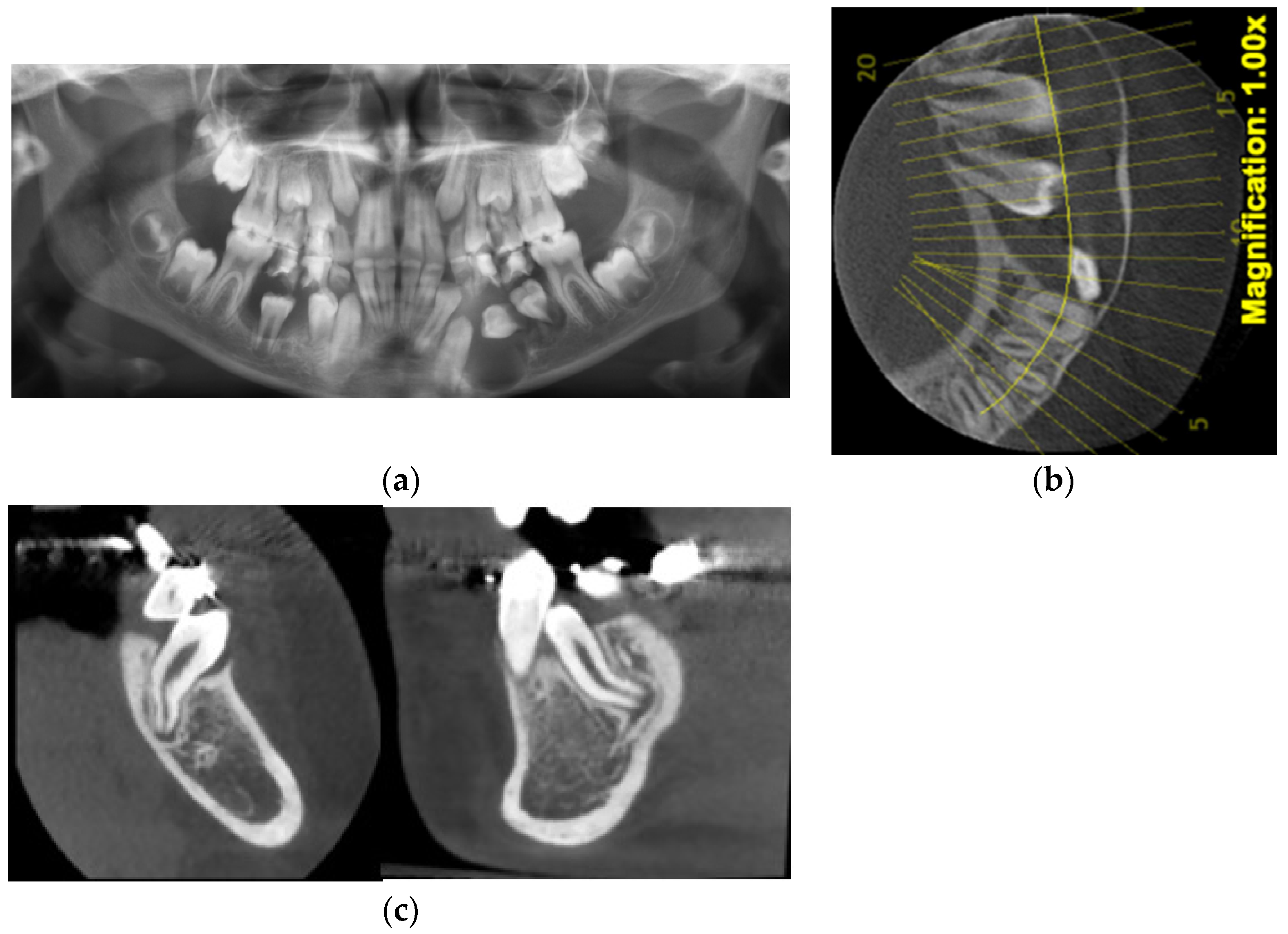
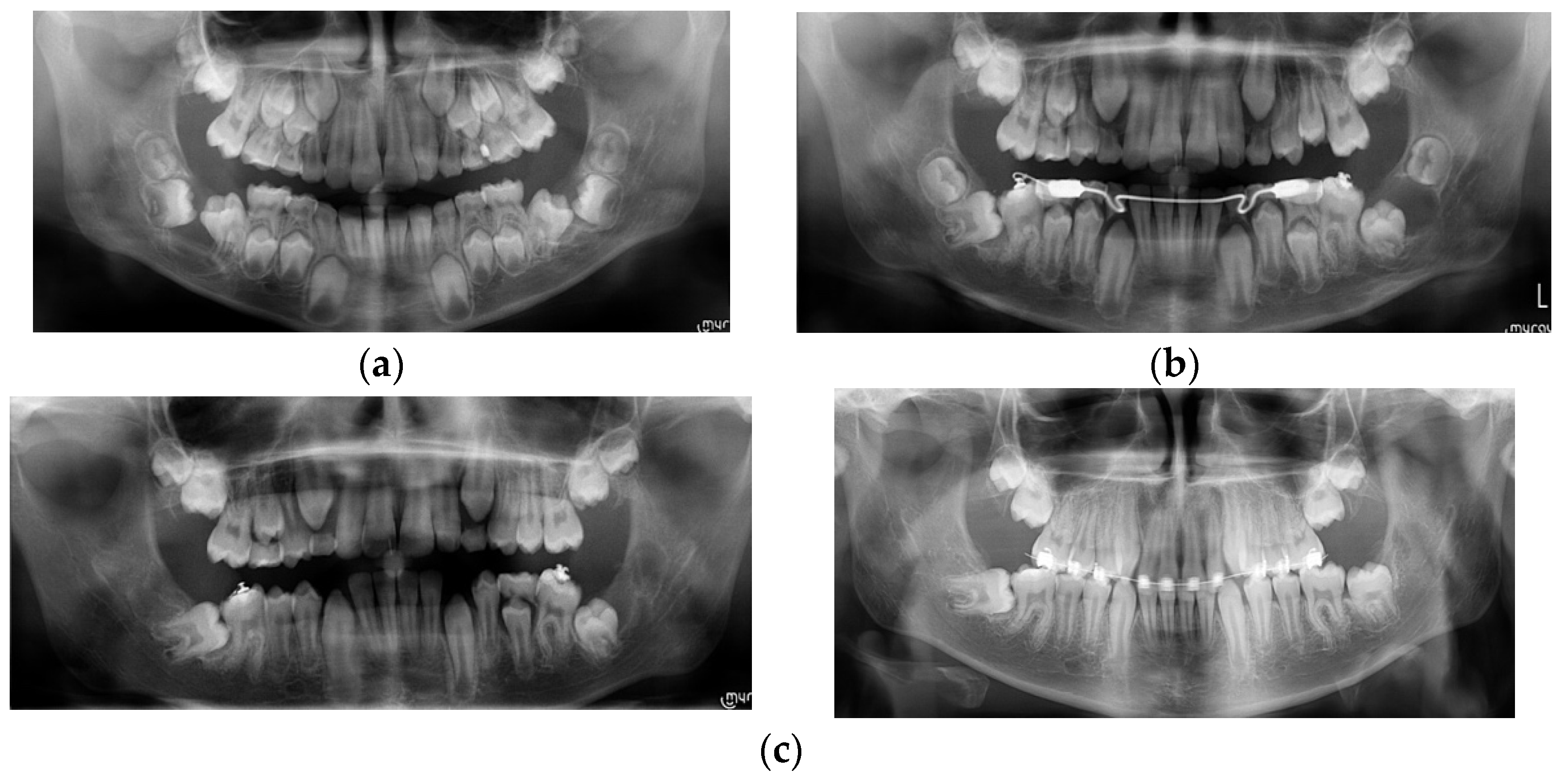
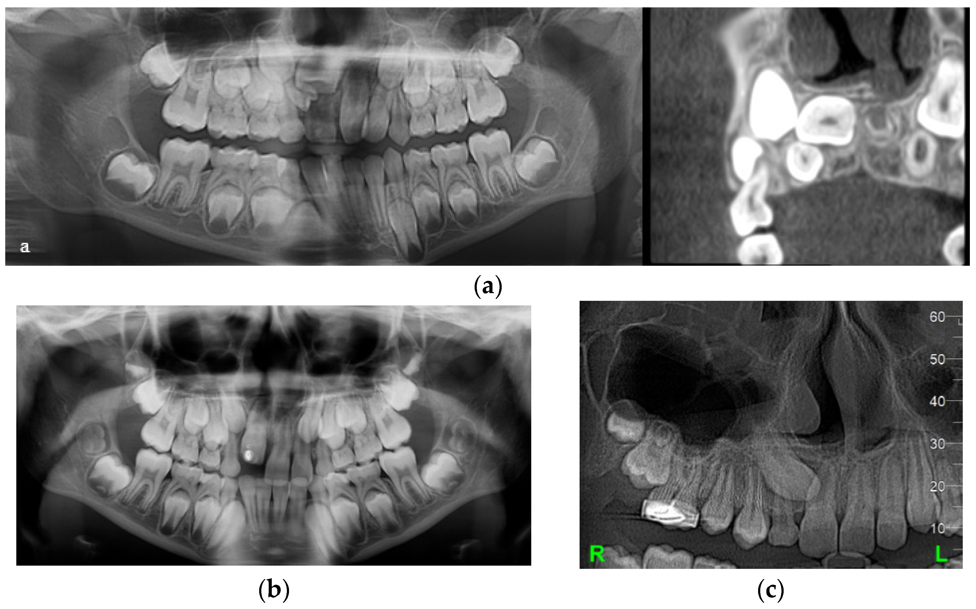
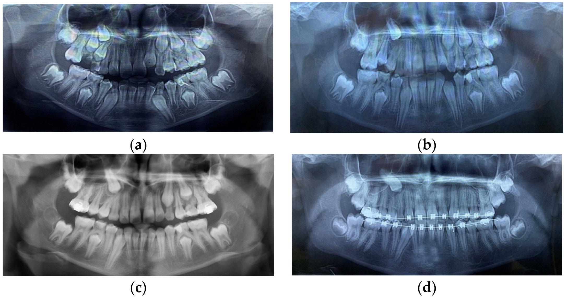
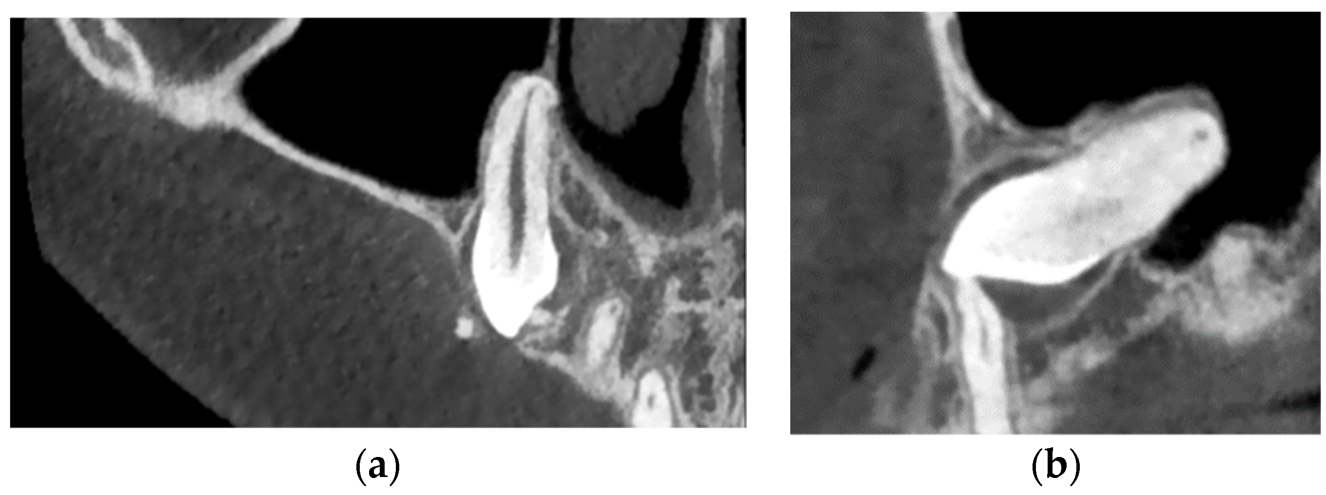
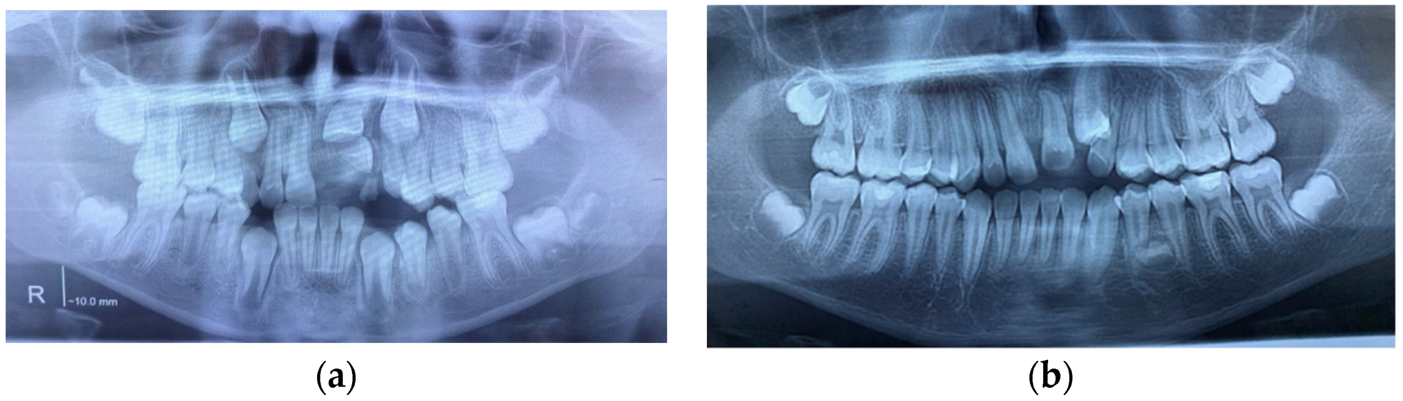
This retrospective case series study was performed on five patients (two females and three males) from a private practice aged between 8 and 10 years old. In accordance with the Helsinki Declaration of 1975, as revised in 2008, informed consent was asked of all patients for their participation in the study. The patients had sought orthodontic treatment, either for different reasons and the cyst, and subsequently, the root DL were detected at the routine X-ray exam or for the absence of a permanent tooth that must have erupted at that age. The inclusion criteria were the presence of root DL of at least one tooth and a DC of developmental origin, including the dilacerated tooth or another tooth nearby. The exclusion criteria were as follows: syndromic patients, other types of cysts, history of trauma, and previous orthodontic treatment. All types of dilacerations were included, as described in the introduction.
Both DLs and DCs were initially detected and diagnosed by an X-ray exam, especially panoramic X-ray, while differential diagnosis and more precise details were obtained with the aid of CBCT using the VGI Evo scanner (NewTom, Verona, Italy) with a field of view (FOV) of 15 × 12 cm.
The treatment objective for DC in all cases was to preserve the cyst-associated tooth and to marsupialise the cyst with or without orthodontic traction of the tooth. The use of orthodontic traction depends on the size of the cyst, the position of the tooth, tooth depth, and stage of root development [57].
The study aimed to assess the cause–effect relationship between DCs and root DLs.
Case 1
A girl aged ten was brought by her parents for orthodontic treatment due to aesthetic reasons. She was in average mixed dentition with all first premolars intraorally, except for the upper right one, where the deciduous molars presented caries and massive coronal destructions, which might be the causes of the delayed eruption of the first premolar.
At a routine X-ray exam, it was noted that the right upper canine with a DC was in a very high position in the bone, mesially inclined, in transposition with the upper first premolar and in closed contact with its apical half of the root, which was already dilacerated, with a mesioangulation (Figure 1a). This position could be a risk factor for canine impaction. Because of the lack of space and crowding, after the extraction of right deciduous molars (to facilitate the eruption of the premolars), the first step of the treatment plan was maxillary expansion, which could have also been a preventive method of canine impaction.
After maxillary expansion, the first premolar partially erupted, but the second one was impacted due to the lack of space and both distally inclined and completely formed roots. The roots of the premolars had different types of DLs, from a slight flexion of the second premolar’s root to a true dilacerated root of the first premolar. The canine remained in the same position while the DC was still growing (Figure 1b).
To obtain more space for upper premolars and canines, the patient wore a low pull headgear to distalize the upper first molar and then fixed appliances to fulfil the objective of the orthodontic treatment. Because the canine did not change its position (Figure 1c), marsupialization of the cyst was tried, and consequently, the canine started to erupt between the roots of the first premolar. At this moment, it has been decided to incline the roots of the first premolar distally to prevent root resorption by the canine.
Regarding the cause–effect association between DC and root DL, in this case, DC could be the cause of root DL (taking into consideration the initial high and mesioinclined position of the canine, in transposition with the first premolar, along with its DC that had continued to grow, in closed contact with the apical half of the first premolar root, the DLs of first premolar root as the flexion of the second premolar root). This could be explained by the pressure applied by the cyst on that area of dense bone structure. The last panoramic X-ray (Figure 1d) showed a root DL of the second upper left premolar. Thus, the DL could have another cause, namely a genetic one, as a dental anomaly pattern (DAP) or associated dental anomaly (ADA), in which many dental anomalies coexist, as in this case, the canine impaction, transposition, DC, and root DL. The root DL of the impacted canine could also be seen on the CBCT (Figure 2a,b).
Case 2
A 10-year-old boy sought orthodontic treatment for esthetic reasons due to the absence of the upper left central incisor, and malalignment of upper front teeth, and was without a history of trauma. The patient had permanent dentition with the persistence of the right deciduous canine and left lateral incisors, macrodontia and class 1 Angle malocclusion. A panoramic X-ray revealed the impaction of the left central incisor due to a supernumerary impacted tooth blocking the eruption path of the central incisor. A DC was associated with the impacted left upper incisor, and its root was not in the axis of the crown. The space for the upper left canine was closed because the lateral incisor was displaced in a distal position by the supernumerary tooth, the canine bulging buccally (Figure 3a).
The objective of the orthodontic treatment was to preserve all the permanent teeth, as the replacement of any frontal teeth is very challenging despite the successful techniques in periodontology and implantology. The treatment started with surgery, including extraction of the supernumerary tooth and marsupialization of its DCs. A removable appliance was worn to maintain the space and eventually for orthodontic traction of the impacted teeth. The eruption process of the central incisor was monitored periodically by X-ray exams, and it was found that the central incisor erupted very slowly, with a tendency for impaction, and the decision was made to apply orthodontic traction to bring it into the oral cavity. At the same time, the canine changed its eruption path in a mesial direction, in transposition with the lateral incisor. The abnormal shape of the canine, with a big cingulum, like a supernumerary cusp, root DL of the central incisors and a new tooth bud between left mandibular premolars (Figure 3b), could be seen on the panoramic X-ray. In this clinical case, the root DL was seen only in the impacted central incisor associated with the DC, and the cause of DL could be the lack of space for the root to develop in a normal direction. This case is another case of dental anomaly pattern (DAP) or associated dental anomaly (ADA)—macrodontia, impaction, supernumerary teeth, DL and DC, transposition, an early eruption of permanent teeth, and DL might also have a genetic origin.
Case 3
A 10-and-a-half-year-old girl was referred by her dentist after unexpectedly finding two big DCs associated with the mandibular premolars on an X-ray. The patient had a severe class II div 1 malocclusion, being a thumb-sucker, and an early mixed dentition due to a delayed eruption. All mandibular deciduous molars had fillings and root canal treatments that could advocate for DCs of inflammatory origin. The panoramic X-ray revealed on the right side a DC, associated with the second premolar, and on the left side both premolars are included in another DC, which was bilocular (Figure 4a). The first left premolar was in a horizontal position with the crown facing labially and root lingually (Figure 4b). The eruption of the left lateral incisor and canine was blocked by the lack of space, due to a supernumerary erupted mandibular incisor.
A lingual arch was placed to maintain the position of the first mandibular molars. Extraction of deciduous mandibular molars and the marsupialization of the cysts were performed. After surgery, all impacted teeth started their eruption in an appropriate position. Although the left first premolar changed its position to a normal one, there was not enough space for its eruption in addition to having a root DL (Figure 4c).
The left lateral incisors and canine also had small degrees of root DL. As was stated previously, the cause of the DCs could be the pulp infection of the deciduous molars, but there were differences between the two DCs and local conditions. The right DC was of inflammatory origins, and after the marsupialization, the premolar started to erupt in its normal position. On the left side, the cyst is bilocular, and the inflammatory origin could not be the only etiological factor. There are also other dental anomalies, supernumerary teeth, and the different degrees of root DL affect not only the tooth associated with the DC but also other teeth from that area. This clinical case is a DAP case.
Case 4
A 9-year-old boy came for orthodontic treatment because of the protrusion of his upper incisors. A trainer was recommended for him, but he could not manage it. The patient had a class II div 1 Angle malocclusion, mixed dentition with a delayed eruption of the permanent teeth, and the absence of the first mandibular molars. On the panoramic X-ray, it was noted that the rest of the permanent teeth on the right side (including the third molars), the first and second mandibular molars were impacted and associated with a sizeable DC, with the development of the roots delayed, and the first molar was dilacerated. On the left side, the situation was almost the same, with both the first and second molars being impacted and associated with DC. The roots of the molars were in an early stage of development, uncorrelated with the patient’s age (Figure 5a). As in all previous cases, the treatment objective regarding the DC-associated impacted teeth was to try to preserve the teeth. Therefore, the cyst’s marsupialization was done, followed by monitoring of the patients. Six months later, although the first permanent molars started to erupt slowly, orthodontic traction from a bonded attachment was applied to bring the teeth intraorally (Figure 5b). The DC of the left second molar continued to grow, blocking the molar’s eruption and the development of its roots. On the right side, the second molar became more horizontally positioned in the bone under the crown of the first molar. We decided on another marsupialization of the cyst-associated second left molar and extraction of both mandibular third molars. After four months, the left second molar was erupting in an optimal vertical position. The right second molar was still almost horizontally impacted, without any bony defect, due to DC (Figure 5c). At that moment, both first mandibular molars were in the oral cavity, well aligned in the lower arch, but not in the appropriate one, because of the roots DL. There was also a slight root flexion of the right mandibular canine.
The aforementioned is a rare case of bilateral mandibular DC-associated first and second molars, impaction, and root DL of the first molar. The DCs could be the cause of a root DL because the tooth did not erupt, and the roots were constrained to develop in a closed area of dense bone. Another factor that could determine the first molar’s root DL was the pressure applied by DCs of the second molar’s and probably the third molar’s buds. Because the root DL also affected the canine, it could also be considered the genetic etiology of root DL in a DAP patient.
Case 5
An 8-year-old boy was brought by his parents for a second opinion. His parents’ concern was the absence of the upper right incisors. He would have been wearing a removable appliance for maxillary expansion, but he could not accept it. The patient had a class III Angle malocclusion and a delayed mixed dentition without central and lateral upper right incisors. The panoramic X-ray showed that the upper right central incisor was impacted in a very high position, almost horizontally, with the root in the palatal bone. There was also a congenitally missing lateral incisor and a supernumerary tooth, partially below the central incisor. The CBCT also revealed a DC-associated upper central incisor (Figure 6a).
Along with the supernumerary tooth extraction, the marsupialization of the DC was performed, and the central incisor started its eruption. After a while, a second surgical intervention was done to bond an attachment for orthodontic traction of the central incisor, which had an abnormal mesiodistal width—gigantism. Because of its size, the space for the missing right lateral incisor was closed, and there was not enough space for the canine, which remained impacted, included in a DC. The new panoramic X-ray showed the root DL of all the incisors, being more evident on the left side (Figure 6b). For the left central incisor, the root DL could be explained by the closed contact of DC-associated right central incisor DC with its root at different vertical levels, as seen on the CBCT. For establishing the treatment plan, the long-term prognosis of the right canine had to be taken into consideration with the aid of a new CBCT. It was noted on the CBCT that the canine is impacted, included in a bigger DC than initially seen, and with root DL (Figure 6c). The case described above was a DAP of genetic origin, impaction of the central incisor, congenitally missing lateral incisor associated with canine impaction, and DCs including impacted teeth. The root DL of the incisors and right canine could be caused in this case not only by DC and deep impaction of the central incisors, but also by genetic causes.

3. Results

Table 1 summarizes the radiological findings of each case, which are the basis for diagnosing DC and DL.
Table 2 shows the cases where the DLs could be caused by DCs, due to their proximity. A “Y” indicates the presence of both DL and DC in the same area, either situated on the same arch or on both arches but close to each other.

4. Discussion

Although DL and DCs have been reported in the literature, mainly as case reports, the cause–effect relationship between has scarcely been debated. Root DL is of great importance to orthodontics because dilacerated roots are harder to move orthodontically, have a higher risk of tooth impaction and root resorption, and impede the favorable insertion of miniscrews [58].
The cause of DL is still unknown, but many theories have been proposed, one of them being DC-associated with the dilacerated tooth or in closed contact with it. The purpose of the case series retrospective stud was to analyze if the DCs could be the single cause of root DLs. The patients included in this study were randomly selected, excluding patients with trauma or syndromic origin as etiology.
Generally, both tooth DL and DCs are diagnosed unexpectedly at a routine X-ray exam. Therefore, a detailed 3D imaging investigation, CBCT, is mandatory to establish the appropriate treatment plan and reduce the severity of the case. Accordingly, all the patients included in the study underwent panoramic X-rays and CBCTs.
In accordance with other studies, in the present case series study, DL had no sex predilection, occurred anywhere along the length of the root, and affected any teeth [36]. A recent study showed that root DLs in incisors, canines, and premolars are most common in the apical third of the roots. DL within the middle third of the root is more frequent in molars, whereas DL within the coronal third of the root is most commonly seen in third molars [16]. Crown DLs are less common than root DLs, and they usually occur in maxillary permanent incisors because of their proximity to primary incisors [23,24]. Our findings have shown that in cases where the DL occurred in the cyst-associated tooth, the middle or coronal third of the root is affected (cases 3 and 5). In one of five cases, bilateral DL occurs in mandibular first molars.
Without a consensus among researchers, the etiology of DL is not fully understood, thus many etiologic factors are considered, such as trauma, scar formation, a developmental anomaly of the primary tooth germ, facial clefting, advanced root canal infections, ectopic development of the tooth germ, lack of space, and the effect of anatomic structures (cortical bone of the maxillary sinus, mandibular canal, or nasal fossa), which might deflect the epithelial diaphragm, presence of an adjacent cyst, tumor, or odontogenic hamartoma, mechanical interference with the eruption from an ankylosed primary tooth that does not resorb, tooth transplantation, extraction of primary teeth, and genetic factors [7,12,15,16,17,21,23,58,59,60,61,62,63,64,65,66,67]. The involvement of the genetic factor can also be enhanced by the fact that the identification of the master genes, from the gene regulatory system, responsible for induction and tooth regeneration should be taken into consideration to obtain an appropriate architectural design of a bioengineered tooth [68]. There is also another hypothesis regarding DL etiology affecting the middle and apical part of the root–tooth root dilacerations due to the bone gradients of plasticity present in alveolar bone, which could be affected by the rate of eruption, jaw rotation, length of the root, length of the eruption path, delayed eruption, transverse uprighting of molar teeth during eruption, and root resorption [45,55].
The present study aimed to find if the presence of DC, mentioned in the literature as an etiologic factor, could be the single cause of tooth DL. The results show that in all five cases, the presence of a DC is associated with root DLs either in the adjacent teeth or in the cyst-associated tooth. Consequently, DC could be considered the cause of DL. In all of these clinical situations, there was a lack of space in the affected area caused by another etiologic factor, such as a supernumerary tooth, impaction of an adjacent tooth, or a delayed eruption. Bone density should also be taken into consideration as another etiologic factor (cases 3 and 4), together with jaw rotation (5), closed proximity of different anatomical structures (sinus-case 1; nasal cavity-case2,5) and length of the eruption path (case 1). At least one of the following dental anomalies was noted in every case: supernumerary tooth, transposition, impaction, congenitally missing tooth, delayed eruption, and in cases 1, 3 and 4, root DLs also affected other teeth away from the area of the DC-associated tooth. Thus, another etiologic factor could be the genetic origin of DL.
While considering the limitations of the present case series study, it could be stated that except for trauma, the etiology of DC is multifactorial, and associated with dental anomaly patterns of a genetic origin. DC could cause tooth DL in association with other local or genetic factors.
As a dental anomaly pattern, early treatment for DLs is important to reduce the complexity of the treatment and save the teeth. Sometimes classic DC coexists with other serious conditions, such as keratocyst or ameloblastoma. DCs may have a multilocular aspect, probably due to their growth in different density areas [50,51]. For particular cases, a histological exam is mandatory for differential diagnosis and should be considered for choosing the treatment modality, such as marsupialization or cystectomy (cyst enucleation).
The main objective of the treatment of DC should be the preservation of the tooth associated with DC (a conservative treatment), which also depends on the depth of the impaction, the position of the impacted tooth, and to reduce, if possible, the severity of the tooth DLs because of the consequences of root DL, not only for orthodontic treatment but also for endodontic and surgical treatments [52,53]. Initially, in all cases from our study, marsupialization of the DC was performed, followed by spontaneous eruption of the tooth or orthodontic traction, as was described for each clinical case.

5. Conclusions

In conclusion, despite the limitations of this study, the association of DC and root DL might be of genetic origins within dental anomaly patterns.

Author Contributions

Conceptualization, A.M.E.; methodology, D.F.; software, F.B.; validation, D.F., M.G. and M.D.; formal analysis, D.F. and E.B.N.; investigation, A.M.E. and M.D.; resources, F.B.; data curation, F.B.; writing—original draft preparation, A.M.E.; writing—review and editing, E.B.N.; visualization, M.G.; supervision, A.M.E. and D.F.; project administration, A.M.E. All authors have equal contribution with the first author. All authors have read and agreed to the published version of the manuscript.

Funding

This research received no external funding.

Institutional Review Board Statement

Not applicable.

Informed Consent Statement

Informed consent was obtained from all subjects involved in the study.

Conflicts of Interest

The authors declare no conflict of interest.

Abbreviations

DLroot dilaceration
DCdentigerous cyst
3Dthree dimensional
CBCTcone-beam computed tomography
DAPdental anomaly pattern
ADAassociated dental anomaly

References

  1. Tomes, J. A Course of Lectures on Dental Physiology and Surgery. Am. J. Dent. Sci. 1848, 8, 313–350. [Google Scholar] [PubMed]
  2. BSI 4492; Glossary of Dental Terms. British Standards Institute: London, UK, 1983.
  3. Shafer, W.G.; Hine, M.K.; Levy, B.M. A Textbook of OralPathology; WB Saunders Co.: Philadelphia, PA, USA, 1983; pp. 308–311. [Google Scholar]
  4. Tiecke, R.W.; Stuteville, O.H.; Calandra, J.C. Pathologic Physiology of Oral Disease; CV Mosby Co.: St Louis, MI, USA, 1959. [Google Scholar]
  5. Hamasha, A.A.; Al-Khateeb, T.; Darwazeh, A. Prevalence of dilaceration in Jordanian adults. Int. Endod. J. 2002, 35, 910–912. [Google Scholar] [CrossRef] [PubMed]
  6. Chohayeb, A.A. Dilaceration of permanent upper lateral incisors: Frequency, direction, and endodontic treatment implications. Oral. Surg. Oral. Med. Oral. Pathol. 1983, 55, 519–520. [Google Scholar] [CrossRef]
  7. Andreasen, J.O.; Sundström, B.; Ravn, J.J. The effect of traumatic injuries to primary teeth on their permanent successors. I. A clinical and histologic study of 117 injured permanent teeth. Scand. J. Dent. Res. 1971, 79, 219–283. [Google Scholar] [CrossRef] [PubMed]
  8. Andreasen, J.O.; Flores, M.T. Injuries to Developing Teeth. In Textbook and Color Atlas of Traumatic Injuries to the Teeth; Andreasen, J.O., Andreasen, F.M., Andreasen, L., Eds.; Blackwell Munksgaard: Oxford, UK, 2007; pp. 542–576. [Google Scholar]
  9. Becker, A. Maxillary central incisors. In The Orthodontic Treatment of Impacted Teeth; Becker, A., Ed.; Wiley-Blackwell: Hoboken, NJ, USA, 2012; pp. 70–109. [Google Scholar]
  10. Bimstein, E. Root dilaceration and stunting in two unerupted primary incisors. ASDC J. Dent. Child. 1978, 45, 223–225. [Google Scholar] [PubMed]
  11. Neville, B.W.; Damm, D.D.; Allen, C.M.; Bouquot, J.E. Oral and Maxillofacial Pathology, 2nd ed.; WB Saunders: Philadelphia, PA, USA, 2002; pp. 86–88. [Google Scholar]
  12. Yeung, K.H.; Cheung, R.C.; Tsang, M.M. Compound odontoma associated with an unerupted and dilacerated maxillary primary central incisor in a young patient. Int. J. Paediatr. Dent. 2003, 13, 208–212. [Google Scholar] [CrossRef]
  13. Rengaswamy, V. Bilateral dilaceration of maxillary central incisors. Oral. Surg. Oral. Med. Oral. Pathol. 1979, 47, 200. [Google Scholar] [CrossRef]
  14. Edmondson, H.D.; Crabb, J.J. Dilaceration of both upper central incisor teeth: A case report. J. Dent. 1975, 3, 223–224. [Google Scholar] [CrossRef]
  15. Lin, L.; Dowden, W.E.; Langeland, K. Bilateral dilaceration. J. Endod. 1982, 8, 85–87. [Google Scholar] [CrossRef]
  16. Malcić, A.; Jukić, S.; Brzović, V.; Miletić, I.; Pelivan, I.; Anić, I. Prevalence of root dilaceration in adult dental patients in Croatia. Oral. Surg. Oral. Med. Oral. Pathol. Oral. Radiol. Endod. 2006, 102, 104–109. [Google Scholar] [CrossRef]
  17. Seow, W.K.; Perham, S.; Young, W.G.; Daley, T. Dilaceration of a primary maxillary incisor associated with neonatal laryngoscopy. Pediatr. Dent. 1990, 12, 321–324. [Google Scholar] [PubMed]
  18. Kilpatrick, N.M.; Hardman, P.J.; Welbury, R.R. Dilaceration of a primary tooth. Int. J. Paediatr. Dent. 1991, 1, 151–153. [Google Scholar] [CrossRef] [PubMed]
  19. Kearns, H.P. Dilacerated incisors and congenitally displaced incisors: Three case reports. Dent. Update 1998, 25, 339–342. [Google Scholar]
  20. Maragakis, M.G. Crown dilaceration of permanent incisors following trauma to their primary predecessors. J. Clin. Pediatr. Dent. 1995, 20, 49–52. [Google Scholar] [PubMed]
  21. Chadwick, S.M.; Millett, D. Dilaceration of a permanent mandibular incisor. A case report. Br. J. Orthod. 1995, 22, 279–281. [Google Scholar] [CrossRef] [PubMed]
  22. Feldman, B.S. Tooth with a 'tail'. A case report of a dilacerated mandibular incisor. Br. J. Orthod. 1984, 11, 42–43. [Google Scholar] [CrossRef] [PubMed]
  23. Matsuoka, T.; Sobue, S.; Ooshima, T. Crown dilaceration of a first premolar caused by extraction of its deciduous predecessor: A case report. Endod. Dent. Traumatol. 2000, 16, 91–94. [Google Scholar] [CrossRef]
  24. Asokan, S.; Rayen, R.; Muthu, M.S.; Sivakumar, N. Crown dilaceration of maxillary right permanent central Incisor—A case report. J. Indian Soc. Pedod. Prev. Dent. 2004, 22, 197–200. [Google Scholar]
  25. Agnihotri, A.; Marwah, N.; Dutta, S. Dilacerated unerupted central incisor: A case report. J. Indian Soc. Pedod. Prev. Dent. 2006, 24, 152–154. [Google Scholar] [CrossRef]
  26. Prabhakar, A.R.; Reddy, V.V.; Bassappa, N. Duplication and Dilaceration of a Crown with Hypercementosis of the Root Following Trauma: A Case Report. Quintessence Int. 1998, 29, 655–657. [Google Scholar]
  27. Meadow, D.M.; Needleman, H.L. Dilaceration of the mandibular permanent incisor teeth: Two case reports. Pediatr. Dent. 1981, 3, 276–278. [Google Scholar] [PubMed]
  28. Yen, H.T.; Diau, M.H.; Tsai, J.W.; Roan, R.T. Dilaceration of a central incisor with iatrogenic root perforation—A case report. Gaoxiong Yi Xue Ke Xue Za Zhi 1991, 7, 386–390. [Google Scholar] [PubMed]
  29. McNamara, T.G.; McNamara, C.M. Dilaceration of a mandibular incisor: Case report. Singap. Dent. J. 2000, 23, 29–31. [Google Scholar]
  30. Maia, R.L.; Vieira, A.P. Auto-transplantation of central incisor with root dilaceration. Technical note. Int. J. Oral. Maxillofac. Surg. 2005, 34, 89–91. [Google Scholar] [CrossRef] [PubMed]
  31. Glenn, F.B.; Stanley, H.R., Jr. Dilaceration of a mandibular permanent incisor. Report of a case. Oral. Surg. Oral. Med. Oral. Pathol. 1960, 13, 1249–1252. [Google Scholar] [CrossRef]
  32. Ngeow, W.C. An unusual dilacerated root of a second maxillary molar. J. Ir. Dent. Assoc. 1996, 42, 51–52. [Google Scholar]
  33. Edler, R. Dilaceration of upper central and lateral incisors. A case report. Br. Dent. J. 1973, 135, 331–332. [Google Scholar] [CrossRef]
  34. Lowe, P.L. Dilaceration caused by direct penetrating injury. Br. Dent. J. 1985, 159, 373–374. [Google Scholar] [CrossRef]
  35. Thongudomporn, U.; Freer, T.J. Prevalence of dental anomalies in orthodontic patients. Aust. Dent. J. 1998, 43, 395–398. [Google Scholar]
  36. Eversole, L.R. Clinical Outline of Oral Pathology: Diagnosis and Treatment, 3rd ed.; BC Decker Inc.: Hamilton, ON, Canada, 2002; p. 350. [Google Scholar]
  37. Jafarzadeh, H.; Abbott, P.V. Dilaceration: Review of an endodontic challenge. J. Endod. 2007, 33, 1025–1030. [Google Scholar] [CrossRef]
  38. Smith, D.M.; Winter, G.B. Root dilaceration of maxillary incisors. Br. Dent. J. 1981, 150, 125–127. [Google Scholar] [CrossRef] [PubMed]
  39. McNamara, T.; Woolfe, S.N.; McNamara, C.M. Orthodontic management of a dilacerated maxillary central incisor with an unusual sequela. J. Clin. Orthod. 1998, 32, 293–297. [Google Scholar] [PubMed]
  40. Walia, P.S.; Rohilla, A.K.; Choudhary, S.; Kaur, R. Review of Dilaceration of Maxillary Central Incisor: A Mutidisciplinary Challenge. Int. J. Clin. Pediatr. Dent. 2016, 9, 90–98. [Google Scholar] [CrossRef] [PubMed]
  41. Von Gool, A.V. Injury to the permanent tooth germ after trauma to the deciduous predecessor. Oral. Surg. Oral. Med. Oral. Pathol. 1973, 35, 2–12. [Google Scholar] [CrossRef]
  42. Bassetti, M.A.; Kuttenberger, J.; Novak, J.; Bassetti, R.G. Die follikuläre Zyste: Zwei unterschiedliche Behandlungsmöglichkeiten, präsentiert anhand zweier Fallbeispiele [The dentigerous cyst: Two different treatment options illustrated by two cases]. Swiss. Dent. J. 2019, 129, 193–203. (In German) [Google Scholar] [PubMed]
  43. Hu, N.N.; Li, Y.Y.; Wang, J.W.; Hou, Z.J.; Yang, X.C. A case report of multiple dentigerous cyst of mandible and review of literature. Shanghai Kou Qiang Yi Xue 2019, 28, 110–112. (In Chinese) [Google Scholar] [PubMed]
  44. Dhupar, A.; Yadav, S.; Dhupar, V.; Mittal, H.C.; Malik, S.; Rana, P. Bi-maxillary dentigerous cyst in a non-syndromic childReview of literature with a case presentation. J. Stomatol. Oral. Maxillofac. Surg. 2017, 118, 45–48. [Google Scholar] [CrossRef]
  45. Daley, T.D.; Wysocki, G.P.; Pringle, G.A. Relative incidence of odontogenic tumors and oral and jaw cysts in a Canadian population. Oral. Surg. Oral. Med. Oral. Pathol. 1994, 77, 276–280. [Google Scholar] [CrossRef]
  46. Ziccardi, V.B.; Eggleston, T.I.; Schneider, R.E. Using fenestration technique to treat a large dentigerous cyst. J. Am. Dent. Assoc. 1997, 128, 201–205. [Google Scholar] [CrossRef]
  47. Shetty, R.M.; Dixit, U. Dentigerous Cyst of Inflammatory Origin. Int. J. Clin. Pediatr. Dent. 2010, 3, 195–198. [Google Scholar] [CrossRef]
  48. Grewal, H.K.; Batra, R. Non Syndromic Bilateral Dentigerous Cysts: A case report. Int. J. Dent. Clin. 2010, 2, 49–51. [Google Scholar]
  49. Terauchi, M.; Akiya, S.; Kumagai, J.; Ohyama, Y.; Yamaguchi, S. An analysis of dentigerous cysts developed around a mandibular third molar by panoramic radiographs. Dent. J. 2019, 7, 13. [Google Scholar] [CrossRef]
  50. Zhang, L.L.; Yang, R.; Zhang, L.; Li, W.; MacDonald-Jankowski, D.; Poh, C.F. Dentigerous cyst: A retrospective clinicopathological analysis of 2082 dentigerous cysts in British Columbia, Canada. Int. J. Oral. Maxillofac. Surg. 2010, 39, 878–882. [Google Scholar] [CrossRef] [PubMed]
  51. Martinelli-Kläy, C.P.; Martinelli, C.R.; Martinelli, C.; Macedo, H.R.; Lombardi, T. Unusual Imaging Features of Dentigerous Cyst: A Case Report. Dent. J. 2019, 7, 76. [Google Scholar] [CrossRef] [PubMed]
  52. Hyomoto, M.; Kawakami, M.; Inoue, M.; Kirita, T. Clinical conditions for eruption of maxillary canines and mandibular premolars associated with dentigerous cysts. Am. J. Orthod. Dentofacial. Orthop. 2003, 124, 515–520. [Google Scholar] [CrossRef]
  53. Ghandour, L.; Bahmad, H.F.; Bou-Assi, S. Conservative Treatment of Dentigerous Cyst by Marsupialization in a Young Female Patient: A Case Report and Review of the Literature. Case Rep. Dent. 2018, 2018, 7621363. [Google Scholar] [CrossRef]
  54. Bansal, S.; Kaur, H.; Bansal, R.N.; Goyal, P. An idiosyncratic post-traumatic tetrad: Compound odontome, dentigerous cyst, impaction, and double-dilaceration. Quintessence Int. 2014, 45, 885–889. [Google Scholar] [CrossRef]
  55. Tanaka, O.M.; Meira, T.M.; Batista, R.A.C.; Willems, G.; Baggio, G.L.; Couto, S.P.H. Marsupialization of a Large Radicular Cyst with Extensive Maxillary Tooth Displacement: Eight-Year Follow-Up. J. Dent. Child. 2019, 86, 64–68. [Google Scholar]
  56. Osório, L.B.; Ferrazzo, V.A.; Serpa, G.; Ferrazzo, K.L. Apicotomy as treatment for failure of orthodontic traction. Case Rep. Dent. 2013, 2013, 168232. [Google Scholar] [CrossRef]
  57. Aoki, N.; Ise, K.; Inoue, A.; Kosugi, Y.; Koyama, C.; Iida, M.; Baba, J.; Iwai, T.; Mitsudo, K. Multidisciplinary approach for treatment of a dentigerous cyst—Marsupialization, orthodontic treatment, and implant placement: A case report. J. Med. Case Rep. 2018, 12, 305. [Google Scholar] [CrossRef]
  58. Cao, D.; Shao, B.; Izadikhah, I.; Xie, L.; Wu, B.; Li, H.; Yan, B. Root dilaceration in maxillary impacted canines and adjacent teeth: A retrospective analysis of the difference between buccal and palatal impaction. Am. J. Orthod. Dentofacial. Orthop. 2021, 159, 167–174. [Google Scholar] [CrossRef] [PubMed]
  59. Gorlin, R.J.; Goldman, H.M. Thoma’s Oral Pathology, 6th ed.; Mosby: St Louis, MI, USA, 1970; pp. 104–106. [Google Scholar]
  60. Kalra, N.; Sushma, K.; Mahapatra, G.K. Changes in developing succedaneous teeth as a consequence of infected deciduous molars. J. Indian Soc. Pedod. Prev. Dent. 2000, 18, 90–94. [Google Scholar]
  61. Stewart, D.J. Dilacerate unerupted maxillary central incisors. Br. Dent. J. 1978, 145, 229–233. [Google Scholar] [CrossRef] [PubMed]
  62. Walton, R.E.; Torabinejad, M. Principles and Practice of Endodontics, 3rd ed.; WB Saunders: Philadelphia, PA, USA, 2002; pp. 176–179. [Google Scholar]
  63. Atwan, S.M.; Turner, D.; Khalid, A. Early intervention to remove mesiodens and avoid orthodontic therapy. Gen. Dent. 2000, 48, 166–169. [Google Scholar] [PubMed]
  64. Dayi, E.; Gürbüz, G.; Bilge, O.M.; Ciftcioğlu, M.A. Adenomatoid odontogenic tumour (adenoameloblastoma). Case report and review of the literature. Aust. Dent. J. 1997, 42, 315–318. [Google Scholar] [CrossRef] [PubMed]
  65. Angelos, G.M.; Smith, D.R.; Jorgenson, R.; Sweeney, E.A. Oral complications associated with neonatal oral tracheal intubation: A critical review. Pediatr. Dent. 1989, 11, 133–140. [Google Scholar] [PubMed]
  66. Proffit, W.R.; Fields, H.W., Jr.; Ackerman, J.L.; Bailey, L.J.; Camilla, T.J.F. Contemporary Orthodontics, 3rd ed.; Mosby: St Louis, MI, USA, 2000; Volume 78, p. 124. [Google Scholar]
  67. Monsour, F.N.; Adkins, K.F. Responses of periodontal tissues and cementum following transplantation of teeth. J. Oral. Maxillofac. Surg. 1984, 42, 441–446. [Google Scholar] [CrossRef]
  68. Olaru, M.; Sachelarie, L.; Calin, G. Hard Dental Tissues Regeneration—Approaches and Challenges. Materials 2021, 14, 2558. [Google Scholar] [CrossRef]
Figure 1. (a) Initial panoramic X-ray showing the upper right canine in a very high position and transposition with the first premolar. (b,c) Different types of root DL affecting right premolars. No improvement regarding the position of the right canine after maxillary expansion. (d) The upper left second premolar also has a root DL.
Applsci 12 09854 g001
Figure 2. (a) The right maxillary canine also has a root DL, presenting a “hook” bend towards the mesial in the apical third of the root. (b) “Bull’s eye” phenomena highlighted in the sagittal section.
Applsci 12 09854 g002
Figure 3. (a) Impacted upper left central incisor blocked by a DC surrounding its crown and a supernumerary tooth. (b) Abnormal shape of the canine, root DL of the upper left central incisor, and a new tooth bud developing in the lower arch.
Applsci 12 09854 g003
Figure 4. (a) Initial panoramic X-ray revealed large DCs on both sides of the mandible. (b) CBCT image showing the labiolingual orientation of the lower left first and second premolars. (c) CBCT showing root DL of the left first premolar.
Applsci 12 09854 g004
Figure 5. (a) Bilateral impaction of lower first and second molars associated with DCs. All four molars presenting abnormal root development, uncorrelated with the age of the patient. (b) Orthodontic traction applied from a lingual arch to aid the eruption of the lower first molars. (c) Progress records at four months after the removal of both mandibular third molars.
Applsci 12 09854 g005
Figure 6. (a) Impaction of the central incisor, congenitally missing lateral incisor, and a supernumerary tooth were noticed on the initial panoramic X-ray and CBCT. (b) Panoramic X-ray showing the root DL of all the incisors. (c) CBCT—upper right impacted canine presenting a DC.
Applsci 12 09854 g006
Table 1. Summary of the radiological findings of the cases described above—the association between DL and other dental anomalies of genetic origin.
Case
Number
Summary of X-ray Findings
Case 1
  • Impacted upper right canine.
  • Right upper canine with a DC, in transposition with the upper first premolar and in closed contact with its apical half of the root.
  • The roots of the premolars had different types of DLs, from a slight flexion of the second premolar’s root to a true dilacerated root of the first premolar.
  • The DC was continually growing.
  • Root DL of the second upper left premolar.
Case 2
  • Impacted left central incisor.
  • Impacted supernumerary tooth.
  • Impacted upper left canine.
  • Macrodontia.
  • A DC was associated with the impacted left central incisor and its root was not in the axis of the crown.
  • Root DL of the left central incisors.
  • Abnormal shape of the canine, with a big cingulum, like a supernumerary cusp.
  • A new tooth bud between the left mandibular premolars.
Case 3
  • Two big DCs associated with the mandibular premolars.
  • Right side a DC-associated with the second premolar.
  • Left side, both premolars are included in another DC, which was bilocular.
  • The first left premolar was in a horizontal position with the crown facing labially and root lingually.
  • The eruption of the left lateral incisor and canine was blocked by the lack of space, due to a supernumerary erupted mandibular incisor.
  • All mandibular deciduous molars had fillings and root canal treatments that could advocate for DCs of inflammatory origin.
  • Although the left first premolar changed its position to a normal one, there was not enough space for its eruption, in addition to having a root DL.
  • The left lateral incisors and canine also had small degrees of root DL.
  • There were differences between the two DCs and local conditions.
  • That right DC is of inflammatory origin, and after the marsupialization, the premolar started to erupt in its normal position.
  • On the left side, the cyst is bilocular, and the inflammatory origin could not be the only etiological factor.
Case 4
  • On the right side, the first and second mandibular molars (including the third molars) were impacted and associated with a sizeable DC, with the development of the roots delayed and the first molar being dilacerated.
  • On the left side, the situation was almost the same, with both the first and second molars being impacted and associated with DC.
  • The DC of the left second molar continued to grow, blocking the molar’s eruption and the development of its roots.
  • At that moment, both first mandibular molars were in the oral cavity, well aligned in the lower arch, but not in the appropriate one, because of the roots’ DL.
  • Impaction and root DL of the right/left first molar.
  • The DCs could be the cause of root DL because the tooth did not erupt, and the roots were constrained to develop in a closed area of dense bone.
  • Another factor that could determine the first molar’s root DL was the pressure applied by DCs of the second molar’s and likely the third molar’s buds.
Case 5
  • The upper right central incisor was impacted in a very high position, almost horizontally, with the root in the palatal bone.
  • DC-associated upper right central incisor.
  • Congenitally missing lateral incisor and a supernumerary tooth, partially below the central incisor.
  • Because of the gigantism of the central incisor, the space for the missing right lateral incisor was closed, and there was not enough space for the canine, which remained impacted, included in a DC.
  • Root DL of all the incisors, being more evident on the left side.
  • For the left central incisor, the root DL could be explained by the closed contact of the DC-associated right central incisor, with its root at different vertical levels seen on the CBCT.
The right canine is impacted, included in a bigger DC than initially seen and with root DL.
Table 2. Association between DCs and DLs.
Case No./Position of DLs or DCs1 Right DL1 Left DL23 Right DC3 Left DC4 Right
DC
4 Left DC5 Right DL5 Left DL
DLYYYNYYYYY
DCYNYYYYYYY 1
Legend: “No.”: number; “DL”: root dilaceration; “DC”: dentigerous cyst; “Y”: yes; “N”: no. 1 In case 5, although the DCs are situated in the right quadrant, the left central incisor is close to the DC of the right central incisor, therefore, in the table, a “Y” signals an association between the DL in the left quadrant and the DC in the right quadrant.
Publisher’s Note: MDPI stays neutral with regard to jurisdictional claims in published maps and institutional affiliations.

