Applications of Artificial Intelligence in Endobronchial Ultrasound for Lung Cancer Diagnosis and Staging: A Scoping Review

 ,
,
Simple Summary
Abstract
1. Introduction
2. Methods
2.1. Study Design
2.2. Research Question
2.3. Eligibility Criteria
2.4. Search Strategy
2.5. Study Selection
2.6. Data Extraction and Synthesis of Results
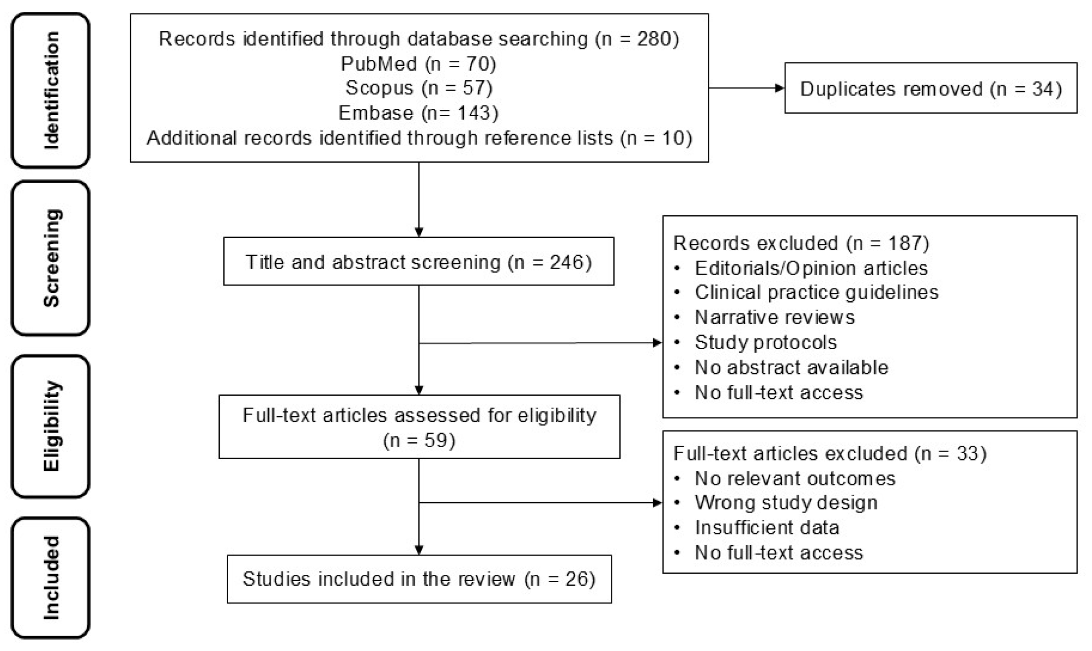
3. Results
4. Discussion
Limitations and Strengths
5. Conclusions
Supplementary Materials
Author Contributions
Funding
Institutional Review Board Statement
Informed Consent Statement
Data Availability Statement
Conflicts of Interest
References
- Smolarz, B.; Łukasiewicz, H.; Samulak, D.; Piekarska, E.; Kołaciński, R.; Romanowicz, H. Lung Cancer-Epidemiology, Pathogenesis, Treatment and Molecular Aspect (Review of Literature). Int. J. Mol. Sci. 2025, 26, 2049. [Google Scholar] [CrossRef] [PubMed]
- Deshpand, R.; Chandra, M.; Rauthan, A. Evolving trends in lung cancer: Epidemiology, diagnosis, and management. Indian J. Cancer 2022, 59, S90–S105. [Google Scholar] [CrossRef]
- Lynge, E.; Andersen, B.; Bojesen, S.E.; Petersen, L.K.; Bech, M.; Bøgsted, M.; Dam, M.S.; Egstrand, S.; Hvass, A.M.F.; Kobylecki, C.J.; et al. Future of Cancer Screening Working Group. Challenges in the future of cancer screening. Int. J. Cancer 2026, 158, 2013–2022. [Google Scholar] [CrossRef]
- Bouchard, N.; Daaboul, N. Lung Cancer: Targeted Therapy in 2025. Curr. Oncol. 2025, 32, 146. [Google Scholar] [CrossRef] [PubMed]
- Lam, S.; Baldwin, D.R.; Devaraj, A.; Field, J.; Henschke, C.I.; Heuvelmans, M.A.; Huber, R.M.; Jones, C.; Borondy-Kitts, A.; Ching Li, M.S.; et al. A Game-Changing 20 Years: Progress and Future Directions in Lung Cancer Screening. J. Thorac. Oncol. 2026, 21, 235–252. [Google Scholar] [CrossRef] [PubMed]
- Qi, Y.J.; Zhang, J.; Salcedo Lobera, E.; Song, Q.Y.; Zhong, R.H.; Kontogianni, K.; Huang, Z.S.; Ariza-Prota, M.; Gupta, N.; Madan, M.; et al. Endobronchial ultrasound-guided mediastinal biopsies for the diagnosis of mediastinal diseases: A network meta-analysis. Pulmonology 2026, 32, 2593067. [Google Scholar] [CrossRef]
- Fan, Y.; Zhang, A.M.; Wu, X.L.; Huang, Z.S.; Kontogianni, K.; Sun, K.; Fu, W.L.; Wu, N.; Kuebler, W.M.; Herth, F.J.F. Transbronchial needle aspiration combined with cryobiopsy in the diagnosis of mediastinal diseases: A multicentre, open-label, randomised trial. Lancet Respir. Med. 2023, 11, 256–264. [Google Scholar] [CrossRef]
- Tuta-Quintero, E.; Giraldo-Cadavid, L.F.; Sanmiguel-Reyes, C.; Navia, M.E.; Cardenas, R.; Bastidas, A.; Mora, A.; Páez-Espinel, N.; Viola, L.; Suárez, M.; et al. Optimizing diagnostic yield in pulmonary lesions: Impact of combined sampling tools and EBUS-TBNA during radial EBUS. Ther. Adv. Respir. Dis. 2025, 19, 17534666251357699. [Google Scholar] [CrossRef]
- Ohannesian, V.A.; Falcão, L.; Ishizuka, B.M.; Menezes, I.R.; Han, M.L.; Suruagy-Motta, R.F.O.; Maximiano, M.L.B.; Cordeiro, D.M.H.; Baptista, J.M.; Mariussi, M.; et al. The diagnostic accuracy of artificial intelligence models in detecting lymph node metastases in lung cancer using endobronchial ultrasound (EBUS) images: A bivariate meta-analysis. Clin. Imaging 2025, 128, 110633. [Google Scholar] [CrossRef]
- Winiarski, S.; Radziszewski, M.; Wiśniewski, M.; Cisek, J.; Wąsowski, D.; Plewczyński, D.; Górska, K.; Korczyński, P. Integrating Artificial Intelligence in Bronchoscopy and Endobronchial Ultrasound (EBUS) for Lung Cancer Diagnosis and Staging: A Comprehensive Review. Cancers 2025, 17, 2835. [Google Scholar] [CrossRef]
- Chen, J.; Li, J.; Zhang, C.; Zhi, X.; Wang, L.; Zhang, Q.; Yu, P.; Tang, F.; Zha, X.; Wang, L.; et al. Deep learning for detection and diagnosis of intrathoracic lymphadenopathy from endobronchial ultrasound multimodal videos: A multi-center study. Cell Rep. Med. 2025, 6, 102243. [Google Scholar] [CrossRef] [PubMed]
- Yong, S.H.; Lee, S.H.; Oh, S.I.; Keum, J.S.; Kim, K.N.; Park, M.S.; Chang, Y.S.; Kim, E.Y. Malignant thoracic lymph node classification with deep convolutional neural networks on real-time endobronchial ultrasound (EBUS) images. Transl. Lung Cancer Res. 2022, 11, 14–23. [Google Scholar] [CrossRef]
- Lin, C.K.; Wu, S.H.; Chua, Y.W.; Fan, H.J.; Cheng, Y.C. TransEBUS: The interpretation of endobronchial ultrasound image using hybrid transformer for differentiating malignant and benign mediastinal lesions. J. Formos. Med. Assoc. 2025, 124, 28–37. [Google Scholar] [CrossRef]
- Arksey, H.; O’Malley, L. Scoping studies: Towards a methodological framework. Int. J. Soc. Res. Methodol. 2005, 8, 19–32. [Google Scholar] [CrossRef]
- Levac, D.; Colquhoun, H.; O’Brien, K.K. Scoping studies: Advancing the methodology. Implement. Sci. 2010, 5, 69. [Google Scholar] [CrossRef] [PubMed]
- Aromataris, E.; Munn, Z. JBI Manual for Evidence Synthesis; JBI: New York, NY, USA, 2020. [Google Scholar]
- Tricco, A.C.; Lillie, E.; Zarin, W.; O’Brien, K.K.; Colquhoun, H.; Levac, D.; Moher, D.; Peters, M.D.J.; Horsley, T.; Weeks, L.; et al. PRISMA Extension for Scoping Reviews (PRISMA-ScR): Checklist and Explanation. Ann. Intern. Med. 2018, 169, 467–473. [Google Scholar] [CrossRef] [PubMed]
- Ouzzani, M.; Hammady, H.; Fedorowicz, Z.; Elmagarmid, A. Rayyan—A web and mobile app for systematic reviews. Syst. Rev. 2016, 5, 210. [Google Scholar] [CrossRef] [PubMed]
- Grudniewicz, A.; Nelson, M.; Kuluski, K.; Lui, V.; Cunningham, H.V.; XNie, J.; Colquhoun, H.; Wodchis, W.P.; Taylor, S.; Loganathan, M.; et al. Treatment goal setting for complex patients: Protocol for a scoping review. BMJ Open 2016, 6, e011869. [Google Scholar] [CrossRef]
- Ervik, Ø.; Tveten, I.; Hofstad, E.F.; Langø, T.; Leira, H.O.; Amundsen, T.; Sorger, H. Automatic Segmentation of Mediastinal Lymph Nodes and Blood Vessels in Endobronchial Ultrasound (EBUS) Images Using Deep Learning. J. Imaging. 2024, 10, 190. [Google Scholar] [CrossRef]
- Ervik, Ø.; Rødde, M.; Hofstad, E.F.; Langø, T.; Leira, H.O.; Amundsen, T.; Sorger, H. Explainability of a Deep Learning Model for Mediastinal Lymph Node Station Classification in Endobronchial Ultrasound (EBUS). Bioengineering 2026, 13, 198. [Google Scholar] [CrossRef]
- Chen, C.H.; Lee, Y.W.; Huang, Y.S.; Lan, W.R.; Chang, R.F.; Tu, C.Y.; Chen, C.Y.; Liao, W.C. Computer-aided diagnosis of endobronchial ultrasound images using convolutional neural network. Comput. Methods Programs Biomed. 2019, 177, 175–182. [Google Scholar] [CrossRef]
- Patel, Y.S.; Gatti, A.A.; Farrokhyar, F.; Xie, F.; Hanna, W.C. Clinical utility of artificial intelligence-augmented endobronchial ultrasound elastography in lymph node staging for lung cancer. JTCVS Tech. 2024, 27, 158–166. [Google Scholar] [CrossRef]
- Guberina, M.; Herrmann, K.; Pöttgen, C.; Guberina, N.; Hautzel, H.; Gauler, T.; Ploenes, T.; Umutlu, L.; Wetter, A.; Theegarten, D.; et al. Prediction of malignant lymph nodes in NSCLC by machine-learning classifiers using EBUS-TBNA and PET/CT. Sci. Rep. 2022, 12, 17511. [Google Scholar] [CrossRef]
- Wang, H.; Nakajima, T.; Shikano, K.; Nomura, Y.; Nakaguchi, T. Classification of peripheral pulmonary lesions in Endobronchial ultrasonography image using a multi-branch framework and voting ensemble. Comput. Biol. Med. 2025, 197, 111064. [Google Scholar] [CrossRef] [PubMed]
- Xing, J.; Li, C.; Wu, P.; Cai, X.; Ouyang, J. Optimized fuzzy K-nearest neighbor approach for accurate lung cancer prediction based on radial endobronchial ultrasonography. Comput. Biol. Med. 2024, 171, 108038. [Google Scholar] [CrossRef]
- Ito, Y.; Nakajima, T.; Inage, T.; Otsuka, T.; Sata, Y.; Tanaka, K.; Sakairi, Y.; Suzuki, H.; Yoshino, I. Prediction of Nodal Metastasis in Lung Cancer Using Deep Learning of Endobronchial Ultrasound Images. Cancers 2022, 14, 3334. [Google Scholar] [CrossRef]
- Lan, H.; Chen, P.; Wang, C.; Chen, C.; Yao, C.; Jin, F.; Wan, T.; Lv, X.; Wang, J. A Multiscale Connected UNet for the Segmentation of Lung Cancer Cells in Pathology Sections Stained Using Rapid On-Site Cytopathological Evaluation. Am. J. Pathol. 2024, 194, 1712–1723. [Google Scholar] [CrossRef] [PubMed]
- Ishiwata, T.; Inage, T.; Aragaki, M.; Gregor, A.; Chen, Z.; Bernards, N.; Kafi, K.; Yasufuku, K. Deep learning-based prediction of nodal metastasis in lung cancer using endobronchial ultrasound. JTCVS Tech. 2024, 28, 151–161. [Google Scholar] [CrossRef]
- Chen, Z.; Feng, J.; Wei, X.; He, Q.; Li, S.; Zhong, C.; Luo, J. Multi-modal features for intelligent differential diagnosis of solitary pulmonary tumors by using endobronchial ultrasonography images. In Proceedings of the 2023 IEEE International Ultrasonics Symposium (IUS), Montreal, QC, Canada, 3–8 September 2023; pp. 1–4. [Google Scholar] [CrossRef]
- Churchill, I.F.; Gatti, A.A.; Hylton, D.A.; Sullivan, K.A.; Patel, Y.S.; Leontiadis, G.I.; Farrokhyar, F.; Hanna, W.C. An Artificial Intelligence Algorithm to Predict Nodal Metastasis in Lung Cancer. Ann. Thorac. Surg. 2022, 114, 248–256. [Google Scholar] [CrossRef]
- Lin, C.K.; Chang, J.; Huang, C.C.; Wen, Y.F.; Ho, C.C.; Cheng, Y.C. Effectiveness of convolutional neural networks in the interpretation of pulmonary cytologic images in endobronchial ultrasound procedures. Cancer Med. 2021, 10, 9047–9057. [Google Scholar] [CrossRef] [PubMed]
- Khomkham, B.; Lipikorn, R. Pulmonary Lesion Classification Framework Using the Weighted Ensemble Classification with Random Forest and CNN Models for EBUS Images. Diagnostics 2022, 12, 1552. [Google Scholar] [CrossRef]
- Yu, K.L.; Tseng, Y.S.; Yang, H.C.; Liu, C.J.; Kuo, P.C.; Lee, M.R.; Huang, C.T.; Kuo, L.C.; Wang, J.Y.; Ho, C.C.; et al. Deep learning with test-time augmentation for radial endobronchial ultrasound image differentiation: A multicentre verification study. BMJ Open Respir. Res. 2023, 10, e001602. [Google Scholar] [CrossRef]
- Oh, J.E.; Chung, H.S.; Gwon, H.R.; Park, E.Y.; Kim, H.Y.; Lee, G.K.; Kim, T.S.; Hwangbo, B. Prediction of Lymph Node Metastasis in Lung Cancer Using Deep Learning of Endobronchial Ultrasound Images With Size on CT and PET-CT Findings. Respirology 2025, 30, 515–522. [Google Scholar] [CrossRef]
- Hotta, T.; Kurimoto, N.; Shiratsuki, Y.; Amano, Y.; Hamaguchi, M.; Tanino, A.; Tsubata, Y.; Isobe, T. Deep learning-based diagnosis from endobronchial ultrasonography images of pulmonary lesions. Sci. Rep. 2022, 12, 13710. [Google Scholar] [CrossRef] [PubMed]
- Wang, C.W.; Khalil, M.A.; Lin, Y.J.; Lee, Y.C.; Huang, T.W.; Chao, T.K. Deep Learning Using Endobronchial-Ultrasound-Guided Transbronchial Needle Aspiration Image to Improve the Overall Diagnostic Yield of Sampling Mediastinal Lymphadenopathy. Diagnostics 2022, 12, 2234. [Google Scholar] [CrossRef]
- Patel, Y.S.; Gatti, A.A.; Farrokhyar, F.; Xie, F.; Hanna, W.C. Artificial Intelligence Algorithm Can Predict Lymph Node Malignancy from Endobronchial Ultrasound Transbronchial Needle Aspiration Images for Non-Small Cell Lung Cancer. Respiration 2024, 103, 741–751. [Google Scholar] [CrossRef]
- Wang, H.; Nakajima, T.; Shikano, K.; Nomura, Y.; Nakaguchi, T. Diagnosis of Lung Cancer Using Endobronchial Ultrasonography Image Based on Multi-Scale Image and Multi-Feature Fusion Framework. Tomography 2025, 11, 24. [Google Scholar] [CrossRef] [PubMed]
- Amante, E.; Ghyselinck, R.; Thiberville, L.; Trisolini, R.; Guisier, F.; Delchevalerie, V.; Dumas, B.; Frénay, B.; Duparc, I.; Mazellier, N.; et al. Human and Deep Learning Predictions of Peripheral Lung Cancer Using a 1.3 mm Video Endoscopic Probe. Respirology 2025, 30, 861–870. [Google Scholar] [CrossRef]
- Ervik, Ø.; Rødde, M.; Hofstad, E.F.; Tveten, I.; Langø, T.; Leira, H.O.; Amundsen, T.; Sorger, H. A New Deep Learning-Based Method for Automated Identification of Thoracic Lymph Node Stations in Endobronchial Ultrasound (EBUS): A Proof-of-Concept Study. J. Imaging 2025, 11, 10. [Google Scholar] [CrossRef]
- Ozcelik, N.; Ozcelik, A.E.; Bulbul, Y.; Oztuna, F.; Ozlu, T. Can artificial intelligence distinguish between malignant and benign mediastinal lymph nodes using sonographic features on EBUS images? Curr. Med. Res. Opin. 2020, 36, 2019–2024. [Google Scholar] [CrossRef] [PubMed]
- Tang, F.; Zha, X.K.; Ye, W.; Wang, Y.M.; Wu, Y.F.; Wang, L.N.; Lyu, L.P.; Lyu, X.M. Artificial intelligence-assisted endobronchial ultrasound for differentiating between benign and malignant thoracic lymph nodes: A meta-analysis. BMC Pulm. Med. 2025, 25, 303. [Google Scholar] [CrossRef]
- Cold, K.M.; Vamadevan, A.; Laursen, C.B.; Bjerrum, F.; Singh, S.; Konge, L. Artificial intelligence in bronchoscopy: A systematic review. Eur. Respir. Rev. 2025, 34, 240274. [Google Scholar] [CrossRef]
- Petrick, N.; Chen, W.; Delfino, J.G.; Gallas, B.D.; Kang, Y.; Krainak, D.; Sahiner, B.; Samala, R.K. Regulatory considerations for medical imaging AI/ML devices in the United States: Concepts and challenges. J. Med. Imaging 2023, 10, 051804. [Google Scholar] [CrossRef]
- Suleman, M.U.; Mursaleen, M.; Khalil, U.; Saboor, A.; Bilal, M.; Khan, S.A.; Subhani, M.A.; Hussnain, M.A.; Tabassum, S.N.; Tahir, M. Assessing the generalizability of artificial intelligence in radiology: A systematic review of performance across different clinical settings. Ann. Med. Surg. 2025, 87, 8803–8811. [Google Scholar] [CrossRef]
- Matthews, G.A.; McGenity, C.; Bansal, D.; Treanor, D. Public evidence on AI products for digital pathology. npj Digit. Med. 2024, 7, 300. [Google Scholar] [CrossRef]
- Ogut, E. Artificial Intelligence in Clinical Medicine: Challenges Across Diagnostic Imaging, Clinical Decision Support, Surgery, Pathology, and Drug Discovery. Clin. Pract. 2025, 15, 169. [Google Scholar] [CrossRef]
- Echeverri-Hoyos, J.; Echeverri-Franco, J.A.; Bonilla, N.; Monsalve-Morales, G.; Quintero-Tuta, E. Applications of Artificial Intelligence in Endobronchial Ultra-Sound for Lung Cancer Diagnosis and Staging: A Scoping Review; OSF: Peoria, IL, USA, 2026. [Google Scholar] [CrossRef]
- Collins, G.S.; Moons, K.G.M.; Dhiman, P.; Riley, R.D.; Beam, A.L.; Van Calster, B.; Ghassemi, M.; Liu, X.; Reitsma, J.B.; van Smeden, M.; et al. TRIPOD+AI statement: Updated guidance for reporting clinical prediction models that use regression or machine learning methods. BMJ 2024, 385, e078378. [Google Scholar] [CrossRef] [PubMed]
- Liu, X.; Cruz Rivera, S.; Moher, D.; Calvert, M.J.; Denniston, A.K.; Grupo de Trabajo SPIRIT-AI y CONSORT-AI. Directrices para presentación de informes de ensayos clínicos sobre intervenciones con inteligencia artificial: Extensión CONSORT-AI. Rev. Panam. Salud Publica 2024, 48, e13. [Google Scholar] [CrossRef] [PubMed]
- Mongan, J.; Moy, L.; Kahn, C.E., Jr. Checklist for Artificial Intelligence in Medical Imaging (CLAIM): A Guide for Authors and Reviewers. Radiol. Artif. Intell. 2020, 2, e200029. [Google Scholar] [CrossRef] [PubMed]


| Author (Year) | AI Type/Model | Clinical Objective | Clinical Application | Sample | Main Results | Limitations |
|---|---|---|---|---|---|---|
| Øyvind Ervik et al. (2024) [20] | U-Net (convolutional neural network for segmentation) | Automatic segmentation of mediastinal lymph nodes and blood vessels in EBUS images | Real-time assistance during EBUS-TBNA for anatomical identification and bronchoscopist support | 1161 images from 40 patients (1307 lymph nodes and 800 vessels annotated; 134 images in test set) | Simultaneous segmentation with Dice scores of 0.71 (lymph nodes) and 0.76 (vessels); detection rate of 98% for lymph nodes and 64% for vessels; near real-time processing (~55 ms/image), supporting clinical feasibility | Small sample size, single-center study, lower performance for vessels, and slight limitations for optimal real-time speed |
| Øyvind Ervik et al. (2026) [21] | CNN (DenseNet-121) + Grad-CAM (explainable AI) | Classification of mediastinal lymph node stations in EBUS images | Automated anatomical navigation with model explainability to support bronchoscopists and training | 35,527 images from 75 patients (3131 images in test set) | Overall accuracy of 63.1% (precision 62.8%, sensitivity 59.0%, F1-score 59.1%); Grad-CAM demonstrated that model attention frequently overlapped with clinically relevant anatomical structures; substantial inter-observer agreement in interpretability assessment (81.6%, kappa 0.529) | Moderate classification performance limits immediate clinical adoption, variability across lymph node stations, and reliance on expert interpretation for validation of explainability |
| Chen CH et al. (2019) [22] | CNN (fine-tuned CaffeNet) + SVM (hybrid model with transfer learning) | Differentiation between benign and malignant pulmonary lesions using EBUS images | Intraoperative diagnostic support for lesion characterization | 164 cases (56 benign, 108 malignant; evaluated with 5-fold cross-validation) | Hybrid CNN-SVM model achieved accuracy 85.4%, sensitivity 87.0%, specificity 82.1%, and AUC 0.87; significantly outperformed CNN alone and handcrafted feature-based methods; transfer learning and asymmetric data augmentation improved performance | Limited dataset size, class imbalance requiring augmentation strategies, and lack of external validation |
| Patel YS et al. (2024) [23] | NeuralSeg (deep learning model for segmentation and elastography analysis) | Assessment of lymph node malignancy using EBUS elastography | Selection of suspicious lymph nodes prior to biopsy (EBUS-TBNA guidance) | 187 lymph nodes (from 124 patients; prospective single-center study) | Diagnostic accuracy 70.6%, sensitivity 43.0%, specificity 90.7%, PPV 77.3%, NPV 68.5%, and AUC 0.82; AI-based stiffness area ratio (SAR) and CLNS were significant predictors of malignancy, supporting its role as a decision-support tool | Low sensitivity limiting detection of malignant nodes, single-center design, and dependence on predefined elastography thresholds |
| Lin CK et al. (2025) [13] | Hybrid Transformer (TransEBUS: CNN + Transformer + two-stream multimodal + MoCo) | Classification of benign vs. malignant mediastinal lesions from EBUS-TBNA videos | Automated multimodal EBUS video analysis integrating grayscale, Doppler, and elastography for diagnostic support | 330 EBUS videos from 150 patients (50 lesions in independent test set) | Accuracy 82%, sensitivity 84.2%, specificity 80.7%, and AUC 0.88; outperformed conventional CNN/3D models and achieved performance comparable to experienced clinicians; demonstrated real-time capability (~0.19 s per clip) and effective multimodal feature integration | Single-center dataset, lack of external validation, and potential overfitting due to limited data and homogeneous acquisition settings |
| M. Guberina et al. (2022) [24] | Random Forest + MLP (with comparison to logistic regression) | Prediction of lymph node metastasis (EBUS positivity) using PET/CT and clinical features | Complementary tool to PET/CT for radiotherapy planning and nodal staging | 675 lymph node stations from 180 patients with stage III NSCLC | High sensitivity (94.5%) comparable to expert readers; AUC > 0.93 for all models (MLP best); lower misclassification rates than standard PET/CT assessment; combined models further improved sensitivity (~96–97%) while reducing false negatives | Restricted to stage III NSCLC population, single-center dataset, and performance dependent on feature engineering (PET/CT-derived variables) |
| H. Wang et al. (2025) [25] | Multi-branch deep learning framework (ensemble of CNNs with voting and optimization strategies) | Classification of peripheral pulmonary lesions (benign vs. malignant) using Radial Probe-EBUS images | Computer-aided diagnosis for peripheral bronchoscopy (Radial Probe-EBUS-guided procedures) | 95 patients with Radial Probe-EBUS videos (multi-frame image dataset derived from videos; 4-fold cross-validation) | Best performance achieved with AUC 0.80, accuracy 0.78, sensitivity 85%, and specificity 72%; multi-branch ensemble with Bayesian optimization outperformed single-branch and prior methods, improving robustness to class imbalance | Single-center study, limited sample size, lack of external validation, and challenges in model interpretability |
| J. Xing et al. (2024) [26] | Optimized Fuzzy KNN with feature selection (bECMRFO-FKNN) | Prediction of lung cancer malignancy using R-EBUS, CT, and clinical-biochemical features | Decision support tool for lung cancer diagnosis integrating multimodal clinical data | 156 patients undergoing R-EBUS-guided biopsy | Very high performance: accuracy 99.38%, sensitivity 100%, specificity 98.89%, and MCC 98.82%; outperformed classical ML models and other feature selection approaches; robust feature selection enhanced discrimination | Single-center retrospective dataset, relatively small sample size, potential overfitting due to extremely high performance, lack of external validation |
| Y. Ito et al. (2022) [27] | CNN (Xception-based AI-CAD) | Prediction of lymph node metastasis using EBUS-TBNA ultrasound images | Support for lymph node selection and diagnostic assessment during EBUS-TBNA | 91 patients, 166 lymph nodes (6444 images) | High specificity (90.2% CV; 95.0% hold-out) and good overall accuracy (87.9% hold-out), but low sensitivity in cross-validation (37.3%); better performance in hold-out (sensitivity 76.9%), indicating usefulness for confirming metastasis | Moderate sample size, variability between validation methods, low sensitivity in cross-validation, single-center study |
| S. H. Yong et al. (2022) [12] | CNN (modified VGG16 with GAP and custom loss function) | Classification of malignant lymph nodes in EBUS images | Real-time assistance during EBUS-TBNA | 2394 images from 888 lymph nodes (310 patients) | Moderate accuracy (75.8%), sensitivity 72.7%, specificity 79.0%, and AUC 0.80; real-time performance (~63 images/second) with improved results over standard architectures | Intermediate performance, reliance on manual lymph node annotation, single-center study, variability across model configurations |
| H. Lan et al. (2024) [28] | CUNet3+ (fully convolutional network) | Cytological cell segmentation in ROSE-stained images | Automation of rapid cytopathological analysis during EBUS procedures | 130 ROSE cytology images (augmented to >50,000 patches) | Excellent segmentation performance (F1 = 0.9604, Dice = 0.9150); high accuracy (0.9834); outperformed cytopathologists and demonstrated fast inference (~0.116 s/image); maintained strong performance in external validation (F1 ≈ 0.91) | Small original dataset, heavy reliance on data augmentation, predominantly single-center training data, evaluation partly based on cropped/selected images |
| T. Ishiwata et al. (2024) [29] | CNN (SqueezeNet, transfer learning) | Prediction of lymph node metastasis from EBUS images | Support for nodal sampling decision-making during EBUS-TBNA | 53 patients, 90 lymph nodes (balanced dataset; 3060+ image frames) | Very high diagnostic performance with SqueezeNet (accuracy, sensitivity, specificity, PPV, NPV all ≈ 96.7% using Adam optimizer); significantly outperformed SVM baseline; demonstrated feasibility of automated frame extraction from EBUS videos | Small sample size after selection, single-center retrospective design, no external validation, potential instability in training (Adam), and occasional mislocalization of relevant regions (Grad-CAM inconsistency) |
| Z. Chen et al. (2023) [30] | CNN (VGG19) + radiomics + clinical features (multimodal model) | Characterization of solitary pulmonary tumors using EBUS images | Complementary differential diagnosis integrating imaging and clinical data | EBUS dataset with manually selected ROIs (5-fold cross-validation) | Good diagnostic performance with multimodal fusion (AUC 85.14%, accuracy 80.55%, sensitivity 80.14%, specificity 81.69%, and F1-score 80.88%); performance improved with feature fusion and selection compared to single/dual modalities | Manual ROI delineation and clinician-dependent feature extraction (time-consuming), lack of full automation, and potential challenges for clinical integration of multimodal pipelines |
| I. F. Churchill et al. (2022) [31] | NeuralSeg (U-Net-based CNN segmentation + logistic regression) | Prediction of lymph node metastasis from EBUS images | Prioritization and risk stratification of suspicious lymph nodes during EBUS-TBNA | 406 lymph nodes (298 derivation + 108 prospective validation) | High specificity (90.79%) and good negative predictive value (75.92%), supporting its role in ruling out malignancy; overall accuracy 72.87%; improved performance when combined with clinician scoring (accuracy 84.3% and NPV 90.22%) | Moderate sensitivity with notable false-negative rate, dependence on segmentation quality, and need for integration with clinical assessment to improve performance |
| C. K. Lin et al. (2021) [32] | CNN (ResNet101 for classification + HRNet for segmentation) | Analysis of lung cytology images during ROSE | Rapid cytological diagnosis and malignant cell localization during EBUS procedures | 97 patients, 499 cytologic images (patch-based augmentation to 7486 patches) | Very high performance in patch-based classification (accuracy, sensitivity, and specificity all 98.8%); strong image-level accuracy (95.5%) and patient-level sensitivity (100%); effective segmentation with HRNet (mIoU 89.2%), enabling precise localization of malignant cells | Lower specificity at image/patient level, small test cohort, class imbalance handling required, and lack of prospective external validation |
| B. Khomkham et al. (2022) [33] | Ensemble (Random Forest + CNN + DenseNet169 multi-model framework) | Classification of pulmonary lesions (benign vs. malignant) | AI-assisted diagnosis using multimodal EBUS data (images + clinical features) | 200 patients/200 EBUS images (124 malignant, 76 benign; augmented training set to 602 images) | High diagnostic performance with ensemble approach (accuracy 95%, sensitivity 100%, specificity 86.7%, and AUC 0.933); improved performance through integration of radiomics, clinical data, and image-based models | Small dataset with reliance on augmentation, limited generalizability, potential misclassification in small lesions (few patches), and no external validation |
| K. L. Yu et al. (2023) [34] | CNN (EfficientNet-B0) + test-time augmentation (TTA) + fine-tuning | Differentiation of benign vs. malignant lesions in rEBUS images | Automated interpretation of rEBUS images for diagnostic support | Multicenter retrospective study (3 centers; training: 260 patients; external cohorts: 190 and 35 lesions; >1100 images total) | Good performance in internal validation (AUC 0.88, sensitivity 0.85, and specificity 0.97); moderate performance in external validation (AUC 0.65–0.75), improved with TTA + fine-tuning (AUC up to 0.82 and accuracy ~0.79–0.80) | Reduced performance across centers, need for external fine-tuning, variable sensitivity, and limited generalizability |
| J. E. Oh et al. (2025) [35] | ResNet18 + multimodal integration (EBUS + ROI + CT + PET-CT) | Prediction of mediastinal lymph node metastasis | Multimodal integration for lung cancer staging and nodal assessment | Retrospective study (1454 patients; 2901 EBUS images from 2055 LN stations) | Excellent performance with multimodal model (AUROC 0.914, accuracy 82.3%, sensitivity 84.1%, and specificity 81.1%); significant improvement with PET-CT integration compared to EBUS alone (AUROC 0.870 → 0.914) | Limited sensitivity for false-negative nodes (21.4%), retrospective design, manual ROI annotation required, and high technical complexity for multimodal integration |
| T. Hotta et al. (2022) [36] | CNN (custom architecture) | Differentiation of benign vs. malignant peripheral pulmonary lesions | Diagnostic support in radial EBUS (EBUS-GS) procedures | Retrospective single-center study (213 patients; 171 lesions training, 42 lesions test; 2,421,360 augmented images; 26,674 test images) | Good overall performance (accuracy 83.4%); very high sensitivity (95.3%) with moderate NPV (82.0%); outperformed bronchoscopists in accuracy (83.3% vs. 68.5%) | Low specificity (53.4%) with high false-positive rate; single-center design; heavy reliance on data augmentation; limited lesion-level sample size despite large image dataset |
| C. W. Wang et al. (2022) [37] | Patch-based hierarchical CNN (modified FCN) | Segmentation of metastatic lymph node lesions in EBUS-TBNA cytology | Automated support for ROSE in whole-slide images (WSI) | Retrospective single-center study (122 WSIs from 62 patients; 47 malignant, 75 benign) | High segmentation performance: precision 93.4%, sensitivity 89.8%, Dice coefficient 82.2%, IoU 83.2%; significantly outperformed U-Net, SegNet, and FCN (p < 0.001); efficient WSI processing (<1 min per slide with multi-GPU) | Limited dataset size and single-center design; retrospective nature; limited diversity of cytological patterns; reliance on pixel-level annotations |
| J. Chen et al. (2025) [11] | AI-CEMA (multimodal deep learning with automatic frame selection and LN detection) | Diagnosis of benign vs. malignant intrathoracic lymphadenopathy | Fully automated analysis of Convex Probe-EBUS multimodal videos (B-mode, Doppler, and elastography) with representative frame selection | 1006 LNs (training/retrospective, single center) + 267 LNs (prospective, multicenter) | Strong multimodal performance: AUC 0.889 (retrospective) and 0.849 (prospective); high sensitivity (97.1%) but moderate specificity (52.6%); comparable to expert performance; real-time capability (~23.7 FPS, ~40 ms latency) | Variable generalization across centers; low specificity and misclassification of benign diseases; threshold calibration required; high technical complexity |
| Y. S. Patel et al. (2024) [38] | Ensemble DNN (ResNet152V2 + InceptionV3 + DenseNet201 with MLP fusion) | Prediction of lymph node malignancy in NSCLC | AI-assisted nodal staging using EBUS-TBNA ultrasound images | 2569 LN images from 773 patients (prospective dataset; 80/20 split) | Moderate overall performance: accuracy 80.6%, AUC 0.701; very high specificity (96.9%) and PPV (85.9%), but low sensitivity (43.2%); effective for confirming malignancy rather than screening | Low sensitivity limits detection of malignant cases; class imbalance; moderate AUC; requires larger datasets and further optimization before clinical adoption |
| H. Wang et al. (2025) [39] | M3-Net (multi-branch deep learning with attention-based feature fusion) | Diagnosis of lung cancer from EBUS images | Computer-aided diagnosis (CAD) system for peripheral lung lesions using EBUS-TBLB | 95 patients (82 malignant, 13 benign); 1140 EBUS images extracted from videos | Moderate performance with best AUC ≈ 0.79 and improved results through multi-feature fusion (up to +8% AUC vs. single modality); performance enhanced using weighted loss and multi-scale inputs | Small and highly imbalanced dataset; single-center retrospective design; moderate overall performance |
| E. Amante et al. (2025) [40] | Deep learning (ResNet-50-based CNN on Iriscope video frames) | Prediction of malignancy in peripheral pulmonary nodules (rEBUS + Iriscope) | Decision-support tool for bronchoscopists, particularly less experienced operators | 61 patients (PPL < 20 mm); 62,072 video frames | Moderate performance with balanced accuracy ~71.5%, sensitivity ~68% and specificity ~75% (optimal 45-frame window); outperformed junior physicians but remained inferior to experts; demonstrates value as an assistive tool | Small single-center retrospective cohort; limited sample size; variability depending on window size; performance inferior to expert bronchoscopists |
| Ø. Ervik et al. (2025) [41] | DNN (MobileNetV3 + LSTM for spatiotemporal analysis) | Automatic classification of thoracic lymph node stations | Real-time anatomical navigation support during EBUS-TBNA | 28,134 EBUS images/56 patients | Moderate performance with accuracy 59.5% (stateful mode) vs. 54.6% (stateless); improved performance using temporal information; real-time feasibility demonstrated (≈0.65 s per prediction) | Small cohort; limited accuracy for clinical deployment; variability across lymph node stations; early exploratory stage |
| N. Ozcelik et al. (2020) [42] | ANN based on textural features (ROI and LN) | Differentiation of benign vs. malignant mediastinal lymph nodes | Diagnostic support using quantitative texture analysis in EBUS images | 345 images (300 training/45 testing) | Acceptable performance: accuracy up to 85% (LN pattern) and AUC 0.782; lower performance in ROI (accuracy ~64%) | Small dataset, manual ROI segmentation, retrospective design, and limited generalizability |
| Dimension of Heterogeneity | Categories | Description | Representative Studies |
|---|---|---|---|
| Imaging modality | CP-EBUS | Convex probe EBUS for mediastinal lymph node evaluation and staging | [11,20,21,27,31,35] |
| RP-EBUS | Radial probe EBUS for peripheral pulmonary lesions | [25,26,34,36,40] | |
| ROSE/Cytology | Rapid on-site cytological evaluation and whole-slide imaging | [28,32,37] | |
| Input data type | Static images | Selected frames or regions of interest from EBUS | [20,22,27,29,42] |
| Video-based | Full EBUS video sequences or temporal data | [13,25,40,41] | |
| Multimodal | Integration of EBUS with CT, PET-CT, or clinical variables | [11,24,30,33,35] | |
| Validation strategy | Internal validation | Cross-validation or random split within same dataset | [22,25,30,39] |
| External validation | Independent test datasets from different cohorts | [11,31,34] | |
| Multicenter validation | Data from multiple institutions | [11,34] | |
| Clinical objective | Diagnosis | Benign vs. malignant lesion classification | [22,13,33,36,39] |
| Staging | Lymph node metastasis prediction | [24,27,31,35,38] | |
| Navigation | Lymph node station classification/anatomical guidance | [21,41] | |
| Segmentation/Cytology | Lesion, lymph node or cell segmentation | [20,28,37] |
Disclaimer/Publisher’s Note: The statements, opinions and data contained in all publications are solely those of the individual author(s) and contributor(s) and not of MDPI and/or the editor(s). MDPI and/or the editor(s) disclaim responsibility for any injury to people or property resulting from any ideas, methods, instructions or products referred to in the content. |
© 2026 by the authors. Licensee MDPI, Basel, Switzerland. This article is an open access article distributed under the terms and conditions of the Creative Commons Attribution (CC BY) license.
Share and Cite
Echeverri-Hoyos, J.; Echeverri-Franco, J.A.; Bonilla, N.; Monsalve-Morales, G.; Tuta-Quintero, E. Applications of Artificial Intelligence in Endobronchial Ultrasound for Lung Cancer Diagnosis and Staging: A Scoping Review. Curr. Oncol. 2026, 33, 287. https://doi.org/10.3390/curroncol33050287
Echeverri-Hoyos J, Echeverri-Franco JA, Bonilla N, Monsalve-Morales G, Tuta-Quintero E. Applications of Artificial Intelligence in Endobronchial Ultrasound for Lung Cancer Diagnosis and Staging: A Scoping Review. Current Oncology. 2026; 33(5):287. https://doi.org/10.3390/curroncol33050287
Chicago/Turabian StyleEcheverri-Hoyos, Jacobo, Jaime A. Echeverri-Franco, Nicole Bonilla, Gustavo Monsalve-Morales, and Eduardo Tuta-Quintero. 2026. "Applications of Artificial Intelligence in Endobronchial Ultrasound for Lung Cancer Diagnosis and Staging: A Scoping Review" Current Oncology 33, no. 5: 287. https://doi.org/10.3390/curroncol33050287
APA StyleEcheverri-Hoyos, J., Echeverri-Franco, J. A., Bonilla, N., Monsalve-Morales, G., & Tuta-Quintero, E. (2026). Applications of Artificial Intelligence in Endobronchial Ultrasound for Lung Cancer Diagnosis and Staging: A Scoping Review. Current Oncology, 33(5), 287. https://doi.org/10.3390/curroncol33050287