Article

Mutational Landscape and Treatment Response in Extensive-Stage Small-Cell Lung Cancer: A Single-Center Real-World Analysis

Department of Pathology, Shanghai Chest Hospital, Shanghai Jiao Tong University School of Medicine, Shanghai 200025, China
*
Authors to whom correspondence should be addressed.
These authors contributed equally to this work.
Curr. Oncol. 2026, 33(5), 256; https://doi.org/10.3390/curroncol33050256
Submission received: 15 March 2026 / Revised: 21 April 2026 / Accepted: 27 April 2026 / Published: 29 April 2026

Simple Summary

Small-cell lung cancer is a highly aggressive form of lung cancer, especially when diagnosed at an advanced stage. While combining chemotherapy with immunotherapy has become a standard treatment, not all patients benefit equally. In this real-world study, we compared the effectiveness of chemoimmunotherapy versus chemotherapy alone in 170 patients and analyzed whether genetic mutations are linked to clinical features. We found that adding immunotherapy significantly improved the tumor objective response rate. Patients with adrenal gland metastasis had worse outcomes. We also identified specific gene mutations associated with metastasis to different organs (e.g., brain, pleura, adrenal). By linking genetic profiles to clinical outcomes, this study provides insights for developing personalized treatment strategies in SCLC.

Abstract

Objectives: Small-cell lung cancer (SCLC) is an aggressive malignancy often diagnosed at the extensive stage (ES-SCLC). While chemoimmunotherapy (CIT) has emerged as a first-line option, SCLC’s “cold” immune profile limits broad efficacy. This study evaluates the real-world clinical efficacy of CIT versus chemotherapy (CT) alone and analyzes the association between gene mutation characteristics and clinical indicators. Methods: We retrospectively analyzed 170 patients with ES-SCLC treated at a single center between January 2020 and January 2024. Patients were categorized by first-line treatment (CIT vs. CT). Subgroup analyses were conducted to evaluate treatment response. Genomic profiling was integrated for a subset of patients to identify associations between mutation signatures and clinicopathological factors. Results: Of the 115 patients (67.6%) who received CIT and 55 (32.4%) who received CT, the CIT group achieved a significantly higher objective response rate (76.5% vs. 56.4%). Median progression-free survival was numerically but not significantly longer in the CIT group (6.0 vs. 5.8 months). Adrenal metastasis was identified as an independent adverse prognostic factor. Genomic analysis revealed site-specific correlations: MYC mutations with pleural metastasis, NTRK3 with brain metastasis, ALK with adrenal metastasis, and NTRK1 with intrapulmonary metastasis. Additionally, smokers showed higher mutation frequencies in SMAD4 and PIK3CA. Conclusions: CIT significantly improves initial response rates in ES-SCLC compared to CT alone. Baseline adrenal metastasis serves as a poor prognostic indicator. Distinct genomic mutation signatures are associated with clinical characteristics, suggesting potential pathways for personalized treatment strategies.

1. Introduction

Small-cell lung cancer (SCLC) is an aggressive, smoking-related neuroendocrine malignancy that comprises about 15% of lung cancers. It typically demonstrates rapid progression and early dissemination, leading to a diagnosis of extensive-stage (ES) disease in approximately 70% of patients, as defined by the Veterans Administration Lung Study Group (VALG) staging system. This clinical presentation contributes to a very poor five-year survival rate. For decades, platinum-doublet chemotherapy has been the cornerstone of first-line treatment for ES-SCLC. While offering high initial response rates, its efficacy is notoriously transient due to the rapid emergence of multidrug resistance. Consequently, median progression-free survival (PFS) and overall survival (OS) have historically plateaued at 5–6 and 9–10 months, respectively [1]. This inevitable rapid relapse, coupled with the highly heterogeneous nature of the tumor, highlights a critical unmet clinical need for novel therapeutic strategies and predictive biomarkers to improve long-term outcomes in this devastating disease.
The incorporation of immune checkpoint inhibitors (ICIs) into first-line regimens marks a significant recent therapeutic shift. Landmark phase III clinical trials such as IMpower133 and CASPIAN demonstrated that adding a PD-L1 inhibitor—specifically atezolizumab or durvalumab—to representative platinum–etoposide chemotherapy significantly improved median PFS and OS compared to chemotherapy alone [2,3]. Based on these pivotal studies, chemoimmunotherapy (CIT) has become the new standard of care for ES-SCLC. Nevertheless, the absolute survival benefit from immunotherapy remains modest, and long-term outcomes for most patients are still unfavorable. Furthermore, clinical trials enforce strict enrollment criteria, frequently excluding patients with poor performance status, untreated brain metastases, or complex comorbidities. Therefore, understanding the true effectiveness of CIT versus traditional chemotherapy in more diverse, representative clinical settings is urgently needed, making real-world evaluations indispensable [4,5]. Leveraging real-world data, this study systematically characterized and compared the efficacy of CIT versus standard chemotherapy in ES-SCLC patients, while performing a comprehensive evaluation of baseline clinicopathological features. Because the therapeutic landscape for ES-SCLC is hindered by high genomic heterogeneity and a lack of reliable predictive biomarkers, we also delineated the genomic landscape and co-mutation patterns in SCLC. By investigating their correlations with clinical phenotypes and treatment responses, this study aims to provide critical insights into disease prognosis and facilitate the development of more personalized and improved therapeutic approaches.

2. Patients and Methods

2.1. Patients

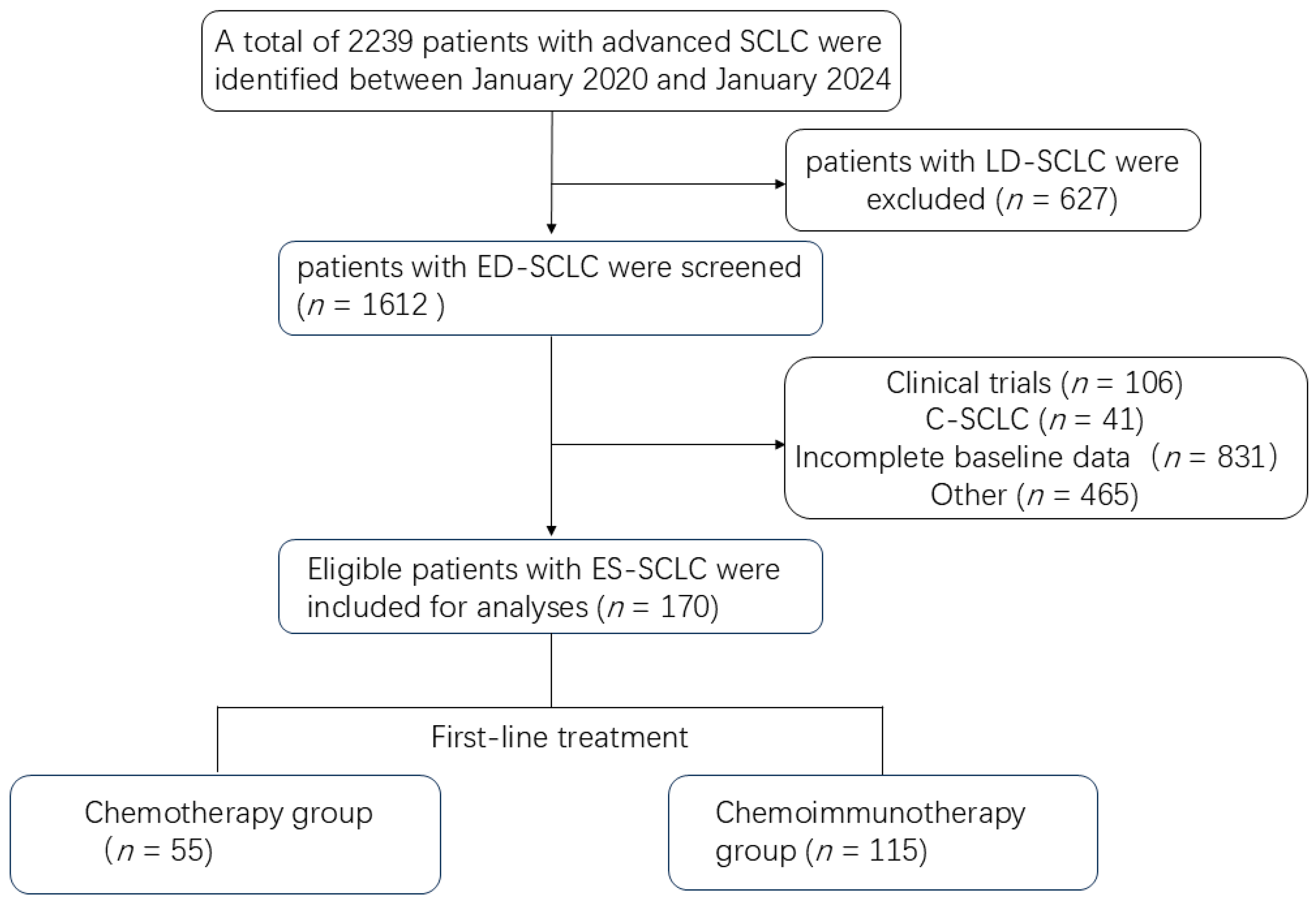
This was a single-center, retrospective cohort study. Data were collected from patients diagnosed and treated for ES-SCLC at Shanghai Chest Hospital between January 2020 and January 2024. The inclusion criteria were as follows: (1) all SCLC cases were diagnosed per WHO criteria using immunohistochemistry for neuroendocrine (CD56, SYN, ChgA, INSM1), epithelial (CK), and proliferative (Ki-67) markers, with TTF-1 supporting pulmonary origin; (2) histologically and radiologically confirmed SCLC with at least one measurable lesion; (3) classified as ES-SCLC according to the Veterans Administration Lung Study Group (VALG) staging system; and (4) received first-line treatment with either chemotherapy or chemoimmunotherapy and completed at least two cycles (21 days per cycle) of therapy. Patients were excluded for any of the following: (1) incomplete demographic or treatment data; (2) a history of other concurrent malignant tumors; or (3) receipt of investigational drugs. A total of 170 patients who met all the criteria were ultimately enrolled in the final cohort. The patient screening process and outcomes are detailed in Figure 1.

2.2. Data Collection and Assessment

Patients were stratified into two cohorts based on the inclusion of immunotherapy in their first-line treatment: the CIT and the CT. Collected data encompassed demographic and baseline clinical characteristics, including sex, age, smoking history, anatomical location of the primary tumor, disease stage and metastatic sites at diagnosis, PD-L1 expression level, immunohistochemistry gene mutation status, and PFS. The chemotherapeutic agents used primarily included etoposide, carboplatin, and nedaplatin. The immune checkpoint inhibitors involved comprised PD-L1 inhibitors (atezolizumab, durvalumab) and PD-1 inhibitors (pembrolizumab, tislelizumab, toripalimab, and camrelizumab).
This study defined PFS as the primary endpoint, calculated from the commencement of first-line therapy until radiologically confirmed disease progression or death from any cause. The data cutoff for PFS and survival follow-up was 30 July 2024. Secondary endpoints comprised the objective response rate (ORR) and disease control rate (DCR), both determined by the investigators based on Response Evaluation Criteria in Solid Tumors (RECIST 1.1) guidelines. Specifically, ORR was defined as the combined incidence of complete response (CR) and partial response (PR). DCR was defined as the proportion of patients exhibiting a best objective response of CR, PR, or stable disease (SD).

2.3. Statistical Analysis

All statistical analyses in this study were performed using SPSS software (version 25.0). Differences in continuous variables were evaluated using a t-test or the Wilcoxon rank-sum test. Categorical variables were compared using the chi-square test or Fisher’s exact test, as appropriate. Univariate and multivariate analyses were conducted using the Cox proportional hazards regression model to evaluate the association between various variables and PFS, with results presented as hazard ratios (HRs) and their 95% confidence intervals (CIs). Survival curves were generated using the Kaplan–Meier method and compared with the log-rank test. All tests were two-sided, and a p value < 0.05 was considered statistically significant.

2.4. Ethics Statement

Prior to initiation, this study obtained formal approval from the Ethics Committee of Shanghai Chest Hospital. All subsequent procedures and data collection activities strictly adhered to the ethical standards and protocols authorized by the committee.

3. Results

3.1. Baseline Characteristics of ES-SCLC Patients

170 eligible ES-SCLC patients were identified from 2239 pathology records screened between January 2020 and January 2024. The baseline characteristics are presented in Table 1. The enrolled population was predominantly male (139, 81.8%) and had a smoking history (114, 67.1%), with central (123, 72.4%) and stage IV (124, 72.9%) tumors being most common. At initial diagnosis, metastases were frequently observed in the bone (55, 32.4%), liver (33, 19.4%), and brain (30, 17.6%); metastases to lymph nodes, adrenal glands, lungs, and pleura constituted 14%, 14%, 13%, and 12% of cases, respectively. Immunohistochemical analysis demonstrated high positive rates for CD56 (98.24%), cytokeratin (CK) (98.24%), TTF-1 (90.00%), synaptophysin (SYN) (95.65%), and INSM1 (83.08%). Conversely, chromogranin A (ChgA) positivity was low (13.66%). The proliferation marker Ki-67 showed an average positivity of 77.78% across all cases. The highest expression was 95%, and the lowest was 40%. For first-line treatment, 55 patients received EC/EP chemotherapy (CT group) and 115 received chemoimmunotherapy (CIT group). In the CIT group, most patients received a PD-L1 inhibitor (n = 97), while 16 received a PD-1 inhibitor; five patients in this group were administered two cycles of induction chemotherapy prior to CIT. The patient selection flowchart is shown in Figure 1.

3.2. Survival Analysis

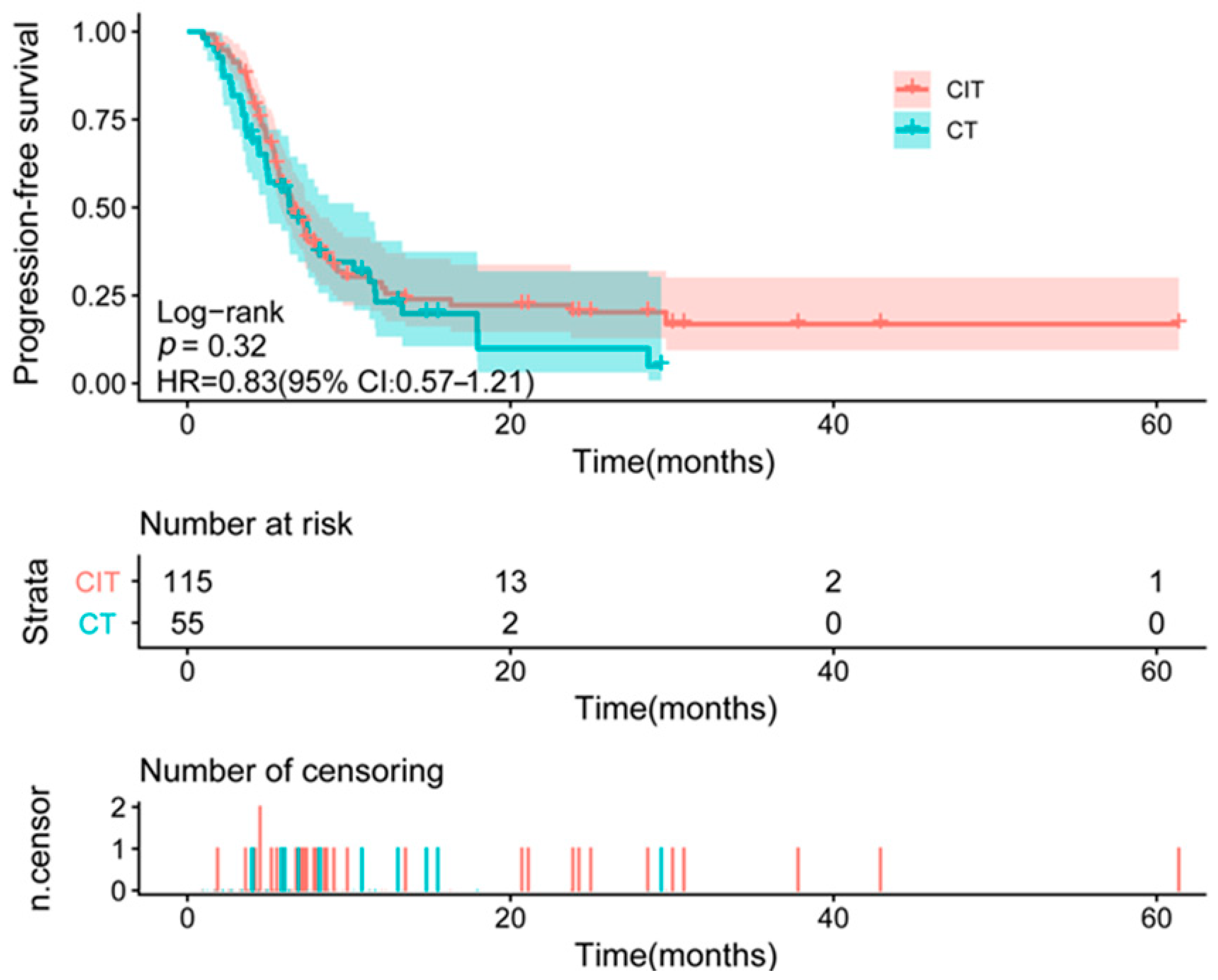
After a median follow-up of 21.3 months (95% CI: 20.0–24.5), with assessments by CT scan every 2–3 months, 119 patients had experienced disease progression. The median PFS was 14.5 months (95% CI: 10.1–18.5) in the CT group (41/55 events) and 15.0 months (95% CI: 13.5–17.3) in the CIT group (78/115 events). The corresponding HR for progression or death was 0.83 (95% CI: 0.57–1.21) for CIT versus CT, which was not statistically significant (Figure 2).
Objective tumor responses are summarized in Table 2. In the CT group, PR, SD, and PD were observed in 31 (56.4%), 18 (32.7%), and 6 (10.9%) patients, respectively. Conversely, the CIT group demonstrated higher rates of PR in 88 patients (76.5%) and SD in 22 patients (19.1%), with only 5 (4.4%) experiencing PD. The confirmed ORR was significantly higher in the CIT group than in the CT group (76.5% [95% CI, 67.9–83.3] vs. 56.4% [95% CI, 43.3–68.6]; p = 0.007). The DCR also favored the CIT group (95.7% [95% CI, 90.2–98.1] vs. 89.1% [95% CI, 78.2–94.9]; p = 0.10).
Univariate Cox regression identified liver metastasis and adrenal metastasis as significant adverse prognostic factors for PFS (p < 0.05), while no significant associations were found for other clinical variables, including age, gender, smoking history, tumor location, disease stage, PD-L1 expression, and metastatic involvement in other common sites (Table 3). On multivariate analysis, adrenal metastasis was confirmed as an independent predictor of poorer PFS (Table 4).

3.3. The Mutational Landscape of ES-SCLC

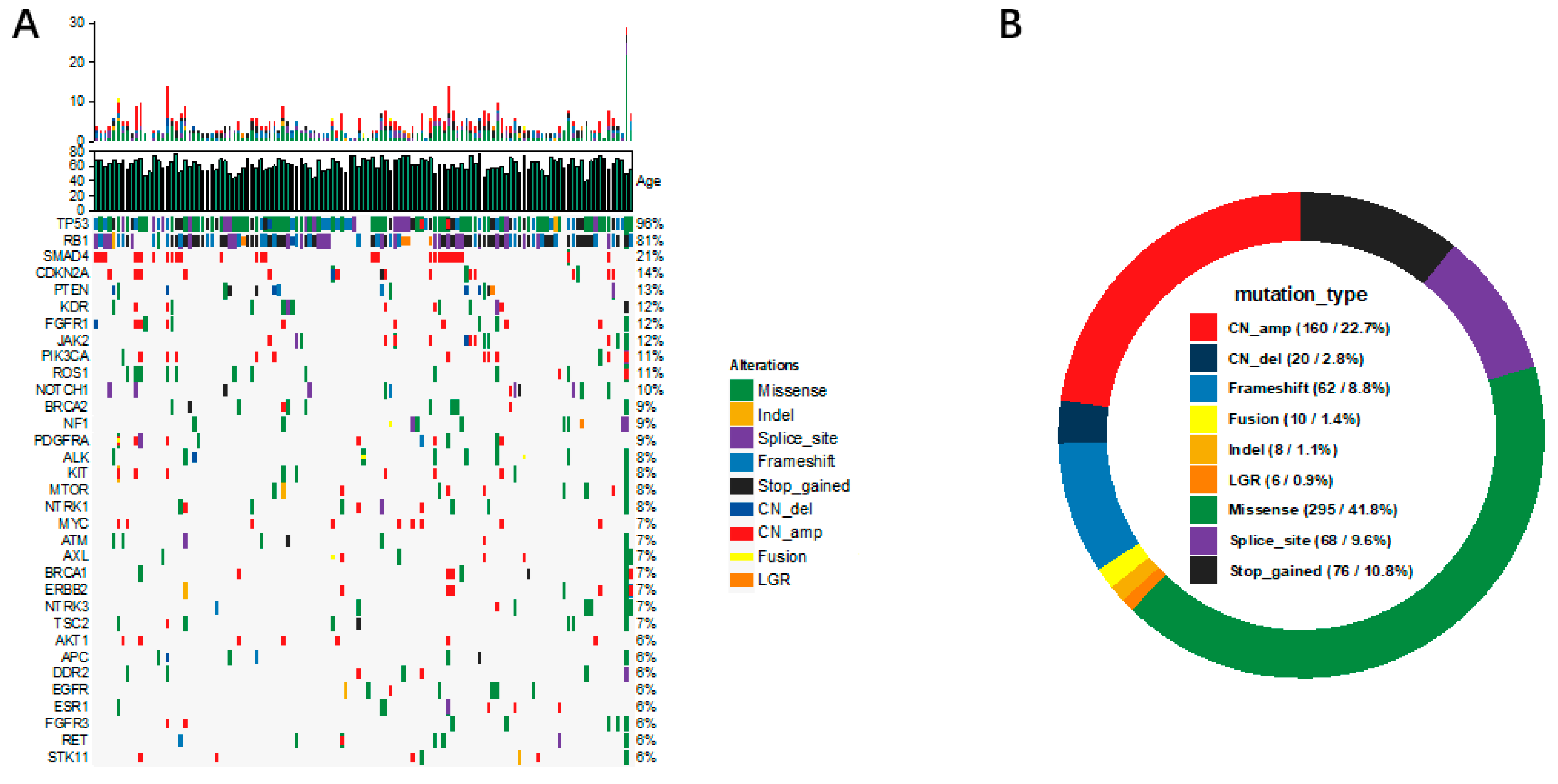
Of the 170 enrolled patients, 121 had available next-generation sequencing data based on a 68-gene lung cancer panel. Further analysis was conducted on genes with a mutation frequency >5%. The dual inactivation of the tumor suppressor genes RB1 and TP53 constitutes the genetic cornerstone of SCLC pathogenesis. As shown in Figure 3A, TP53 and RB1 mutations were the most prevalent in our cohort, with mutation frequencies of 95.8% (116/121) and 80.9% (98/121), respectively, consistent with rates reported in previous literature [6]. Other recurrently mutated genes, in descending order of frequency, included SMAD4 (21.4%, 26/121), CDKN2A (14.0%, 17/121), PTEN (13.2%, 16/121), KDR (12.3%, 15/121), FGFR1 and JAK2 (11.5%, 14/121), PIK3CA and ROS1 (10.7%, 13/121), NOTCH1 (9.9%, 12/121), BRCA2, NF1, and PDGFRA (9.0%, 11/121), as well as ALK, KIT, MTOR, and NTRK1 (8.2%, 10/121). The spectrum of the most frequent genetic alterations comprised missense mutations (41.8%), amplifications (22.7%), stop-gain variants (10.8%), splice-site mutations (9.6%), and frameshift mutations (8.8%) (Figure 3B).

3.4. Co-Occurrence and Mutual Exclusivity of Somatic Alterations in SCLC

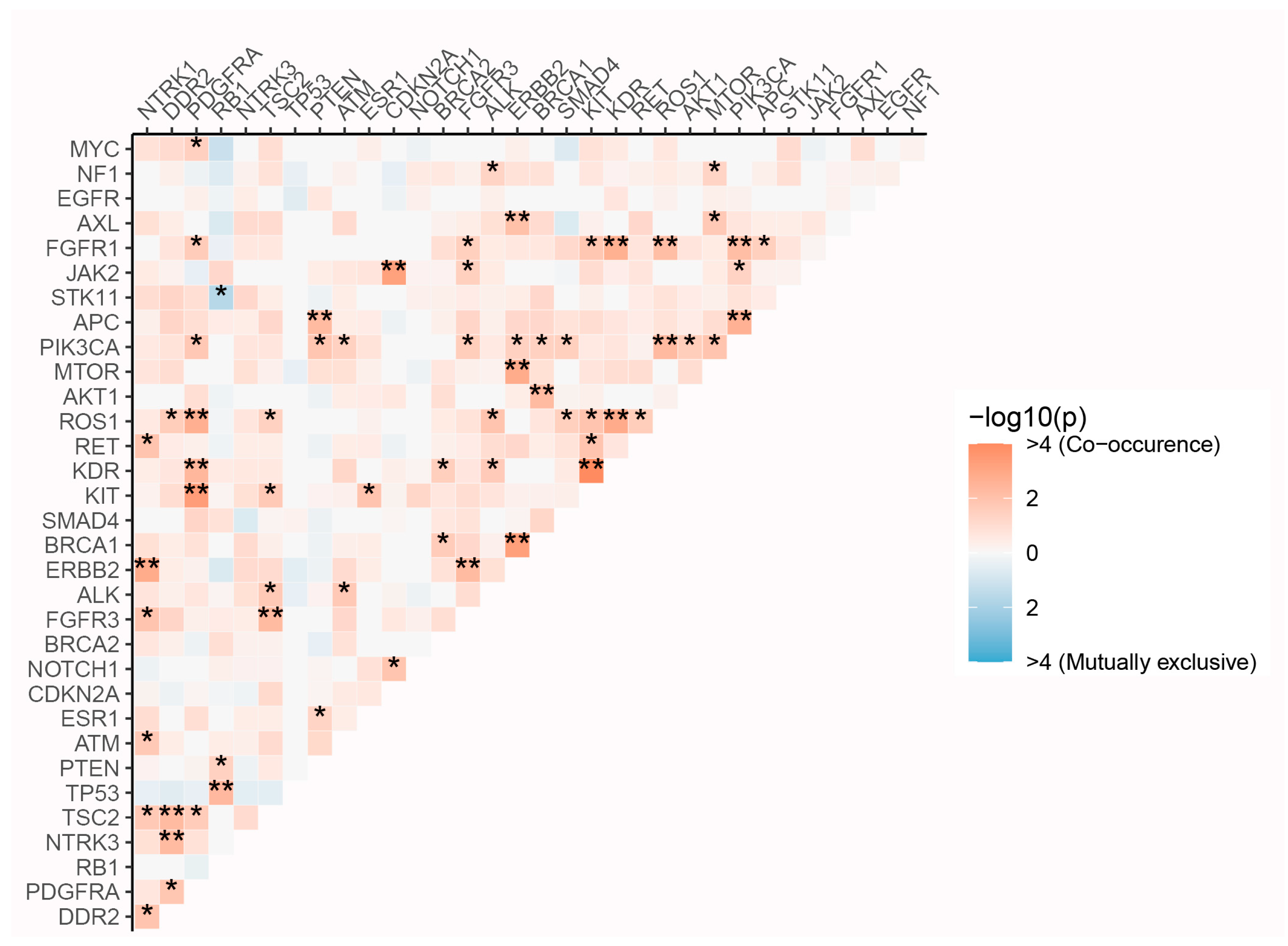
In our subsequent analysis of co-occurring genomic alterations in SCLC, we identified several mutations that are significantly co-mutated, suggesting that these lesions cooperate to promote a malignant phenotype (Figure 4). For instance, our analysis confirmed the co-occurrence of TP53 and RB1 mutations; their simultaneous inactivation is known to lead to uncontrolled cell proliferation, thereby promoting SCLC development [7].
Notably, the significant co-mutated genes were not randomly distributed but were highly enriched within several core biological pathways. Primarily, synergistic activation of the PI3K/AKT/mTOR pathway was observed. PIK3CA acted as a central hub in the network, co-mutating with ten distinct genes, including upstream receptor tyrosine kinases (RTKs) such as ERBB2 and FGFR3; other pathway members like AKT1 and MTOR; the tumor suppressor PTEN (via inactivation); and DNA damage response (DDR) genes, including ATM and BRCA1. PTEN, a key negative regulator of the PI3K-AKT-mTOR signaling pathway, promotes transcriptional heterogeneity and can consequently drive resistance to immunotherapy upon its loss [8]. The concurrent presence of PIK3CA mutations and PTEN loss may represent a dual activation mechanism, enhancing PI3K/AKT/mTOR pathway activity and serving as a common driver of tumor progression [9,10].
Moreover, analysis revealed cross-talk between the DNA damage repair and RTK/PI3K signaling pathways, characterized by specific co-occurring genetic pairs: PIK3CA with both ATM and BRCA1, AKT1 with BRCA1, and ERBB2 with ATM. As the most commonly mutated DDR gene, the co-mutation of ATM with PIK3CA potentially indicates a link to increased genomic instability.
Our genomic profiling uncovered co-mutated BRCA1 and BRCA2, pointing to a constitutive homologous recombination deficiency (HRD) in a subset of patients [11]. HRD is a well-validated predictive biomarker that confers heightened sensitivity to PARP inhibitors and can also forecast response to platinum-based chemotherapy [12].
Mutual exclusivity is a well-documented phenomenon in cancer genomics, where large-scale sequencing studies consistently show that driver oncogene mutations tend to be mutually exclusive, though the biological basis for this pattern is not fully elucidated [13]. We identified a striking pattern of mutual exclusivity between STK11 and RB1 mutations in SCLC. This finding is particularly significant given that RB1 inactivation is a near-universal, defining feature of SCLC, while STK11 (LKB1) loss—a known regulator of cell cycle and polarity—is well-documented in non-small-cell lung cancer (NSCLC) [14]. This novel observation may provide a clue for refining the molecular classification of SCLC. Overall, this work reveals a complex interactome in SCLC that extends beyond TP53/RB1.
The landscape of co-occurring and mutually exclusive genetic events highlighted here offers a valuable framework for elucidating pathogenic mechanisms and revealing new therapeutic opportunities.

3.5. Correlation of Somatic Mutations with Clinicopathologic Features in SCLC

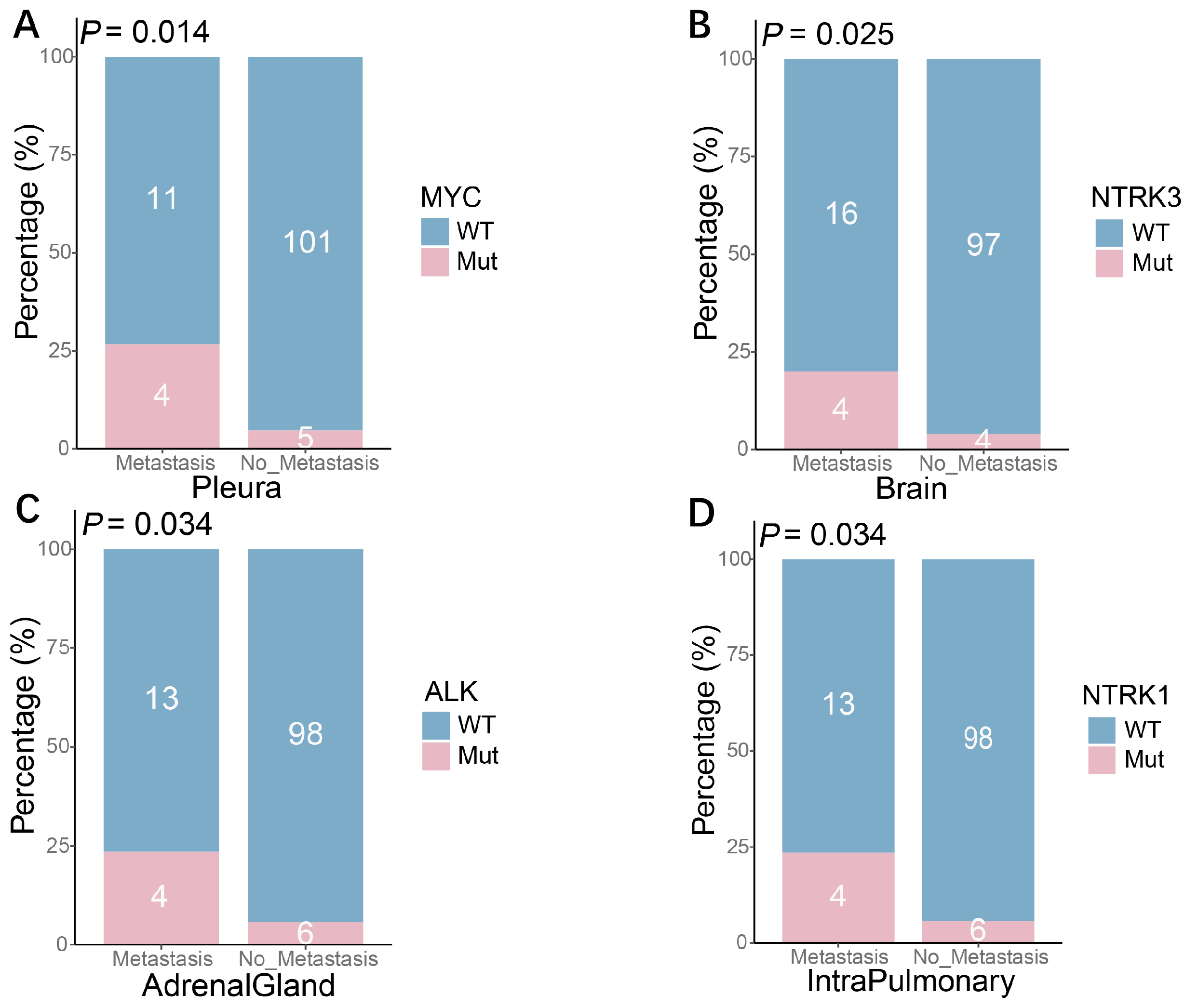
Metastatic spread is a hallmark of SCLC and a key determinant of its poor prognosis; however, the specific genetic alterations that underlie this aggressive behavior remain poorly defined. To explore this, we examined potential associations between somatic mutations and specific metastatic patterns. Our analysis revealed several notable associations: MYC mutations were more frequent in patients with pleural metastases (26.7% vs. 4.7%; p = 0.014). Similarly, NTRK3 mutations were observed more commonly in cases with brain metastases (20.0% vs. 4.0%; p = 0.025), while ALK and NTRK1 mutations were associated with adrenal (23.5% vs. 5.8%; p = 0.034) and intra-pulmonary metastases (23.5% vs. 5.8%; p = 0.034), respectively. These results indicate that mutations in MYC, NTRK3, ALK, and NTRK1 are significantly associated with distinct metastatic patterns in SCLC, suggesting their potential involvement in organ-specific dissemination (Figure 5).
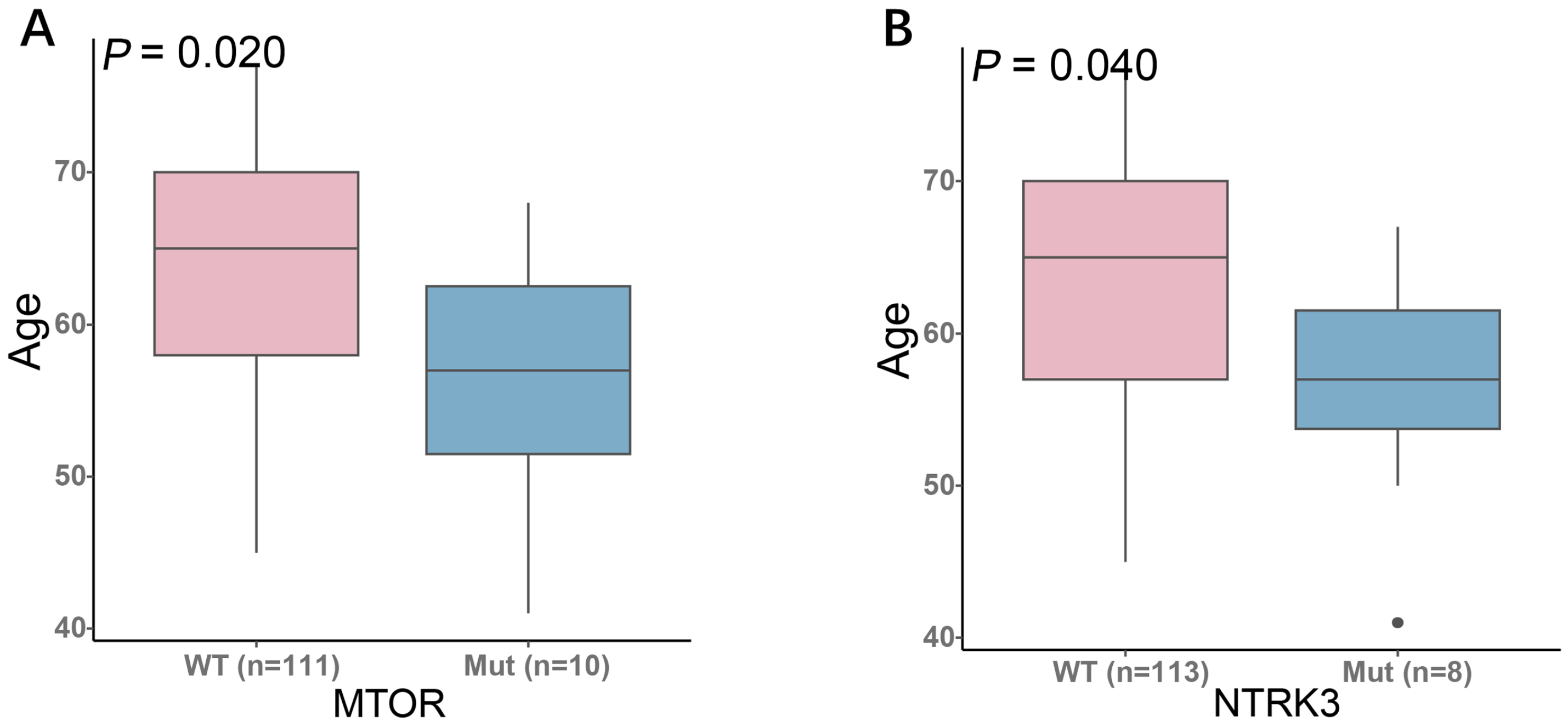
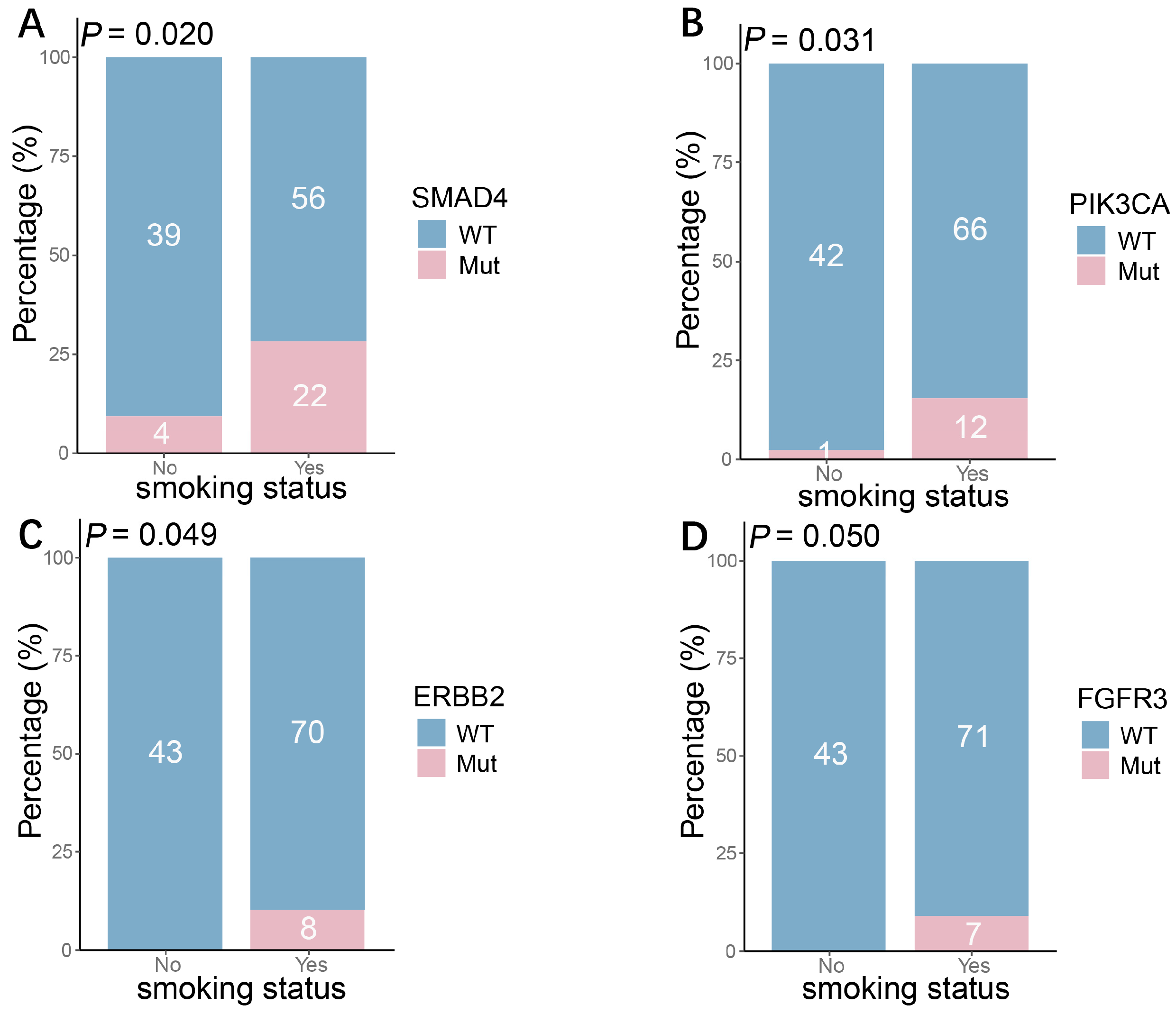
We next investigated the associations between gene mutations and other clinical features. Analysis by age group revealed that mutations in MTOR and NTRK3 were significantly enriched in younger patients, suggesting a potential age-related molecular pathogenesis in SCLC (Figure 6). Furthermore, we evaluated the relationship between smoking history and the genomic landscape. Smokers exhibited significantly higher mutation frequencies in SMAD4 (p = 0.020), PIK3CA (p = 0.031), ERBB2 (p = 0.049), and FGFR3 (p = 0.050) compared to non-smokers. Notably, SMAD4 mutations were observed in 75% of smokers, highlighting a strong association with smoking history (Figure 7).

4. Discussion

SCLC represents the most aggressive subtype of lung cancer, characterized by rapid progression, high relapse rates, and an exceedingly poor prognosis, with a five-year overall survival of only 10% [15]. Approximately 70% of SCLC cases are diagnosed as ES-SCLC, with the majority of patients presenting with systemic metastasis at initial diagnosis. For decades, platinum-based doublet chemotherapy has remained the standard first-line treatment for ES-SCLC. Although initial responses are often favorable, patients frequently develop rapid resistance, leading to disease progression. The introduction of ICIs has recently transformed the treatment landscape. Currently, PD-L1 inhibitors combined with platinum and etoposide chemotherapy have become the first-line standard of care for ES-SCLC [16]. Landmark trials such as IMpower133 and CASPIAN established the survival benefit of this approach, demonstrating significant improvements in both PFS and OS with the addition of atezolizumab or durvalumab to chemotherapy, respectively [2,6]. These consistent findings underscore the role of immunotherapy as an effective strategy for improving outcomes in ES-SCLC [17]. Building on the efficacy established in these clinical trials, this study further evaluates the real-world clinical performance of CIT using real-world data.
In this real-world cohort, we compared the efficacy of first-line CT (n = 55) versus CIT (n = 115) in 170 patients with SCLC. The median PFS was 14.5 months (95% CI: 10.1–18.5) in the CT group and 15.0 months (95% CI: 13.5–17.3) in the CIT group. Although a numerical extension in median PFS was observed with CIT, the difference was not statistically significant. This aligns with several prior real-world analyses that also reported no significant PFS or OS difference between the two regimens [18]. The attenuated PFS benefit of CIT in real-world settings may be influenced by patient heterogeneity, variations in treatment patterns, or limited follow-up duration. Notably, however, the CIT group demonstrated superior objective and disease control response rates (ORR, DCR), suggesting a more pronounced initial antitumor effect. The failure of this response advantage to translate into a significant PFS benefit may involve complex factors, including baseline patient characteristics, intrinsic tumor biology, and adaptive changes in the tumor microenvironment, or mechanisms of immune resistance.
Furthermore, univariate Cox regression analysis identified liver metastasis as an independent adverse prognostic factor in SCLC, a finding consistent with multiple large-scale retrospective studies and meta-analyses, reaffirming the clinical significance of liver metastasis as a strong negative prognostic marker [19,20]. Notably, our study also found adrenal metastasis to be an independent prognostic factor. However, evidence regarding the prognostic value of adrenal metastasis remains relatively limited, and the underlying biological mechanisms are not yet fully understood, warranting further investigation.
We performed a systematic genomic analysis to identify co-occurring and mutually exclusive mutation patterns in SCLC, which revealed significant co-alterations among several key genes. For instance, PIK3CA mutations frequently co-occurred with alterations in PTEN, ATM, ERBB2, and BRCA1. These genetic events were not randomly distributed but were highly enriched within several core signaling pathways, most notably revealing a coordinated activation pattern in the PI3K/AKT/mTOR pathway. Within this pathway, PIK3CA appears to function as a hub gene, concurrently altered alongside multiple upstream receptor tyrosine kinases (e.g., ERBB2, FGFR3), intra-pathway components (e.g., AKT1, MTOR), and negative regulators (e.g., PTEN).
Both PIK3CA activating mutations and PTEN loss are well-established driver events in tumor progression. As a key negative regulator of the PI3K-AKT-mTOR signaling pathway [12], the co-occurrence of PTEN loss with PIK3CA mutation may reflect a “dual activation” mechanism that synergistically amplifies oncogenic signaling output. On one hand, PIK3CA-activating mutations or RTK amplification enhance forward signaling; on the other hand, PTEN loss removes intrinsic inhibitory control, thereby cooperatively driving hyperactivation of the pathway and promoting tumor cell proliferation, survival, and metabolic reprogramming. These findings also provide a new perspective for understanding therapy resistance in SCLC. Alterations in the PIK3CA/AKT/PTEN pathway have been linked to resistance to targeted agents such as osimertinib in NSCLC and other malignancies [8,9]. Our data suggest that a similar cooperative pathway activation mechanism may contribute to the development of resistance to chemotherapy or immunotherapy in SCLC, warranting further functional studies for validation.
Additionally, our analysis revealed significant crosstalk between the DDR pathway and RTK/PI3K signaling. Specifically, we identified frequent co-alterations involving key gene pairs, including PIK3CA with ATM or BRCA1, AKT1 with BRCA1, and ERBB2 with ATM. This co-occurrence pattern suggests a potential functional link between growth factor pathway activation and genomic instability in a subset of SCLC cases. As a core component of the DDR pathway, the role of ATM in related malignancies has garnered increasing attention. Supporting this notion, a study in bladder cancer demonstrated that a classifier based on TP53/PIK3CA/ATM mutation status could predict patient responses to ICI therapy [21].
Of particular note was the observed co-occurrence of BRCA1 and BRCA2 mutations, strongly suggesting the presence of HRD. HRD status is not only associated with genomic instability but also serves as an important biomarker predicting sensitivity to PARP inhibitors and platinum-based chemotherapy [22]. Previous studies have indicated that the combination of PARP inhibitors and immune checkpoint inhibitors demonstrates promising efficacy and tolerability in patients with advanced solid tumors [23]. Therefore, SCLC patients with HRD features may represent a potential beneficiary population for PARP inhibitor-targeted therapy.
On the other hand, our study revealed a significant pattern of mutual exclusivity between STK11 and RB1 mutations, suggesting distinct biological dependencies in SCLC pathogenesis. This mutual exclusion may reflect divergent dependencies of SCLC cells on specific signaling pathways: tumors with RB1 inactivation might rely on STK11-mediated metabolic regulation, and vice versa. This phenomenon aligns with observations in large cell neuroendocrine carcinoma (LCNEC), where RB1 mutations and STK11 alterations rarely co-occur [24]. Furthermore, emerging evidence in NSCLC links STK11 mutations to immunotherapy resistance, potentially via metabolic reprogramming that fosters an immunosuppressive microenvironment [25,26]. These insights reinforce the functional significance of STK11 beyond metabolism, implicating it in treatment evasion and tumor aggressiveness.

5. Conclusions

In summary, our integrative analysis of co-occurring and mutually exclusive genetic alterations in SCLC reveals an interactive molecular network that extends beyond the canonical TP53/RB1 inactivation paradigm. These findings deepen the mechanistic understanding of SCLC biology and provide a framework for developing targeted therapeutic approaches. Promising strategies include combined inhibition of the PI3K/AKT/mTOR pathway, PARP inhibitor application guided by HRD status, and subtype-specific treatments based on STK11/RB1 mutual exclusivity. Future studies should explore the functional and clinical relevance of these patterns for precision medicine applications in SCLC.
Our analysis of the real-world SCLC cohort revealed significant associations between specific genetic alterations and distinct patterns of metastatic spread. Notably, we observed that NTRK3 mutations were significantly correlated with brain metastases, suggesting a potential role for the NTRK signaling pathway in the development of SCLC brain metastasis. It is important to emphasize that the NTRK3 alterations identified in this cohort were primarily missense mutations, not the NTRK gene fusions targeted by approved TRK inhibitors such as larotrectinib or entrectinib [27]. While these inhibitors have demonstrated high response rates in various NTRK fusion-positive cancers [28,29], their efficacy against NTRK3 missense mutations in SCLC remains to be investigated. Furthermore, significant correlations were observed between MYC mutations and pleural metastasis, ALK mutations and adrenal metastasis, and NTRK1 mutations and intrapulmonary metastasis, indicating that specific genetic alterations may drive the tropism of tumor cells to particular organs.
Our study also uncovered a significant association between smoking history and specific mutational patterns. Mutations in genes such as SMAD4, PIK3CA, ERBB2, and FGFR3 were significantly enriched in smokers, with SMAD4 mutations being particularly prominent. This aligns with a report from the Japanese National Cancer Center, which indicated that passive smoking induces numerous subclonal mutations with an APOBEC signature, including hotspot mutations in SMAD4 [30]. Research in NSCLC has shown that smoking can induce epigenetic reprogramming of the TGF-β/SMAD3 pathway and that mutations in KRAS and SMAD4 are more frequent in smokers [31,32]. The high frequency of SMAD4 mutations is of particular biological relevance. As a core tumor suppressor within the TGF-β signaling pathway [33], its inactivation might promote epithelial–mesenchymal transition (EMT) and contribute to an immunosuppressive tumor microenvironment, thereby potentially enhancing tumor invasiveness. Conversely, mutations in MTOR and NTRK3 were more frequent in younger patients, suggesting that they may define a molecular subtype associated with age, whose pathogenesis might be more related to endogenous developmental or metabolic pathway dysregulation rather than direct tobacco exposure. This divergence in molecular backgrounds between smoking-associated mutations and those enriched in younger patients further underscores the significant molecular heterogeneity of SCLC across different clinical contexts.
This study is characterized by its analysis of real-world clinical treatment responses and genomic data, providing evidence that SCLC exhibits heterogeneity at both clinical and molecular levels. Yet, the study has several limitations, such as a retrospective design, potential selection bias, and an incomplete overall survival analysis caused by missing follow-up data. Future studies with larger, prospective cohorts are needed to validate these results and to further explore the molecular mechanisms, including associations between protein expression and genomic alterations, and their causal links to treatment outcomes.

Author Contributions

Conceptualization, M.L., Y.H. and C.X.; methodology, M.L., L.G. and R.Z.; software, M.L., S.C. and S.M.; validation, L.G., R.Z., S.C. and C.X.; formal analysis, M.L. and S.C.; investigation, M.L., L.G. and R.Z.; data curation, M.L., L.G., R.Z., S.C. and S.M.; writing—original draft preparation, M.L.; writing—review and editing, M.L., L.G., R.Z., S.C., S.M., C.X. and Y.H.; visualization, M.L.; supervision, Y.H. and C.X.; project administration, L.G., R.Z., S.C. and S.M.; funding acquisition, Y.H. All authors have read and agreed to the published version of the manuscript.

Funding

This work was supported by the National Natural Science Foundation of China (NSFC) (Grant No. 82372669).

Institutional Review Board Statement

The study protocol was approved by the Institutional Ethics Review Board of Shanghai Chest Hospital. IRB approval was obtained on 9 February 2026 (approval code: IS26027). All data were obtained through retrospective review of medical records, and the study was conducted in accordance with the principles of the Declaration of Helsinki. Given the retrospective nature of the research, the requirement for written informed consent was waived.

Informed Consent Statement

Given the retrospective nature of the research, the requirement for written informed consent was waived.

Data Availability Statement

The data presented in this study are available upon request from the corresponding author.

Conflicts of Interest

The authors declare that they have no known competing financial interests or personal relationships that could have appeared to influence the work reported in this paper.

References

  1. Pietanza, M.C.; Byers, L.A.; Minna, J.D.; Rudin, C.M. Small cell lung cancer: Will recent progress lead to improved outcomes? Clin. Cancer Res. 2015, 21, 2244–2255. [Google Scholar] [CrossRef] [PubMed]
  2. Horn, L.; Mansfield, A.S.; Szczęsna, A.; Havel, L.; Krzakowski, M.; Hochmair, M.J.; Huemer, F.; Losonczy, G.; Johnson, M.L.; Nishio, M.; et al. First-Line Atezolizumab plus Chemotherapy in Extensive-Stage Small-Cell Lung Cancer. N. Engl. J. Med. 2018, 379, 2220–2229. [Google Scholar] [CrossRef]
  3. Goldman, J.W.; Dvorkin, M.; Chen, Y.; Reinmuth, N.; Hotta, K.; Trukhin, D.; Statsenko, G.; Hochmair, M.J.; Özgüroğlu, M.; Ji, J.H.; et al. Durvalumab, with or without tremelimumab, plus platinum-etoposide versus platinum-etoposide alone in first-line treatment of extensive-stage small-cell lung cancer (CASPIAN): Updated results from a randomised, controlled, open-label, phase 3 trial. Lancet Oncol. 2021, 22, 51–65. [Google Scholar] [CrossRef]
  4. Dong, X.; Yao, X.; Li, R.; Li, Y.; Li, Y. Comparison of efficacy and safety of first-line immunotherapy combined with chemotherapy in extensive-stage small cell lung cancer, a retrospective study. Sci. Rep. 2025, 15, 40167. [Google Scholar] [CrossRef]
  5. Chang, C.Y.; Wei, Y.F.; Chang, S.C.; Chen, C.Y. Real-World Effectiveness and Safety of Immune Checkpoint Inhibitors Combined with Chemotherapy in Taiwanese Patients with Extensive-Stage Small Cell Lung Cancer. Curr. Oncol. 2025, 32, 472. [Google Scholar] [CrossRef]
  6. Quintanal-Villalonga, A.; Taniguchi, H.; Zhan, Y.A.; Hasan, M.M.; Chavan, S.S.; Meng, F.; Uddin, F.; Manoj, P.; Donoghue, M.T.A.; Won, H.H.; et al. Multiomic Analysis of Lung Tumors Defines Pathways Activated in Neuroendocrine Transformation. Cancer Discov. 2021, 11, 3028–3047. [Google Scholar] [CrossRef]
  7. George, J.; Lim, J.S.; Jang, S.J.; Cun, Y.; Ozretić, L.; Kong, G.; Leenders, F.; Lu, X.; Fernández-Cuesta, L.; Bosco, G.; et al. Comprehensive genomic profiles of small cell lung cancer. Nature 2015, 524, 47–53. [Google Scholar] [CrossRef]
  8. Yehia, L.; Centomo, M.L.; Pandolfi, P.P.; Eng, C. Two’s Company, Three’s a Crowd: Rb1/Trp53/Pten Trifecta and Lung Cancer Molecular and Histopathologic Heterogeneity. J. Thorac. Oncol. 2023, 18, 260–261. [Google Scholar] [CrossRef] [PubMed]
  9. Cheaib, B.; Auguste, A.; Leary, A. The PI3K/Akt/mTOR pathway in ovarian cancer: Therapeutic opportunities and challenges. Chin. J. Cancer 2015, 34, 4–16. [Google Scholar] [CrossRef] [PubMed]
  10. Tan, A.C. Targeting the PI3K/Akt/mTOR pathway in non-small cell lung cancer (NSCLC). Thorac. Cancer 2020, 11, 511–518. [Google Scholar] [CrossRef]
  11. Yusoh, N.A.; Ahmad, H.; Gill, M.R. Combining PARP Inhibition with Platinum, Ruthenium or Gold Complexes for Cancer Therapy. ChemMedChem 2020, 15, 2121–2135. [Google Scholar] [CrossRef] [PubMed]
  12. Wu, S.; Zhang, Y.; Zhang, Y.; Chen, L.H.; Ouyang, H.F.; Xu, X.; Du, Y.; Ti, X.Y. Mutational landscape of homologous recombination-related genes in small-cell lung cancer. Cancer Med. 2023, 12, 4486–4495. [Google Scholar] [CrossRef]
  13. Cisowski, J.; Bergo, M.O. What makes oncogenes mutually exclusive? Small GTPases 2017, 8, 187–192. [Google Scholar] [CrossRef]
  14. Skoulidis, F.; Goldberg, M.E.; Greenawalt, D.M.; Hellmann, M.D.; Awad, M.M.; Gainor, J.F.; Schrock, A.B.; Hartmaier, R.J.; Trabucco, S.E.; Gay, L.; et al. STK11/LKB1 Mutations and PD-1 Inhibitor Resistance in KRAS-Mutant Lung Adenocarcinoma. Cancer Discov. 2018, 8, 822–835. [Google Scholar] [CrossRef] [PubMed]
  15. Sung, H.; Ferlay, J.; Siegel, R.L.; Laversanne, M.; Soerjomataram, I.; Jemal, A.; Bray, F. Global Cancer Statistics 2020: GLOBOCAN Estimates of Incidence and Mortality Worldwide for 36 Cancers in 185 Countries. CA Cancer J. Clin. 2021, 71, 209–249. [Google Scholar] [CrossRef]
  16. McNamee, N.; da Silva, I.P.; Nagrial, A.; Gao, B. Small-Cell Lung Cancer-An Update on Targeted and Immunotherapies. Int. J. Mol. Sci. 2023, 24, 8129. [Google Scholar] [CrossRef]
  17. Cheng, Y.; Han, L.; Wu, L.; Chen, J.; Sun, H.; Wen, G.; Ji, Y.; Dvorkin, M.; Shi, J.; Pan, Z.; et al. Effect of First-Line Serplulimab vs Placebo Added to Chemotherapy on Survival in Patients With Extensive-Stage Small Cell Lung Cancer: The ASTRUM-005 Randomized Clinical Trial. JAMA 2022, 328, 1223–1232. [Google Scholar] [CrossRef]
  18. Lamy, D.; Mouillot, P.; Mariet, A.; Barnestein, R.; Quilot, F.; Fraisse, C.; Ghiringhelli, F.; Bonniaud, P.; Zouak, A.; Foucher, P. Real-world comparison of chemo-immunotherapy and chemotherapy alone in the treatment of extensive-stage small-cell lung cancer. Respir. Med. Res. 2024, 86, 101125. [Google Scholar] [CrossRef]
  19. Fan, L.; Lin, Y.; Fu, Y.; Wang, J. Small cell lung cancer with liver metastases: From underlying mechanisms to treatment strategies. Cancer Metastasis Rev. 2024, 44, 5. [Google Scholar] [CrossRef]
  20. Kaira, K.; Kurata, Y.; Imai, H.; Shiono, A.; Miura, Y.; Hashimoto, K.; Yamaguchi, O.; Mouri, A.; Kagamu, H. Prognostic factors of liver metastases in extensive-stage small cell lung cancer receiving chemo-immunotherapy. Transl. Lung Cancer Res. 2025, 14, 1569–1581. [Google Scholar] [CrossRef] [PubMed]
  21. Pan, Y.H.; Zhang, J.X.; Chen, X.; Liu, F.; Cao, J.Z.; Chen, Y.; Chen, W.; Luo, J.H. Predictive Value of the TP53/PIK3CA/ATM Mutation Classifier for Patients with Bladder Cancer Responding to Immune Checkpoint Inhibitor Therapy. Front. Immunol. 2021, 12, 643282. [Google Scholar] [CrossRef]
  22. Murai, J.; Pommier, Y. BRCAness, Homologous Recombination Deficiencies, and Synthetic Lethality. Cancer Res. 2023, 83, 1173–1174. [Google Scholar] [CrossRef]
  23. Wu, Z.; Tao, H.; Zhang, S.; Wang, X.; Ma, J.; Li, R.; Liu, Z.; Wang, J.; Cui, P.; Chen, S.; et al. Efficacy and safety of anti-PD-1-based therapy in combination with PARP inhibitors for patients with advanced solid tumors in a real-world setting. Cancer Immunol. Immunother. 2021, 70, 2971–2980. [Google Scholar] [CrossRef]
  24. Derks, J.L.; Leblay, N.; Thunnissen, E.; van Suylen, R.J.; den Bakker, M.; Groen, H.J.M.; Smit, E.F.; Damhuis, R.; van den Broek, E.C.; Charbrier, A.; et al. Molecular Subtypes of Pulmonary Large-cell Neuroendocrine Carcinoma Predict Chemotherapy Treatment Outcome. Clin. Cancer Res. 2018, 24, 33–42. [Google Scholar] [CrossRef]
  25. Ricciuti, B.; Garassino, M.C. Precision Immunotherapy for STK11/KEAP1-Mutant NSCLC. J. Thorac. Oncol. 2024, 19, 877–882. [Google Scholar] [CrossRef] [PubMed]
  26. Minor, A.C.; Couser, E.; Eichner, L.J. Targeting LKB1/STK11-mutant cancer: Distinct metabolism, microenvironment, and therapeutic resistance. Trends Pharmacol. Sci. 2025, 46, 722–737. [Google Scholar] [CrossRef]
  27. Repetto, M.; Chiara Garassino, M.; Loong, H.H.; Lopez-Rios, F.; Mok, T.; Peters, S.; Planchard, D.; Popat, S.; Rudzinski, E.R.; Drilon, A.; et al. NTRK gene fusion testing and management in lung cancer. Cancer Treat. Rev. 2024, 127, 102733. [Google Scholar] [CrossRef]
  28. Cocco, E.; Scaltriti, M.; Drilon, A. NTRK fusion-positive cancers and TRK inhibitor therapy. Nat. Rev. Clin. Oncol. 2018, 15, 731–747. [Google Scholar] [CrossRef] [PubMed]
  29. Hsiao, S.J.; Zehir, A.; Sireci, A.N.; Aisner, D.L. Detection of Tumor NTRK Gene Fusions to Identify Patients Who May Benefit from Tyrosine Kinase (TRK) Inhibitor Therapy. J. Mol. Diagn. 2019, 21, 553–571. [Google Scholar] [CrossRef] [PubMed]
  30. Mochizuki, A.; Shiraishi, K.; Honda, T.; Higashiyama, R.I.; Sunami, K.; Matsuda, M.; Shimada, Y.; Miyazaki, Y.; Yoshida, Y.; Watanabe, S.I.; et al. Passive Smoking-Induced Mutagenesis as a Promoter of Lung Carcinogenesis. J. Thorac. Oncol. 2024, 19, 984–994. [Google Scholar] [CrossRef]
  31. Kawaguchi, T.; Koh, Y.; Ando, M.; Ito, N.; Takeo, S.; Adachi, H.; Tagawa, T.; Kakegawa, S.; Yamashita, M.; Kataoka, K.; et al. Prospective Analysis of Oncogenic Driver Mutations and Environmental Factors: Japan Molecular Epidemiology for Lung Cancer Study. J. Clin. Oncol. 2016, 34, 2247–2257. [Google Scholar] [CrossRef] [PubMed]
  32. Bernardo, A.; Díaz-Valdivia, N.; Fernández-Nogueira, P.; Alcaraz, J. Stromagenesis and cancer-associated fibroblast heterogeneity in primary tumors and metastasis: Focus in non-small cell lung cancer. FEBS Open Bio 2025, 15, 1599–1617. [Google Scholar] [CrossRef] [PubMed]
  33. Ahmed, S.; Bradshaw, A.D.; Gera, S.; Dewan, M.Z.; Xu, R. The TGF-β/Smad4 Signaling Pathway in Pancreatic Carcinogenesis and Its Clinical Significance. J. Clin. Med. 2017, 6, 5. [Google Scholar] [CrossRef] [PubMed]
Figure 1. Flowchart of the study. LS-SCLC, limited-stage small-cell lung cancer; ES-SCLC, extensive-stage small-cell lung cancer; C-SCLC, combined small-cell lung carcinoma.
Curroncol 33 00256 g001
Figure 2. Kaplan–Meier curve for progression-free survival in ES-SCLC. HR, hazard ratio; CT, chemotherapy group; CIT, chemoimmunotherapy group.
Curroncol 33 00256 g002
Figure 3. Mutational landscape of SCLC: (A) Genomic alteration profiling. Patterns of the 33 most frequent gene alterations identified in SCLC tumors. Genes are indicated on the left and their alteration frequency on the right. (B) Distribution of mutation types in SCLC.
Curroncol 33 00256 g003
Figure 4. Co-occurrence and mutual exclusivity of genetic alterations in SCLC. * p < 0.05; ** p < 0.01.
Curroncol 33 00256 g004
Figure 5. Correlation of gene mutations with specific metastatic sites in small-cell lung cancer: (A) Higher frequency of MYC mutations in patients with pleural metastasis. (B) Higher frequency of NTRK3 mutations in patients with brain metastasis. (C) Higher frequency of ALK mutations in patients with adrenal metastasis. (D) Higher frequency of NTRK1 mutations in patients with intrapulmonary metastasis.
Curroncol 33 00256 g005
Figure 6. Gene mutations associated with younger age in small-cell lung cancer: (A) Higher frequency of MTOR mutations in younger patients. (B) Higher frequency of NTRK3 mutations in younger patients.
Curroncol 33 00256 g006
Figure 7. Association of gene mutations with smoking status in SCLC: (AD) Mutation frequencies of SMAD4 (A), PIK3CA (B), ERBB2 (C), and FGFR3 (D) were all significantly higher in smokers than in non-smokers.
Curroncol 33 00256 g007
Table 1. Baseline patient demographics and clinical characteristics by treatment group.
Overall
(n = 170)
Chemotherapy Group
(n = 55, %)
Chemoimmunotherapy
(n = 115, %)
p Value
Gender 0.664
Male13946 (83.6)93 (80.9)
Female319 (16.4)22 (19.1)
Age 0.553
Median, range (years)63.5 (62, 66)65 (62, 67)63 (60, 66)
<658626 (47.3)60 (52.2)
≥658429 (52.7)55 (47.8)
SCLC type 0.773
Central12339 (70.9)84 (73.0)
Peripheral4716 (29.1)31 (27.0)
Clinical stage 0.966
IIIa42 (3.6)2 (1.7)
IIIb199 (16.3)10 (8.7)
IIIc234 (7.2)19 (16.5)
IVa4614 (25.5)32 (27.8)
IVb7826 (47.3)52 (45.2)
Smoking history 0.463
Ever11439 (70.9)75 (65.2)
Never5616 (29.1)40 (34.8)
Specimen site 0.558
Lung8127 (49.1)54 (47)
Lymph node8227 (49.1)55 (47.8)
Other a71 (1.8)6 (5.2)
ECOG PS 0.846
0514
115450104
2211
NA936
TPS of PD-L1 0.62
<1% 12838 (69.1)90 (78.3)
1–49%179 (16.3)8 (67)
≥50%00 (0)0 (0)
Unknown258 (14.5)17 (14.7)
Immunohistochemistry
CD56 16754 (98.2)113 (98.3)0.693
CK 16755 (100)112 (97.4)0.307
TTF1 15150 (90.9)101 (87.8)0.033
SYN 8634 (94.4)52 (96.3)0.071
ChgA 2610 (30.3)16 (0.32)0.129
INSM1 7528 (84.8)47 (97.9)0.006
Ki-67 78.377.779.10.416
Metastatic sites
Bone5513 (23.6)42 (36.5)0.094
Liver3310 (18.2)23 (20)0.781
Brain3015 (27.3)15 (13)0.023
lymph nodes247 (12.7)17 (14.8)0.721
Adrenal2410 (18.2)14 (12.2)0.295
Intrapulmonary2210 (18.2)12 (10.4)0.161
Pleura2410 (18.2)14 (12.2)0.374
Other185 (9)13 (11.3)0.663
Treatment
PD-1 inhibitor160 (0)16 (13.9)-
PD-L1 inhibitor970 (0)97 (84.3)
Progression status 0.374
Yes11941 (74.5)78 (67.8)
No5114 (25.5)37 (32.2)
Best response 0.011
PR11931 (56.4)88 (76.5)
SD4018 (32.7)22 (19.1)
PD116 (10.9)5 (4.4)
PD-L1, programmed cell death ligand-1; PD-1, programmed cell death protein 1; TPS, tumor proportion score; CD56, cluster of differentiation 56; CK, cytokeratin; TTF1, thyroid transcription factor-1; SYN, synaptophysin; ChgA, chromogranin A; INSM1, insulinoma-associated protein 1. PR, partial response; SD, stable disease; PD, progressive disease. A t-test was used for the difference in age; the chi-square test for the difference in gender, SCLC type, smoking history, specimen site, metastatic sites, treatment, progression status, and immunohistochemistry; and the Wilcoxon rank-sum test for the difference in clinical stage, TPS of PD-L1, and best response. a Includes hydrothorax, pleura, chest wall mass, and liver.
Table 2. Comparison of efficacy between the CT and CIT groups.
Therapeutic EfficacyChemotherapy Group (n = 55)Chemoimmunotherapy
(n = 115)
p Value
PR, n (%)31 (56.4)88 (76.5)0.011
SD, n (%)18 (32.7)22 (19.1)
PD, n (%)6 (10.9)5 (4.4)
ORR, (%) (95% CI)56.4 (43.3, 68.6)76.5 (67.9, 83.3)0.007
DCR, (%) (95% CI)89.1 (78.2, 94.9)95.7 (90.2, 98.1)0.10
PR, partial response; SD, stable disease; PD, progressive disease; ORR, objective response rate; DCR, disease control rate.
Table 3. Univariate analyses of progression-free survival in ES-SCLC patients.
CharacteristicsHR95% CIp Value
Age, years
≥65 vs. <65
0.97(0.68–1.39)0.86
Gender
Male vs. Female
1.41(0.86–2.32)0.17
Smoking history
Yes vs. No
1.3(0.88–1.94)0.19
Subtyping
central vs. peripheral
0.81(0.54–1.21)0.31
Stage
IV vs. III
1.34(0.89–2.02)0.16
Metastatic sites
Bone 1.33(0.9–1.95)0.15
Liver 1.68(1.10–2.58)0.02
Brain 1.22(0.77–1.92)0.4
lymph nodes 1.21(0.74–1.97)0.45
Adrenal 1.68(1.04–2.72)0.04
Intrapulmonary 1.01(0.77–1.32)0.96
Pleura 1.1(0.81–1.48)0.55
TPS of PD-L1
1–49% vs. <1%
1.14(0.65–1.99)0.66
Treatment mode
CIT vs. CT
0.83(0.57–1.21)0.33
Table 4. Multivariate analyses of progression-free survival in ES-SCLC patients.
CharacteristicsHR95% CIp Value
Age, years
≥65 vs. <65
1.1(0.75–1.62)0.64
Gender
Male vs. Female
1.13(0.55–2.35)0.74
Smoking history
Yes vs. No
1.26(0.70–2.28)0.45
Subtyping
central vs. peripheral
0.74(0.48–1.13)0.16
Stage
IV vs. III
1.1(0.61–1.94)0.77
Metastasis
Bone 1.02(0.61–1.71)0.94
Liver1.56(0.89–2.73)0.12
Brain 1.02(0.6–1.74)0.94
lymph nodes 1.13(0.63–2.0)0.69
Adrenal 1.84(1.06–3.22)0.03
Intrapulmonary 0.751(0.40–1.42)0.38
Pleura 0.79(0.40–1.56)0.5
TPS of PD-L1
1–49% vs. <1%
0.99(0.54–1.82)0.98
Treatment mode
CIT vs. CT
0.9(0.58–1.36)0.6
Disclaimer/Publisher’s Note: The statements, opinions and data contained in all publications are solely those of the individual author(s) and contributor(s) and not of MDPI and/or the editor(s). MDPI and/or the editor(s) disclaim responsibility for any injury to people or property resulting from any ideas, methods, instructions or products referred to in the content.