Merkel Cell Carcinoma of the Thigh Presenting as a Hemorrhagic Mass: A Rare Case Report and Literature Review


Simple Summary
Abstract
1. Introduction
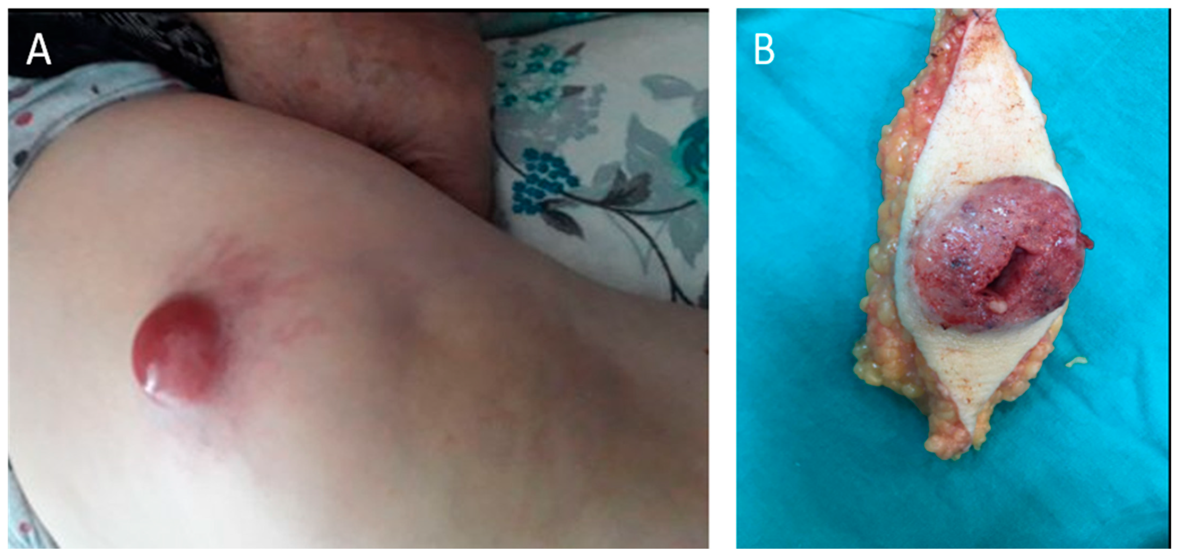
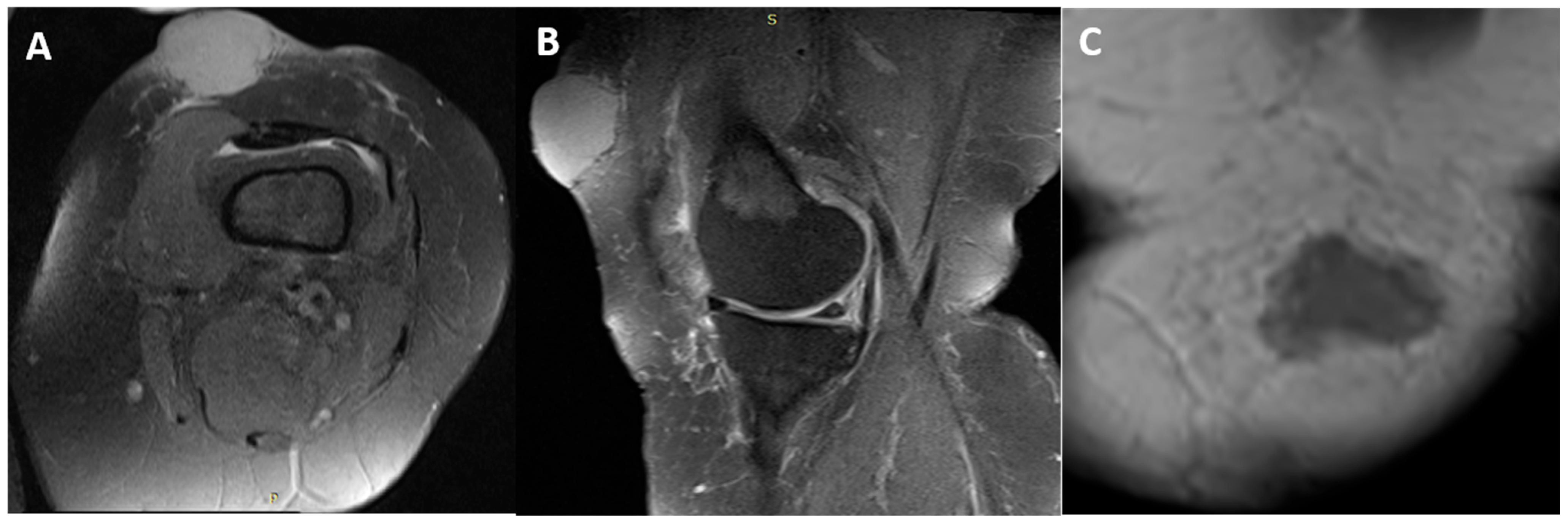
2. Case Presentation
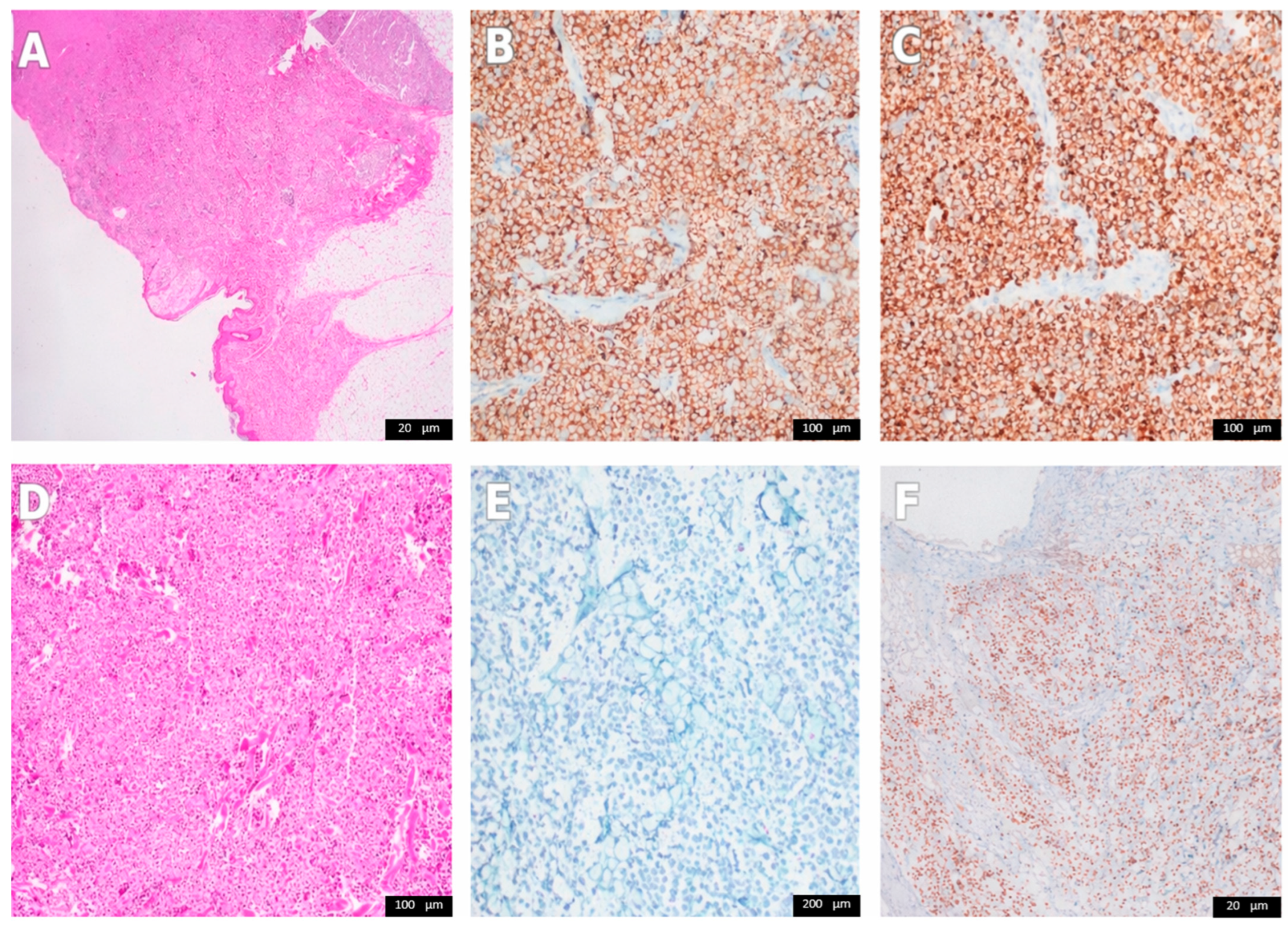
3. Results and Discussion
4. Conclusions
Author Contributions
Funding
Institutional Review Board Statement
Informed Consent Statement
Data Availability Statement
Conflicts of Interest
References
- Lewis, D.J.; Sobanko, J.F.; Etzkorn, J.R.; Shin, T.M.; Giordano, C.N.; McMurray, S.L.; Walker, J.L.; Zhang, J.; Miller, C.J.; Higgins, H.W., 2nd. Merkel Cell Carcinoma. Dermatol. Clin. 2023, 41, 101–115. [Google Scholar] [CrossRef] [Scilit] [PubMed]
- Paulson, K.G.; Park, S.Y.; Vandeven, N.A.; Lachance, K.; Thomas, H.; Chapuis, A.G.; Harms, K.L.; Thompson, J.A.; Bhatia, S.; Stang, A.; et al. Merkel cell carcinoma: Current US incidence and projected increases based on changing demographics. J. Am. Acad. Dermatol. 2018, 78, 457–463.e2. [Google Scholar] [CrossRef] [Scilit] [PubMed]
- Tribble, J.T.; Pfeiffer, R.M.; Brownell, I.; Cahoon, E.K.; Sargen, M.R.; Shiels, M.S.; Luo, Q.; Cohen, C.; Drezner, K.; Hernandez, B.; et al. Merkel Cell Carcinoma and Immunosuppression, UV Radiation, and Merkel Cell Polyomavirus. JAMA Dermatol. 2025, 161, 47–55. [Google Scholar] [CrossRef] [Scilit] [PubMed]
- Heath, M.; Jaimes, N.; Lemos, B.; Mostaghimi, A.; Wang, L.C.; Peñas, P.F.; Nghiem, P. Clinical characteristics of Merkel cell carcinoma at diagnosis in 195 patients: The AEIOU features. J. Am. Acad. Dermatol. 2008, 58, 375–381. [Google Scholar] [CrossRef] [Scilit]
- Feng, H.; Shuda, M.; Chang, Y.; Moore, P.S. Clonal integration of a polyomavirus in human Merkel cell carcinoma. Science 2008, 319, 1096–1100. [Google Scholar] [CrossRef] [Scilit]
- Harms, P.W.; Harms, K.L.; Moore, P.S.; DeCaprio, J.A.; Nghiem, P.; Wong, M.K.K.; Brownell, I.; International Workshop on Merkel Cell Carcinoma Research Working Group. The biology and treatment of Merkel cell carcinoma: Current understanding and research priorities. Nat. Rev. Clin. Oncol. 2018, 15, 763–776. [Google Scholar] [CrossRef] [Scilit]
- Amin, M.B.; Greene, F.L.; Edge, S.B.; Compton, C.C.; Gershenwald, J.E.; Brookland, R.K.; Meyer, L.; Gress, D.M.; Byrd, D.R.; Winchester, D.P. The Eighth Edition AJCC Cancer Staging Manual: Continuing to build a bridge from a population-based to a more “personalized” approach to cancer staging. CA Cancer J. Clin. 2017, 67, 93–99. [Google Scholar] [CrossRef] [Scilit]
- Erdem, B.Y.; Baykal, C.; Ozluk, Y.; Ahmed, M.A.; Kozanoglu, E.; Saip, P.; Buyukbabani, N.; Sari, S.O. Evaluating CK20 and MCPyV Antibody Clones in Diagnosing Merkel Cell Carcinoma. Endocr. Pathol. 2025, 36, 1. [Google Scholar] [CrossRef] [Scilit]
- Tanaka, Y.; Sano, T.; Qian, Z.R.; Hirokawa, M. Expression of adhesion molecules and cytokeratin 20 in merkel cell carcinomas. Endocr. Pathol. 2004, 15, 117–129. [Google Scholar] [CrossRef] [Scilit]
- Cheuk, W.; Kwan, M.Y.; Suster, S.; Chan, J.K. Immunostaining for thyroid transcription factor 1 and cytokeratin 20 aids the distinction of small cell carcinoma from Merkel cell carcinoma, but not pulmonary from extrapulmonary small cell carcinomas. Arch. Pathol. Lab. Med. 2001, 125, 228–231. [Google Scholar] [CrossRef] [Scilit]
- Bobos, M.; Hytiroglou, P.; Kostopoulos, I.; Karkavelas, G.; Papadimitriou, C.S. Immunohistochemical distinction between Merkel cell carcinoma and small cell carcinoma of the lung. Am. J. Dermatopathol. 2006, 28, 99–104. [Google Scholar] [CrossRef] [Scilit] [PubMed]
- Lilo, M.T.; Chen, Y.; LeBlanc, R.E. INSM1 Is More Sensitive and Interpretable than Conventional Immunohistochemical Stains Used to Diagnose Merkel Cell Carcinoma. Am. J. Surg. Pathol. 2018, 42, 1541–1548. [Google Scholar] [CrossRef] [Scilit] [PubMed]
- Leblebici, C.; Yeni, B.; Savli, T.C.; Aydin, O.; Gunes, P.; Cinel, L.; Şimşek, B.Ç.; Yıldız, P.; Tuncel, D.; Kayahan, S. A new immunohistochemical marker, insulinoma-associated protein 1 (INSM1), for Merkel cell carcinoma: Evaluation of 24 cases. Ann. Diagn. Pathol. 2019, 40, 53–58. [Google Scholar] [CrossRef] [Scilit] [PubMed]
- Do, J.; Wang, Y.; Aung, P.P.; Nagarajan, P.; Ning, J.; Curry, J.L.; Ivan, D.; Lenskaya, V.; Torres-Cabala, C.A.; Prieto, V.G.; et al. INSM1: A highly sensitive marker for primary and metastatic Merkel cell carcinoma, superior to SOX11, pancytokeratin, and CK20. Hum Pathol. 2025, 160, 105838. [Google Scholar] [CrossRef] [Scilit]
- Karpinski, P.; Mendez-Pena, J.E.; Wu, C.L.; Akalin, A.; Cornejo, K.M.; Hung, Y.P.; Hoang, M.P. POU4F3 Is a Sensitive and Specific Marker of Merkel Cell Carcinoma. Mod. Pathol. 2025, 38, 100627. [Google Scholar] [CrossRef] [Scilit]
- Starrett, G.J.; Thakuria, M.; Chen, T.; Marcelus, C.; Cheng, J.; Nomburg, J.; Thorner, A.R.; Slevin, M.K.; Powers, W.; Burns, R.T.; et al. Clinical and molecular characterization of virus-positive and virus-negative Merkel cell carcinoma. Genome Med. 2020, 12, 30. [Google Scholar] [CrossRef] [Scilit]
- Horny, K.; Gerhardt, P.; Hebel-Cherouny, A.; Wülbeck, C.; Utikal, J.; Becker, J.C. Mutational Landscape of Virus- and UV-Associated Merkel Cell Carcinoma Cell Lines Is Comparable to Tumor Tissue. Cancers 2021, 13, 649. [Google Scholar] [CrossRef] [Scilit]
- Ricci, C.; Morandi, L.; Righi, A.; Gibertoni, D.; Maletta, F.; Ambrosi, F.; Agostinelli, C.; Uccella, S.; Asioli, S.; Sessa, F.; et al. PD-1 (PDCD1) promoter methylation in Merkel cell carcinoma: Prognostic relevance and relationship with clinico-pathological parameters. Mod. Pathol. 2019, 32, 1359–1372. [Google Scholar] [CrossRef] [Scilit]
- Ricci, C.; Morandi, L.; Ambrosi, F.; Righi, A.; Gibertoni, D.; Maletta, F.; Agostinelli, C.; Corradini, A.G.; Uccella, S.; Asioli, S.; et al. Intron 4-5 hTERT DNA Hypermethylation in Merkel Cell Carcinoma: Frequency, Association with Other Clinico-pathological Features and Prognostic Relevance. Endocr. Pathol. 2021, 32, 385–395. [Google Scholar] [CrossRef] [Scilit]
- Harms, K.L.; Healy, M.A.; Nghiem, P.; Sober, A.J.; Johnson, T.M.; Bichakjian, C.K.; Wong, S.L. Analysis of Prognostic Factors from 9387 Merkel Cell Carcinoma Cases Forms the Basis for the New 8th Edition AJCC Staging System. Ann. Surg. Oncol. 2016, 23, 3564–3571. [Google Scholar] [CrossRef] [Scilit]
- Schmults, C.D.; Blitzblau, R.; Aasi, S.Z.; Alam, M.; Amini, A.; Bibee, K.; Bolotin, D.; Bordeaux, J.; Chen, P.; Contreras, C.M.; et al. NCCN Guidelines® insights: Merkel cell carcinoma, version 1.2024: Featured updates to the NCCN guidelines. J. Natl. Compr. Canc. Netw. 2024, 22, e240002. [Google Scholar] [CrossRef] [Scilit]
- Lugowska, I.; Becker, J.C.; Ascierto, P.A.; Veness, M.; Blom, A.; Lebbe, C.; Migliano, E.; Hamming-Vrieze, O.; Goebeler, M.; Kneitz, H.; et al. Merkel-cell carcinoma: ESMO-EURACAN Clinical Practice Guideline for diagnosis, treatment and follow-up. ESMO Open 2024, 9, 102977. [Google Scholar] [CrossRef] [Scilit] [PubMed]
- Mortier, L.; Villabona, L.; Lawrence, B.; Arance, A.; Butler, M.O.; Beylot-Barry, M.; Saiag, P.; Samimi, M.; Ascierto, P.A.; Spada, F.; et al. Pembrolizumab for the First-Line Treatment of Recurrent Locally Advanced or Metastatic Merkel Cell Carcinoma: Results from the Single-Arm, Open-Label, Phase III KEYNOTE-913 Study. Am. J. Clin. Dermatol. 2024, 25, 987–996. [Google Scholar] [CrossRef] [Scilit] [PubMed]
- Jiang, W.; Xu, J.; Wang, R.; Wang, T.; Shu, Y.; Liu, L. Merkel cell carcinoma of the thigh: Case report and review of the literature. Onco Targets Ther. 2019, 12, 535–540. [Google Scholar] [CrossRef] [Scilit] [PubMed]
- Zhang, Y.; Sun, Y.; Mo, J. Wide local excision for Merkel cell carcinoma of the lower extremity: A case report. Exp. Ther. Med. 2022, 24, 700. [Google Scholar] [CrossRef] [Scilit]
- Limardo, A.; Prats, G.; Ortega, A.; Padín, R. Atypical Presentation of Merkel Cells Carcinoma in Thigh. An Unusual Case. Stud 2017, 2, 555598. [Google Scholar] [CrossRef] [Scilit]
- Guadagni, S.; Chiominto, A.; Mackay, A.R.; Farina, A.; Cappabianca, L.; Puccica, I.; Valiyeva, S.; Clementi, M. Advanced Merkel cell carcinoma of the lower extremity treated with surgery and isolated pelvic and limb perfusion using Melphalan: A case of unexpected long-term survival. Int. J. Surg. Case Rep. 2019, 61, 4–8. [Google Scholar] [CrossRef] [Scilit]
- Howell, R.S.; Rice, J.A.; Sticco, K.; Donovan, V.; Castellano, M.; Gillette, B.; Gorenstein, S. An unusual presentation of Merkel cell carcinoma: A case report. J. Surg. Case Rep. 2018, 2018, rjy185. [Google Scholar] [CrossRef] [Scilit]
- Al Diab, A.I. Merkel Cell Carcinoma of Skin in the Thigh: A Case Report on Metastasis to Groin and Abdominal Wall Without Involvement of Viscera. World Appl. Sci. J. 2013, 24, 641–643. [Google Scholar]


| First Author (Year) | Age/Sex | Primary Site | Presentation/Diagnostic Pitfall | Tumor Size (cm) | Nodal/Stage Details | Stage | Treatment | Outcome/Follow-Up |
|---|---|---|---|---|---|---|---|---|
| Jiang (2019) [24] | 86/M | Right thigh | Red nodule; initially interpreted as “subcutaneous small cell cancer” on outside pathology | 2.5 × 2.0 × 1.2 | T2N1M0; inguinal nodal metastasis on CT (~2.2 cm), no distant mets | T2N1M0 | Excision, then anti-angiogenic therapy (endostar + apatinib) | Partial response at 2 months; PFS 6.5 months; OS 13 months |
| Zhang (2022) [25] | 90/F | Right medial thigh (pretibial/near knee) | Dark red pruritic nodule; MRI showed hypervascular subcutaneous lesion | ~3 × 3 | Inguinal nodes palpable clinically; path nodal evaluation not described | Not reported | Wide local excision with 2 cm margin; patient declined adjuvant radiotherapy | No recurrence at 30 months |
| Limardo (2017) [26] | 62/M | Right thigh (appeared after nodal presentation) | Atypical sequence: initial “inguinal node” mass (occult primary), later violet thigh skin nodule interpreted as primary | 4.5 × 3.5 × 24 | Initial inguinal disease; later thigh primary suspected; CK20/CD56/chromogranin/synaptophysin positive; TTF-1 negative | Not reported | Inguinal lymphadenectomy, radiotherapy (5800 cGy), later thigh resection with flap reconstruction | Disease-free at 24 months |
| Guadagni (2019) [27] | 73/F | Left calcaneal region | Recurrent/in-transit limb nodules after initial surgery; treatment constrained by comorbidity (active hepatitis C, neutropenia) | 1.8 × 0.7; multiple new nodules | Initial SLNB negative; later developed ipsilateral inguinal node involvement during subsequent recurrences | Not reported | Repeated excisions plus isolated pelvic/limb perfusion with melphalan | Alive and disease-free at 56 months |
| Howell (2018) [28] | 68/M | Left buttock (proximal lower extremity region) | Misread as gluteal abscess; foul-smelling drainage; CT showed large lesion + bulky inguinal LAD | Lesion on exam ~10 × 10; CT ~9 × 10.8 × 4.2 cm | Reported as stage 3B; inguinal LAD on imaging | Stage IIIB | Debridement and incisional biopsy followed by definitive excision, radiotherapy, and chemotherapy | Outcome not fully detailed |
| Al Diab (2013) [29] | 38/M | Left thigh (near buttock) | Painless swelling; highlights young age/atypical demographic | Not reported | Not reported | (reported) | Inguinal lymphadenectomy and adjuvant local radiotherapy | Outcome not fully detailed |
| Present case | 54/F | Left suprapatellar thigh | Hemorrhagic/hematoma-like mass; managed initially as suspected soft-tissue tumor | 9 × 4.5 × 3.5 | pT3 pN1b cM0 (stage IIIB); PET-positive ipsilateral inguinal node; dissection 1/4 positive; no extranodal extension | Not reported | Wide local excision including fascia, therapeutic inguinal lymph node dissection, and adjuvant radiotherapy (57 Gy/20 fractions) | No recurrence at 5 years |
Disclaimer/Publisher’s Note: The statements, opinions and data contained in all publications are solely those of the individual author(s) and contributor(s) and not of MDPI and/or the editor(s). MDPI and/or the editor(s) disclaim responsibility for any injury to people or property resulting from any ideas, methods, instructions or products referred to in the content. |
© 2026 by the authors. Licensee MDPI, Basel, Switzerland. This article is an open access article distributed under the terms and conditions of the Creative Commons Attribution (CC BY) license.
Share and Cite
Tepedelenlioğlu, H.E.; Orhan, Ö.; Arıkan, Ş.M.; Esendağlı, G. Merkel Cell Carcinoma of the Thigh Presenting as a Hemorrhagic Mass: A Rare Case Report and Literature Review. Curr. Oncol. 2026, 33, 204. https://doi.org/10.3390/curroncol33040204
Tepedelenlioğlu HE, Orhan Ö, Arıkan ŞM, Esendağlı G. Merkel Cell Carcinoma of the Thigh Presenting as a Hemorrhagic Mass: A Rare Case Report and Literature Review. Current Oncology. 2026; 33(4):204. https://doi.org/10.3390/curroncol33040204
Chicago/Turabian StyleTepedelenlioğlu, Hüseyin Emre, Özlem Orhan, Şefik Murat Arıkan, and Güldal Esendağlı. 2026. "Merkel Cell Carcinoma of the Thigh Presenting as a Hemorrhagic Mass: A Rare Case Report and Literature Review" Current Oncology 33, no. 4: 204. https://doi.org/10.3390/curroncol33040204
APA StyleTepedelenlioğlu, H. E., Orhan, Ö., Arıkan, Ş. M., & Esendağlı, G. (2026). Merkel Cell Carcinoma of the Thigh Presenting as a Hemorrhagic Mass: A Rare Case Report and Literature Review. Current Oncology, 33(4), 204. https://doi.org/10.3390/curroncol33040204