Article

Lessons from a National Liquid Biopsy Program to Provide Cancer Testing and Treatment for Patients with Advanced Solid Tumors †

by
Anna Lapuk
1,2,*,
Benjamin L. S. Furman
2,
Pedro Feijao
1,2,
Ebru Baran
2,
Sonal Brahmbhatt
2,
Betty Chan
2,
Ka Mun Nip
2,
Adrian Kense
2,
Brenda Murphy
2,
Ruth Miller
1,2,
Vincent Funari
2,
Alicja Parker
2,
Melissa K. McConechy
2,
Shaqil Kassam
3,
Arif A. Awan
4,5,6,
Bryan Lo
6,
Daniel Breadner
7,
Barry D. Stein
8 and
David G. Huntsman
2
1
Avitia Inc., Montreal, QC H2S 3H1, Canada
2
Imagia Canexia Health, Vancouver, BC V6R 1P2, Canada
3
Stronach Regional Cancer Centre, Southlake Regional Health Centre, Newmarket, ON L3Y 2P9, Canada
4
Ottawa Hospital Research Institute, Ottawa, ON K1Y 1J8, Canada
5
Division of Medical Oncology, Department of Medicine, The Ottawa Hospital, Ottawa, ON K1H 8M5, Canada
6
Department of Anatomical Pathology, The Ottawa Hospital, Ottawa, ON K1H 7W9, Canada
7
Verspeeten Family Cancer Centre, London Health Sciences Center, London, ON N6A 5W9, Canada
8
Colorectal Cancer Canada, Montreal, QC H3Z 2P9, Canada
*
Author to whom correspondence should be addressed.
This work was conducted at Imagia Canexia Health, the assets of which have been acquired by Avitia Inc.
Curr. Oncol. 2026, 33(1), 18; https://doi.org/10.3390/curroncol33010018
Submission received: 1 November 2025 / Revised: 3 December 2025 / Accepted: 24 December 2025 / Published: 29 December 2025
(This article belongs to the Section Oncology Biomarkers)

Simple Summary

Detection of the patient-specific mutations present in an individual tumor is critical for the selection of the best treatment option for cancer patients. Liquid biopsy (LBx) allows the detection of such mutations in a less invasive manner and is often faster than traditional tissue biopsy. Here we report a successful experience of running an LBx program for Canadian patients with advanced solid tumors. The testing was done over the course of three years for >4000 patients referred from >150 institutions. A total of 97% of patients received high-quality testing results within an average 8 days, which provided oncologists with actionable information for treatment selection. This study has demonstrated the feasibility and growing demand for LBx testing in Canada with the potential to improve patient outcomes, while allowing the healthcare system to operate more efficiently.

Abstract

Personalized cancer treatment depends on the accurate and timely detection of the patient tumor variants. LBx enables minimally invasive tumor mutation profiling. We report results of a pan-Canadian LBx program for patients with advanced solid tumors. Plasma samples were tested at Imagia Canexia Health accredited laboratory using the clinically validated Follow It 38-gene panel. A proprietary platform was used to identify clinically relevant variants in the circulating tumor DNA and report results following accepted international guidelines on clinical significance. A total of 4229 eligible patients submitted samples for LBx testing, and reports for 97% of them were delivered within ~8 days. More than 80% of Canadian oncologists from >150 institutions across 12 provinces (11% from rural centers) participated in the project. The patient cohort consisted mostly of advanced or metastatic lung, breast, and colon cancers. ctDNA mutations were detected in >50% of cases, and clinical trials were recommended for 76% of all participants. Health economics modeling analysis found that Follow It® in combination with tissue biopsy was cost-saving and resulted in an additional 0.1138 QALYs gained relative to tissue biopsy alone. The successful pan-Canadian implementation of a cost-effective, robust LBx testing program demonstrated its sustained demand and feasibility, and its potential economic and health benefits.

1. Introduction

Cancer remains the leading cause of death in Canada, with lung, breast, and colorectal cancers being among the most common newly diagnosed cancers [1]. Targeted therapies available for cancers have shown to improve clinical outcomes [2,3,4,5,6]. The use of these treatments is contingent on relevant biomarker testing, and their tissue-based evaluation is currently the standard of care for many indications. Due to the diversity of oncogenic mechanisms and availability of multiple therapies targeting them, a concurrent evaluation of multiple biomarkers is currently recommended for many solid tumors. For this purpose, the next-generation sequencing (NGS) multi-gene panels have been increasingly recognized as the practical choice [7,8,9,10,11].
Although tissue-based molecular testing has become a mainstay of precision oncology, a number of challenges limit its application. These include availability, lack or insufficiency of tissue material, bone-only disease, difficult-to-biopsy tumor sites, risks associated with tissue collection, and delays in tissue biopsy and molecular testing due to healthcare operational challenges. This is particularly important in Canada, where the level of readiness for genomic-based testing is still low in many provinces, and wait times for surgeries and biopsies are long [12]. This makes the timely reporting of biopsy test results difficult and can increase the average turnaround time between tissue sampling and initiation of treatment to several weeks; it also can lead to suboptimal therapy selection in the absence of molecular information [13,14]. Delays in tissue biopsies have been associated with 17% of patients dying or becoming unsuitable for treatment, and 49.7% not receiving targeted therapies due to delays in biomarker test results [15,16].
Liquid biopsy (LBx) has emerged as an accurate, efficient, and cost-effective tool allowing for minimally invasive tumor mutation profiling. LBx assays can detect mutations in patients’ plasma containing free circulating tumor DNA (ctDNA) and RNA (ctRNA) shed by solid tumors. This information is valuable for therapy selection, as well as monitoring of treatment response, resistance, and disease progression, especially in scenarios when tissue material is limited, unavailable, difficult to obtain safely, or delayed [17]. In Canadian real-world setting, LBx has demonstrated the ability to reduce diagnostic delays and improve access to precision cancer care [18,19,20,21,22,23]. Additionally, LBx can overcome the tissue limitations related to spatial and temporal heterogeneity, providing a more accurate mutational snapshot of a patient’s disease [24]. Despite these benefits and emerging LBx testing recommendations [9], the use of liquid biopsy is still limited across Canada for several reasons, including lack of reimbursement, lack of local molecular testing infrastructure and expertise, and lack of cost-effective, in-house solutions with proven performance.
In response to these challenges, a pilot project named ACTT (Access to Cancer Testing & Treatment), to bring ctDNA testing into the Canadian healthcare system was launched in July 2020, reaching more than 4000 advanced cancer patients over 3 years, who were tested with the LBx assay Follow It®( Imagia Canexia Health, Vancouver, BC, Canada). Follow It® is a high-quality, robust, plasma-based NGS assay developed and rigorously tested for clinical use following AMP/CAP guidelines [25,26]. Follow It® targets actionable genomic mutations in 38 cancer-associated genes, including single-nucleotide variants (SNVs), small insertions and deletions (indels), copy number variants (CNVs), and microsatellite instability (MSI), providing treatment guidance. Designed as a pan-cancer solution for in-house use, Follow It® detects clinically actionable genomic mutations in adult patients with advanced solid cancers, such as lung, breast, and colorectal. This report summarizes the experience of running Follow It® LBx testing in a real-world pan-Canadian setting and provides an early evaluation of its health benefits and cost-effectiveness based on a representative subset of advanced lung cancer patients in a single province.

2. Materials and Methods

Blood was drawn in two 10 mL StreckTM DNA BCTs (Streck, La Vista, NE, USA) and sent to the CAP/CLIA/DAP accredited Imagia Canexia Health laboratory for testing using the clinically validated Follow It® liquid biopsy assay. Plasma was isolated using a double spin protocol, where blood was centrifuged at 1600× g for 15 min at room temperature, and the upper phase was collected and subsequently centrifuged at 3000× g for 10 min at room temperature. Plasma cell-free DNA (cfDNA) was extracted using an optimized Promega Maxwell RSC method, its quality assessed with the Agilent Bioanalyzer High Sensitivity DNA Kit (Agilent Technologies, Santa Clara, CA, USA), and it was quantified using the Qubit™ dsDNA HS Assay Kit (Thermo Fisher Scientific, Waltham, MA, USA) (Supplementary Table S1). Extracted cfDNA was amplified using the multiplex amplicon-based hotspot 30- (v4) or 38-gene (v5) panel and sequenced on Illumina sequencer (Supplementary Table S2A). An in-house developed bioinformatics pipeline and reporting platform were used to identify pathogenic single nucleotide variants (SNVs), including splice site mutations associated with splicing, indels (insertions and deletions), as well as amplification in 9 genes as part of the v5 assay (Supplementary Table S2A–C, Supplementary Figures S1 and S2). The platform has been clinically validated according to CAP/AMP guidelines for NGS assays [25]. Stringent quality criteria and established thresholds for coverage, probability, and tier level of clinical significance were applied in the mutation calling pipeline. The lower limit of detection for SNVs and indels was 0.5% and 0.375% VAF for v4 and v5, respectively, and CNVs were reported based on the strength of evidence provided by the CNV detection pipeline (amplifications or likely amplifications). All indels, complex events, and SNV variants with VAF close to reportable thresholds were manually inspected and validated using the Integrative Genomics Viewer (IGV v2.18.2) [27].
Data visualization was performed using maftools [28]. Pairwise gene–gene mutation interactions were evaluated using the somaticInteractions function from maftools, which applies Fisher’s exact test to mutation matrices to identify significant mutual exclusivity or co-occurrence patterns. Reported p-values are adjusted for multiple testing using the Benjamini–Hochberg false discovery rate (FDR) procedure.
For the cost and health benefit analysis that was focused on non-small cell lung cancer (NSCLC), the model structure was developed, which consists of a decision tree to describe the testing pathway and a Markov model to follow up patients over time (Supplementary Figures S3–S5). The model was parameterized from secondary sources as a result of extensive evidence synthesis activities, and applied for patients in the standard care arm (tissue biopsy alone) and those in a Follow It® arm (tissue biopsy plus Follow It®). Standard of care recommendations regarding tissue biomarker testing that existed at the time of this study were used in the modeling (Table 1) [29]. The primary outcome measure was the quality-adjusted life year (QALY), where one QALY is defined as one year lived in perfect health. The model parameterization and assumptions included NSCLC biomarkers used in standard of care or in Follow It® testing, treatment regimens, cost, and accuracy of tissue biopsy and Follow It®. Only mutations with a clear pathway to targeted therapy and its associated improved patient outcomes were considered (Table 1), with the presence of these mutations in a patient being mutually exclusive [29]. It was also assumed that the identification of any of the included mutations would lead to a patient being offered targeted therapy. Simplified treatment regimens for NSCLC considered for the current modeling are summarized in Table 2, and the costs were informed by the best available secondary sources. Other parameters, retrieved from published literature, included patients’ clinical outcomes on included treatments, feasibility and delays of tissue biopsy (Supplementary Tables S6–S12). The performance of the Follow It® test used in the analysis (sensitivity 100% and specificity 99%) was based on the prior clinical validation of the assay under CAP/CLIA guidelines (data on file), and the testing cost used in the modelling was $1200 CAD, which included institutional overhead.

3. Results

3.1. Nationwide Liquid Biopsy Testing Experience

Canada, by area, is the second-largest country in the world, with population centers scattered across the entire country. Due to the critical importance of timely diagnosis, testing would ideally be done in a local or regional facility. However, due to the condensed time frame of the project ACTT, liquid biopsy testing was performed centrally at Imagia Canexia Health’s CLIA, CAP, and DAP-accredited laboratory in Vancouver, British Columbia. Project collaborators, including Lifelabs, Genolife, Ichor Blood Services, Eastern Ontario Regional Laboratory Association, and hospital systems across Canada, drew blood at the local facilities and shipped blood samples to Imagia Canexia Health for testing. Samples were tested using Follow It® assay [26] and analyzed and reported using a proprietary bioinformatics and clinical reporting platform. Reports covering mutation detection results and their interpretation were issued to ordering physicians via fax. The vast majority of blood specimens were received within 7 days of collection, with ctDNA yield ranging from 0.13 to 246 ng/uL, and a mean of 2.10 ng/uL (±7.07 ng/uL). The yields were similar across all three major cancer types (lung, breast, and colon) with colon samples showing slightly higher average ctDNA concentrations, consistent with published observations [30,31] (Supplementary Table S1).
From July 2020 to August 2023, a total of 4229 eligible patients submitted their samples for liquid biopsy testing using Follow It® assay. For 4103 of those, ctDNA was isolated, and a successful clinical report was produced, indicating an overall testing success rate of 97%. The median turnaround time (TAT) from sample receipt to report send out was 8 days (3–32 days). Oncologists overwhelmingly responded positively to the pilot project, with 82% of all registered oncologists working across 150 institutions ordering the test. The project reached patients in 12 provinces and territories, with the highest participation in Ontario, Quebec, and British Columbia (Figure 1). In the province of Ontario (ON), the main participating centers included Ottawa Hospital, London Health Sciences Center, Niagara Health, Stronach Regional Cancer Centre, Windsor Regional Cancer Program, and Markham Stouffville Hospital.
In the province of Quebec (QC), the University of Montreal Hospital Centre (CHUM), Jewish General Hospital, and University Institute of Cardiology and Respirology of Quebec (IUCPQ) were among the top users. British Columbia (BC) repeated users included BC Cancer Agency (BCCA), Delta, and Richmond Hospitals. The project also exceeded its target of reaching patients in remote and rural areas, with 11% of samples received from outside of major urban centers. Examples include Thunder Bay Regional Health Sciences Centre, Health Sciences North (Sudbury), and Bluewater Health in ON; Regional Hospital Center of Lanaudiere, Centre Hospitalier Regional de Lanaudiere (CHDL) in QC; and BC Cancer—Prince George and Vernon Jubilee Hospital in BC. Other participating rural centers across Canada included Jack Ady Cancer Centre in AB, Cape Breton Cancer Centre in NS, and PEI Cancer Treatment Centre in PEI.

3.2. Patient Cohort and Program Performance

The patient cohort of 4103 samples consisted of advanced or metastatic solid cancers of the lung (2560, 62%), breast (1320, 32%), and colon (151, 4%), with remaining cases coming from a mix of multiple solid tumor types, such as pancreatic, GI, and gynecological cancers (Figure 1). The cohort consisted mostly of stage IV (78%) and III (5%) cancers with a median patient age of 67. All samples were assessed for SNVs and indels, and 2369 samples had additional copy number data for nine genes (v5 panel). Across three major cancer types (breast, lung, colorectal), in ~50% of cases, one or more mutations were identified, reflecting published frequencies of detectable mutations in the plasma of these patients [30,33,34]. In 37% of all samples, at least one tier I/II mutation was detected and targeted treatments were recommended. One or more clinical trials were recommended for 76% of all participants. According to the information provided on sample requisitions, 49% (n = 2053) of patients had no previous molecular testing at the time of liquid biopsy testing. Of those, 54% (n = 1095) were ctDNA mutation-positive, and 36% (n = 729) had one or more tier I/II mutations detected. Altogether, we detected a total of 3695 SNVs and indels across 4103 samples, and 395 CNVs across 2324 samples. Mutation data were summarized across the entire cohort using overlapping content of v4 and v5 panels, excluding SNVs/indels within an additional eight genes unique to v5 (total of 90 mutations from DICER1, FGFR1-3, FOXL2, NTRK1/3, and STK11; data available in Supplementary Table S2B and Supplementary Figure S1).

3.3. Lung Cancer Cohort

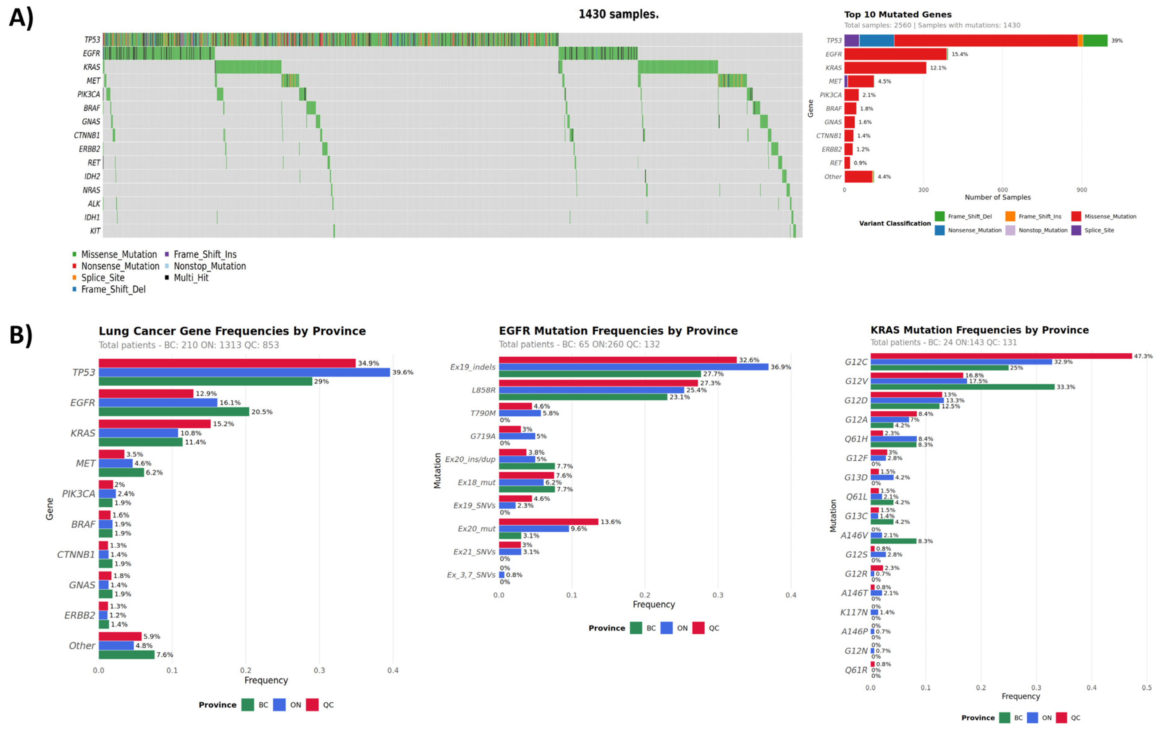
The lung cancer cohort consisted predominantly of NSCLC (90%). Among tested plasma samples (n = 2560), 56% harbored pathogenic mutations in ctDNA, with the most commonly mutated genes being TP53 (36%), EGFR (15%), KRAS (12%), MET (4%), PIK3CA (2%), and BRAF (2%) (Figure 2, Table 3). Tier I/II mutations were detected in 38% of all lung cancer patients. In patients with no previous molecular testing, liquid biopsy detected a total of 640 tier I/II mutations in 543 patients, with top mutated genes being EGFR, KRAS, MET, and BRAF. For those patients who had previous molecular testing, we detected additional ctDNA mutations in key biomarkers. These included EGFR and KRAS mutations in 11% (47/409) and 26% (107/409) of patients, respectively. Interestingly, out of 47 samples with EGFR ctDNA mutations, 14 cases had previously been reported to be PD-L1-positive. PD-L1 expression is an indication for immune-checkpoint inhibitor (ICI) therapy. ICI is less effective among patients harboring driver mutations, such as EGFR, with some evidence showing increased risk of adverse effects [35]. Our findings indicate the importance of timely multi-gene testing for optimal therapy selection.
The most frequently detected EGFR mutations included exon 19 indels (25.3%) and EGFR-L585R (32.7%) (Supplementary Figure S2). These commonly observed oncogenic mutations are associated with response to EGFR tyrosine kinase inhibitor (TKI) treatment [36,37,38,39]. T790M resistance mutation relative frequency was 5.6%. Osimertinib is approved by the FDA and Health Canada for the treatment of advanced NSCLC with the T790M mutation that progressed on prior EGFR TKI, and for first-line treatment of EGFR mutation-positive NSCLC (L858R and exon 19 deletions). KRAS-G12C (37.6%) and KRAS-G12V (18.6%) were the most commonly observed KRAS mutations (Supplementary Figure S2). NSCLC harboring KRAS-G12C mutations have been predicted to respond to sotorasib therapy and have FDA and Health Canada approval [8].
While the overall mutational spectrum in the lung cancer cohort was similar to published data [34], we observed some inter-provincial differences. BC patients showed the highest prevalence of EGFR mutations (21%) followed by KRAS (11.4%), whereas in ON and QC patients, KRAS mutations were observed more frequently relative to EGFR (Table 4, Figure 2). KRAS mutations, particularly G12C, are known to be smoking-related [40,41]. Our observation of relative EGFR and KRAS mutation frequency differences, although marginally statistically significant due to low numbers, correlates with the smoking rates in respective provinces, with BC having the lowest and QC the highest of the three [42].
Gene amplification was assessed in 1870 lung samples, with ~12% of cases showing copy number gains in one of the nine genes. MET was the most frequently amplified (3.6% combined CNVs, Table 5). MET amplifications are present in approximately 1–5% of all NSCLC patients [43,44]. It is a common resistance mechanism to EGFR tyrosine kinase inhibitor (TKI) therapy in NSCLC with or without EGFR mutations, resulting in activation of downstream pathways, and is associated with a poor prognosis. Crizotinib, capmatinib, and tepotinib are suggested for the treatment of patients with high-level MET-amplified NSCLC [45] or MET exon 14 skipping, and other new combinations or novel therapeutic agents are currently under investigation [2,43].

3.4. Breast Cancer Cohort

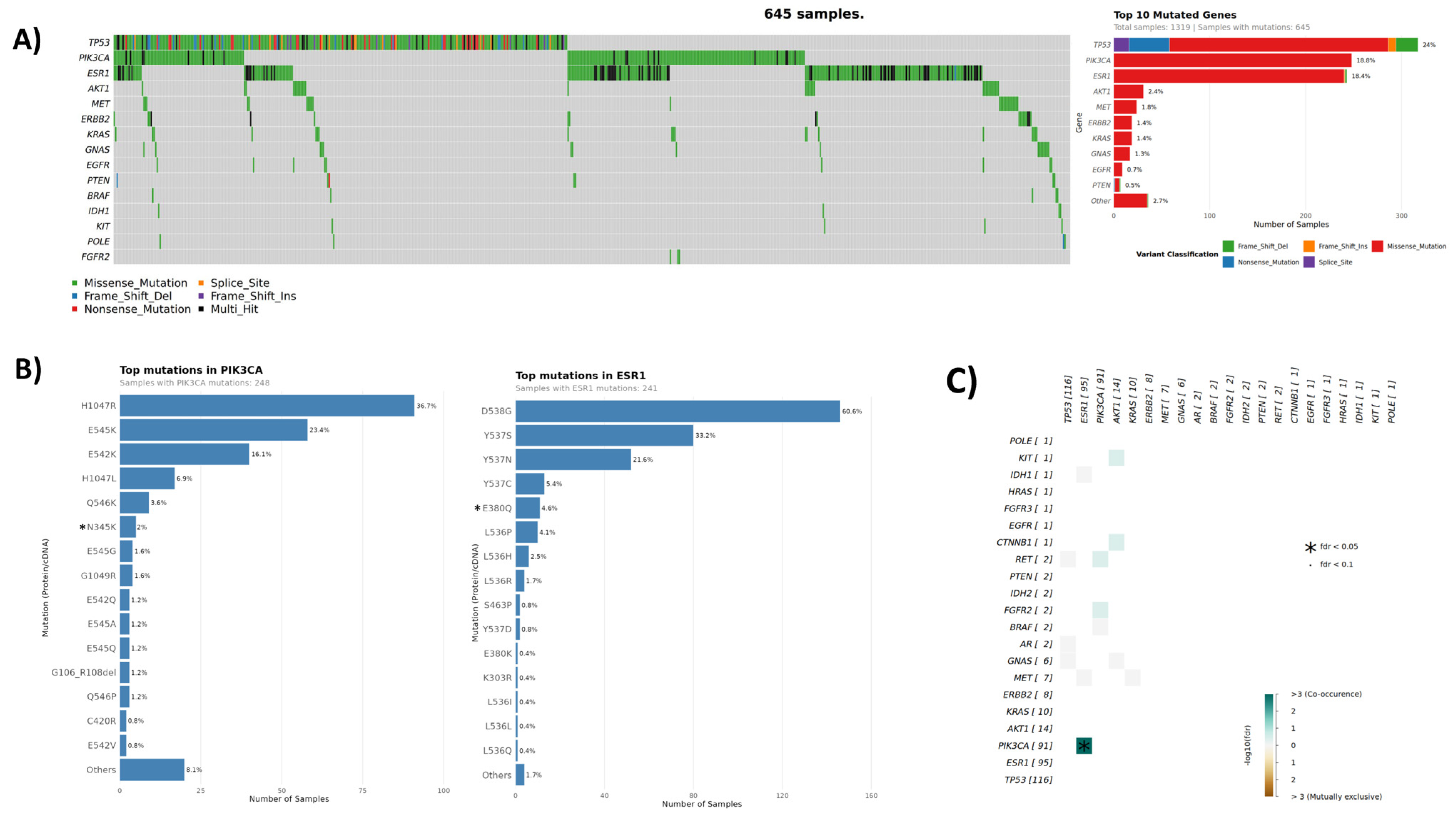
The breast cancer cohort consisted of 1320 samples, 644 (49%) of which harbored pathogenic ctDNA mutations, with the most commonly mutated genes being TP53 (23%), PIK3CA (19%), ESR1 (18%), and AKT1/MET (4%). (Figure 3, Table 3). Tier I/II mutations were detected in 481 (36%) of all breast cancer samples, the majority of which (279/58%) having ≥1 mutation in the PI3K/AKT pathway (PIK3CA, AKT1, and PTEN) (20% total cohort frequency). The most frequent mutations in PIK3CA included exon 20 mutation kinase domain H1047R and exon 9 helical domain mutations E545K/E542K (Figure 3). In the ESR1 gene, frequently observed mutations were D538G, Y537S or Y537N or Y537C, and E380Q. Interestingly, 30% of samples with mutated ESR1 (73/243) harbored ≥2 concurrent ESR1 ctDNA mutations. ESR1 mutations are associated with acquired resistance to antiestrogen therapies, and multiple ESR1 mutations possibly indicate resistance subclones.
In patients with no previous molecular testing (n = 436), LBx detected a total of 232 tier I/II mutations in 156 samples with top mutated genes being ESR1, PIK3CA, AKT1, and ERBB2, providing actionable information in the absence of other molecular testing. In 57% of cases with available previous molecular testing information (508/884), the records indicated the cancer subtype of hormone receptor (ER, PR)-positive with/without HER2-negative status. In this specific subgroup, 49% harbored ctDNA mutations, the majority of which (61%) were PIK3CA and/or ESR1 mutations (Table 6). Mutation interaction analysis identified a significant co-occurrence of PIK3CA and ESR1 mutations (adjusted p-value < 4.02 × 10⁻⁵) that was observed in 6.9% of HR+ samples (35/508, Figure 3), which is similar to published incidences [46]. PI3K/AKT pathway alterations occur frequently in patients with advanced HR-positive breast cancer, collectively affecting up to 50% [47]. Therapies targeting this patient population, such as capivasertib, inavolisib, and alpelisib, are currently approved by FDA and Health Canada, respectively [48,49,50]. ESR1 mutations are a common mechanism of resistance of ER+ breast cancer to endocrine therapies like aromatase inhibitors and tamoxifen. These cancers may retain sensitivity to selective estrogen receptor degraders (SERDs), including FDA-approved elacestrant [51] and imlunestrant [52]. Other SERDs are currently being investigated in clinical trials [53], with LBx-based ESR1 testing serving as a valuable tool to guide treatment [54].
Gene amplification was assessed for 446 breast cancer samples, with ~20% of cases showing copy number gains in one of the nine genes, with FGFR1 being the most frequent alteration (Table 5). FGFR1 amplifications are present in approximately 8.47% of breast carcinoma patients [44] and predict endocrine treatment resistance in HR-positive breast cancer. However, these patients may be sensitive to mTOR inhibitors [55] and are currently under clinical investigation with FGFR inhibitors [56,57,58].
The detection rate of ERBB2 amplification in our breast cancer cohort was low compared to tissue-based rates, which are approximately 15–20%. This can be attributed to a bias in our breast cancer cohort composition and fundamental limitations in CNV detection from cfDNA using small targeted panels. Our breast cancer cohort consisted of only 2.5% of cases with unequivocal HER2-positive status on tissue as indicated by the previous molecular testing. Although the study was open to all advanced cancer patients, such bias can be explained by oncologists’ preferences for obtaining LBx testing for their patients, rendering the breast cancer cohort not fully representative of the advanced breast cancer patient population. Further, it is well established that CNV detection requires a higher tumor fraction and greater genomic coverage than point mutation detection, leading to generally lower sensitivity of LBx assays [59,60,61]. Although our findings underscore a known technical limitation of LBx for CNV detection, we note that the rate of CNV calls in our study was still higher than in a recent comparable study by Nicholas et al. using a similar small NGS panel [23]. Finally, LBx-based HER2 amplification detection in advanced breast cancer patients is challenged by the fact that the primary tissue status may not necessarily reflect that of metastatic disease throughout the body, leading to tissue-plasma discordance [62]. For example, Abraham et al. reported only 30% sensitivity of HER2 amplification detection using Guardant360TM LBx assay in metastatic breast cancer patients with confirmed HER2-positive status in tissue [63].

3.5. Colon Cancer Cohort

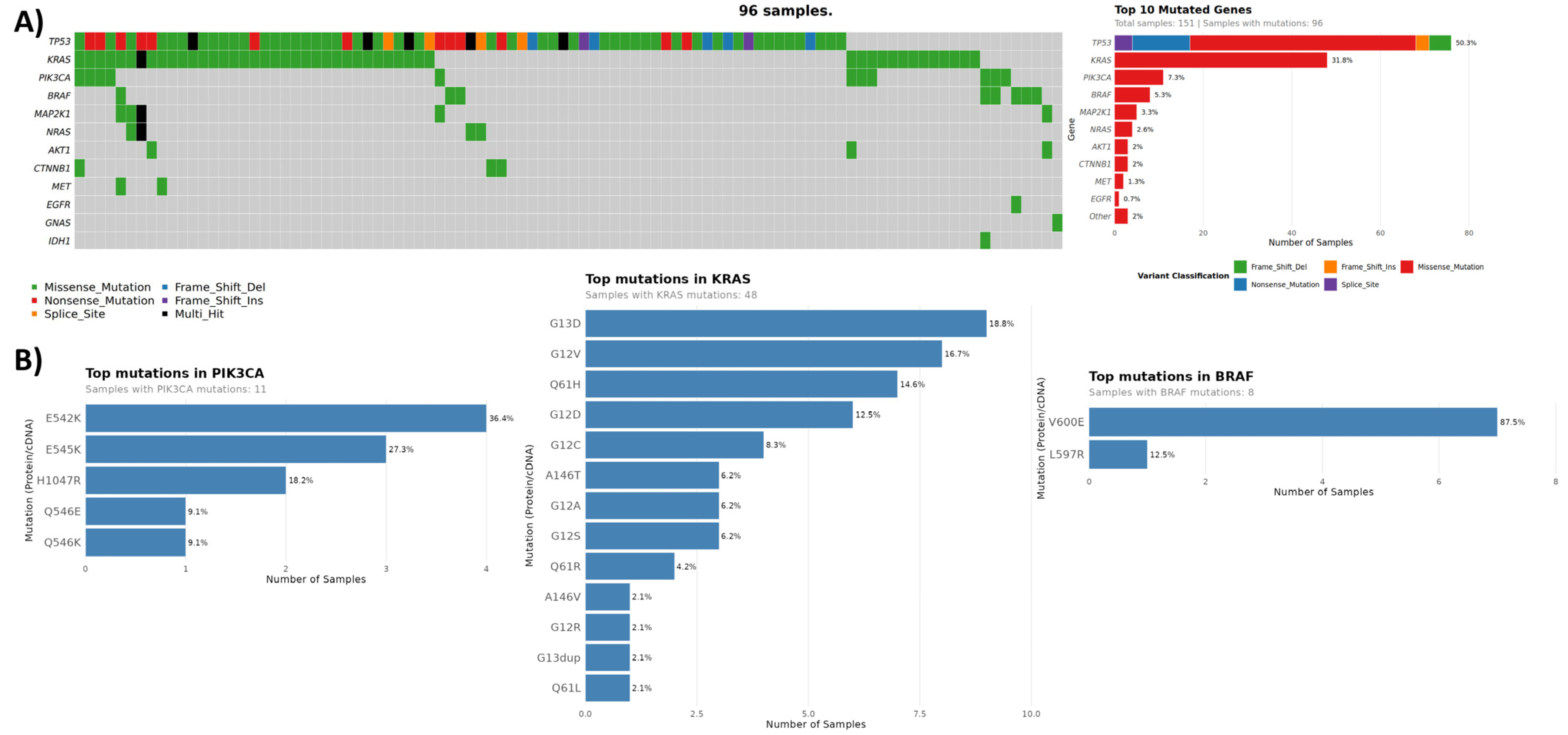
The colon cancer cohort consisted of 151 samples, 96 (64%) of which harbored pathogenic mutations in their ctDNA, with the most commonly mutated genes being TP53 (50%), KRAS (32%), PIK3CA (7%), and BRAF (5%) (Figure 4, Table 3). Tier I/II mutations were detected in 40% of all colorectal cancer patients. In patients with no previous molecular testing, liquid biopsy detected a total of 26 tier I/II mutations in 20 patients, with KRAS and PIK3CA being the top mutated genes. Most frequently observed mutations in KRAS across all colon cancer samples involved G12, G13, and Q61 codons, with a combined prevalence of 29% (44/151) (Figure 4). Consistent with published data [64,65], the most frequently observed PIK3CA mutations included those in exon 9 involving the helical domain of the protein (positions E542-Q546), followed by exon 20 mutation H1047R in the kinase domain with a combined prevalence of 7% (11/151). The predominant BRAF mutation detected was V600E (in 7/8 samples), with a respective cohort prevalence of 5% (7/151). Collectively, mutations in KRAS, PIK3CA, and BRAF, associated with resistance to first-line anti-EGFR monoclonal antibody treatment of mCRC, were detected in 38% of all tested samples (57/151).
Gene amplification was assessed for 26 colon cancer samples, with ~23% of cases showing amplifications in one of the nine genes and MET gain being the most frequent event (Table 5). MET amplifications have been shown to contribute to anti-EGFR resistance in colorectal cancer, and de novo amplifications are one of the major mechanisms of acquired resistance [66,67]. Although the overall prevalence of MET amplifications in colorectal carcinoma is low (0.46% [44]), it is enriched in ctDNA of patients with anti-EGFR refractory disease [68]. These patients may benefit from the use of MET inhibitors, including FDA-approved capmatinib for MET exon 14 alterations [67,69].

3.6. Cost-Effectiveness and Budget Impact Analysis of Follow It® in NSCLC

For LBx technologies to become equally accessible to all Canadians, public funding is a necessity. To help inform provincial payers about the benefits of Follow It® testing, an economic analysis was conducted in collaboration with the Institute of Health Economics (Edmonton, AB, Canada). We aimed to determine the potential for Follow It® to be cost-effective as an addition to tissue biopsy compared to tissue biopsy alone for patients with advanced NSCLC presenting for genetic tumor profiling within the Ontario health care payer market. The modeling was based on 4350 treatment naïve stage IV or incurable stage III non-squamous NSCLC Ontario patients ≥ 60 years of age, with 50% of patients being male.
The results of the modeling are summarized in Table 7. Follow It® would save an average of $18,569 CAD per patient compared to tissue biopsy alone. In terms of health benefit, Follow It® would provide an additional 0.1138 QALYs gained on average per patient. Given that Follow It® would be more effective and less expensive than tissue biopsy, under the current scenario assumptions, it can be said that concurrent Follow It® and tissue biopsy testing dominates tissue biopsy alone.
The budget impact analysis of implementing Follow It® over a 5-year time horizon indicated that increased annual savings will be made over this period, from $74.2 million CAD in year one, to $84.5 million CAD in year 5 (Supplementary Table S13). The majority of the cost savings are due to savings as a result of treatment selection. Of note, although there are increased costs associated with the targeted therapy, these are more than offset by the total savings associated with the overall benefit of improved diagnostic and therapeutic pathway, including, but not limited to, the optimal therapy selection and therefore reduced rates of adverse effects, reduced delays in time to treatment, and the reduced need for costly combinations of chemotherapy and immunotherapy.

4. Discussion

LBx has become an invaluable tool for precision oncology, alleviating some of the challenges associated with tissue testing. This report summarizes the successful implementation of Canada’s first nationwide ctDNA genomic testing program, delivering actionable molecular information within clinically meaningful TAT, and ensuring equitable access to advanced genomic diagnostics for all eligible patients.
The observed high technical success rate of 97% and overwhelming response from the majority of Canadian oncologists across urban and rural centers indicate both the feasibility of establishing liquid biopsy infrastructure on a national scale, and a sustained demand in a rapidly evolving precision oncology space. The assay detected ctDNA mutations in more than half of all patients, with 37% harboring clinically actionable mutations. A total of 76% of patients were matched with clinical trials. Coupled with the clinically meaningful average TAT of 8 days from sample receipt to test results, the project demonstrated the potential to improve patients’ outcomes due to the timely choice of the optimal therapy. Although biomarker testing recommendation is included in NCCN, ASCO, and some of provincial guidelines for breast, colon, and NSCLC, access to molecular testing is still limited across Canadian institutions [70]. In our study, 49% of patients did not have any previous molecular testing. This is in agreement with recent reports on the observed rate of molecular testing of Canadian cancer patients of <30% [14,71]. Additionally, in some cases with limited available information on baseline biomarkers in tissue, Follow it® identified mutations that may interfere with the therapy chosen based on tissue testing alone, such as EGFR ctDNA mutations in lung cancer patients with PD-L1 expression. This underscores the value of concurrent liquid biopsy and tissue testing for multiple actionable biomarkers to enable well-informed treatment decisions.
Several other reports focusing on the clinical benefits of Follow It® testing performed within the ACTT project have been published [19,21,72]. In a study published by Desmeules and co-authors (IUCPQ, QC), 91 patients with advanced NSCLC who had previously undergone single gene tumor tissue genotyping for baseline biomarkers were assessed with LBx using Follow It® via the ACTT project [19]. While for this study cohort Follow It® undoubtedly provided additional information relative to tissue testing alone (47% vs. 18%), translation of this information to clinical practice changing treatment was modest (only 5 pts out of 91 changed therapy). This was due to the regulatory challenges at the time of the study, where access to targeted treatments outside of reimbursed indications was limited. Notably, the median TAT of biomarker information availability was shorter for the centralized Follow It® testing compared to the local tissue testing (10 vs. 13 days). Recently, Breadner and colleagues (Verspeeten Family Cancer Centre, London Health Sciences Centre) reported their experience using ACTT provided Follow It® ctDNA testing as part of the standard work-up for patients with advanced NSCLC. The authors demonstrated significant reduction in (i) time to molecular results (14 vs. 35 days), (ii) time from first respirology/thoracic surgery consult to molecular results (22 vs. 48 days), and (iii) time from medical oncology consultation to initiation of first-line treatment (12 vs. 22 days) [72].
The pilot study of 20 patients of never or light smokers with suspected advanced lung cancer, published by Leighl and colleagues (Princess Margaret Cancer Center, University Health Network), reported the use of Follow It® as part of the pre-diagnostic work-up [21]. Compared to the reference cohorts, the mean TAT for biomarker results and time to treatment initiation (TTT) were significantly shorter for the liquid biopsy arm (17.8 vs. 23.6 days, and 32.6 vs. 62.2 days, respectively). Interestingly, the time to treatment was shortened for all patients participating in the plasma-first approach, even if targetable alterations were not identified. With the observed concordance of 71% between plasma and tissue testing results and clear time savings, the authors concluded that a plasma-first approach can increase detection of therapeutically targetable mutations, especially when tissue DNA is insufficient or unavailable, potentially leading to better patient outcomes.
Demonstrating clinically meaningful outcomes for patients, such as quality-of-life gains and cost-effectiveness, is a critical stepping stone to wide adoption and reimbursement of LBx. Using a model-based evaluation, we assessed the clinical and economic benefit of Follow It® as a clinical-grade, next-generation sequencing targeted panel for somatic tumor testing to predict therapy in advanced NSCLC patients. Considered as an addition to tissue biopsy, and compared to the standard care of tissue biopsy alone, Follow It® was found to be cost-effective, saving approximately $18,569 CAD per patient, and resulted in an additional 0.1138 QALYs gained. Based on the test price of $1200 CAD and its demonstrated accuracy, the assay had the potential to provide significant savings to the healthcare system. Of note, Follow It® testing was performed centrally for this study, and further cost savings can be realized if it is run locally [73,74].
Our study had a number of limitations. Although the Follow It® panel covers most of the actionable biomarkers for major solid tumors, it does not detect fusion events that, although relatively rare, are critical for decision-making in lung and other cancers. Also, the CNV detection using LBx is commonly recognized as a methodological challenge. In our study, the overall rate of CNV detection was higher than that reported in other real-world liquid biopsy studies [23,75], and the spectrum of CNV alterations observed in the NSCLC cohort was consistent with established genomic profiles for this cancer. Nevertheless, the compositional bias of our breast cancer cohort was a critical limitation that precluded a comprehensive evaluation of our assay’s performance for HER2 amplification status, a key biomarker for this malignancy. Further, our cost and health benefit analysis was also constrained by a simplified treatment pathway, an assumption of all biomarker-positive patients receiving targeted treatment, and an evolving landscape of standard-of-care tissue testing practices. Such modeling is recognized to be challenging due to high complexity of actual application of precision oncology principles. Some of the confounding factors include the combination of tests and treatments, patient-level processes and preferences, local vs. centralized testing models, delays in diagnosis and treatment, etc. [74,76]. Institutions operate in a constantly evolving regional regulatory environment and often have inequitable access to diagnostic and therapeutic technologies. Despite all of these limitations, our observations were in agreement with a recently published Canadian report [77] demonstrating similar economic and health benefits of using LBx for NSCLC patients. Together with the growing evidence of the Canadian patients’ preference for less invasive biomarker testing [78], our study highlights the feasibility, need, and benefits of the use of LBx to guide timely treatment decisions and improve outcomes.
We note that our analysis was conducted from the perspective of a Canadian provincial single-payer health system, which necessarily influences both the observed clinical intervention implementation processes and the estimated economic outcomes. Although other jurisdictions may operate under different clinical pathways, reimbursement mechanisms, or pricing structures, the core findings—namely the value of coordinated testing pathways and the potential for cost-efficient adoption of liquid biopsy—remain relevant to health systems pursuing value-based care. Nevertheless, adaptation to local governance, payment models, and data infrastructure would be required for direct translation outside of a single-payer context.

5. Conclusions

The need to provide timely diagnostic information for patients and their healthcare providers to select the most effective therapies for their cancer, including the availability of testing closer to home, is increasingly recognized as the priority in Canada. Liquid biopsies have emerged as a promising complement or alternative to tissue biopsy in recent years, which has the potential to provide additional information, reduce turnaround times, decrease suboptimal drug use, and improve treatment selection in oncology, resulting in better outcomes. The Follow It® LBx tool is no exception, with its demonstrated sensitivity and specificity, relatively lower cost, and focus on clinically actionable genomic mutations [26]. Here, we have described the success of running it centrally for Canadian advanced cancer patients, providing high-quality comprehensive information for an informed therapy selection within clinically relevant turnaround times. Local deployment of this test would enable further improvement in overall cost, speed, and accessibility of LBx for patients. The assay has been suggested by the Canada’s Drug Agency (CDA) to have a significant impact on Canada’s healthcare system in the next 1–3 years [79], given its potential to improve patient outcomes and reduce costs. By facilitating timely, cost-effective access to genomic mutation testing, Follow It® is positioned to help Canadian cancer patients receive the best possible care while allowing the healthcare system to operate more efficiently with potential savings.

Supplementary Materials

The following supporting information can be downloaded at: https://www.mdpi.com/article/10.3390/curroncol33010018/s1, References [80,81,82,83,84,85,86,87,88,89,90,91,92,93,94,95,96,97,98,99] are cited in the Supplementary Materials. Table S1: ctDNA concentrations. Table S2: (A) Follow It panel content. (B) List of reported SNV and indel mutations. (C) List of reported CNVs. Tables S3–S5: Mutation frequencies per gene and per province for lung, breast, and colorectal cohorts. Tables S6–S12: Input parameters for the health economics analysis. Table S13: Budget impact analysis. Figure S1: PIK3CA and ESR1 mutation relative frequencies in breast cancer by assay version. Figure S2: Relative mutation frequency in the lung cancer cohort for select genes. Figure S3: The model structure for the testing component of standard of care. Figure S4. The model structure for the testing component of the Follow It® arm. Figure S5: Markov models for patients receiving targeted therapy or chemotherapy.

Author Contributions

Conceptualization, A.L., D.G.H.; data analysis and visualization, B.L.S.F., P.F. and R.M.; writing—original draft preparation, A.L.; writing—review and editing, A.L., S.K., A.A.A., D.B., B.D.S., B.L. and V.F.; clinical testing, E.B., S.B., B.C., K.M.N., A.K., B.M., M.K.M. and D.G.H.; project administration, A.P.; All authors have read and agreed to the published version of the manuscript.

Funding

The work was partially funded by Canada’s Digital Technology Supercluster through the project “Project ACTT—Access to Cancer Testing & Treatment in Response to COVID-19”. Avitia recognizes AstraZeneca Canada for continued financial support of ctDNA testing for NSCLC post completion of the portion of Project ACTT funded by the Canada’s Digital Technology Supercluster.

Institutional Review Board Statement

Ethical review and approval were waived for this study. The research described here was performed using aggregate, anonymized data for cohorts of patients, without any personal health information. The patients’ molecular data was anonymized using “Safe Harbor” method (section 45 CFR §164.514(b)) and therefore no longer is considered “Human Subjects”.

Informed Consent Statement

Informed consent for participation was obtained from all subjects involved in the study.

Data Availability Statement

The original contributions presented in this study are included in the article/Supplementary Material. Further inquiries can be directed to the corresponding author.

Acknowledgments

We thank Sasha van Katwyk (Institute of Health Economics), for insightful discussion and technical assistance with manuscript preparation. D.G.H. is a distinguished scientist at BC Cancer Agency.

Conflicts of Interest

The Follow It test used in this study was provided free of charge by Imagia Canexia Health to all participating sites. Authors A.L., P.F., R.M, B.F., E.B., S.B., B.C., K.M.N., A.K., B.M., M.K.M., and V.F. were previous employees of Imagia Canexia Health. Author D.G.H. was a previous founder, CMO, and consultant of Imagia Canexia Health. Author A.P. was a previous founder and consultant of Imagia Canexia Health. Authors A.L., P.F., and R.M. are employees of Avitia Inc. Authors B.F., A.K., and D.G.H. are involved as consultants in Avitia Inc. Author A.A.A. has received a research grant from Imagia Canexia Health for a sub-study.

References

  1. Brenner, D.R.; Gillis, J.; Demers, A.A.; Ellison, L.F.; Billette, J.-M.; Zhang, S.X.; Liu, J.L.; Woods, R.R.; Finley, C.; Fitzgerald, N.; et al. Projected estimates of cancer in Canada in 2024. Can. Med. Assoc. J. 2024, 196, E615–E623. [Google Scholar] [CrossRef] [Scilit] [PubMed]
  2. Bouchard, N.; Daaboul, N. Lung Cancer: Targeted Therapy in 2025. Curr. Oncol. 2025, 32, 146. [Google Scholar] [CrossRef] [Scilit] [PubMed]
  3. Tan, A.C.; Tan, D.S.W. Targeted Therapies for Lung Cancer Patients with Oncogenic Driver Molecular Alterations. J. Clin. Oncol. 2022, 40, 611–625. [Google Scholar] [CrossRef] [Scilit] [PubMed]
  4. Ohishi, T.; Kaneko, M.K.; Yoshida, Y.; Takashima, A.; Kato, Y.; Kawada, M. Current Targeted Therapy for Metastatic Colorectal Cancer. Int. J. Mol. Sci. 2023, 24, 1702. [Google Scholar] [CrossRef] [Scilit]
  5. Jacobs, A.T.; Martinez Castaneda-Cruz, D.; Rose, M.M.; Connelly, L. Targeted therapy for breast cancer: An overview of drug classes and outcomes. Biochem. Pharmacol. 2022, 204, 115209. [Google Scholar] [CrossRef] [Scilit]
  6. Liu, B.; Zhou, H.; Tan, L.; Siu, K.T.H.; Guan, X.-Y. Exploring treatment options in cancer: Tumor treatment strategies. Signal Transduct. Target. Ther. 2024, 9, 175. [Google Scholar] [CrossRef] [Scilit]
  7. Mosele, F.; Remon, J.; Mateo, J.; Westphalen, C.B.; Barlesi, F.; Lolkema, M.P.; Normanno, N.; Scarpa, A.; Robson, M.; Meric-Bernstam, F.; et al. Recommendations for the use of next-generation sequencing (NGS) for patients with metastatic cancers: A report from the ESMO Precision Medicine Working Group. Ann. Oncol. 2020, 31, 1491–1505. [Google Scholar] [CrossRef] [Scilit]
  8. Cheema, P.K.; Banerji, S.O.; Blais, N.; Chu, Q.S.-C.; Juergens, R.A.; Leighl, N.B.; Sacher, A.; Sheffield, B.S.; Snow, S.; Vincent, M.; et al. Canadian Consensus Recommendations on the Management of KRAS G12C-Mutated NSCLC. Curr. Oncol. 2023, 30, 6473–6496. [Google Scholar] [CrossRef] [Scilit]
  9. Breadner, D.; Hwang, D.M.; Husereau, D.; Cheema, P.; Doucette, S.; Ellis, P.M.; Kassam, S.; Leighl, N.; Maziak, D.E.; Selvarajah, S.; et al. Implementation of Liquid Biopsy in Non-Small-Cell Lung Cancer: An Ontario Perspective. Curr. Oncol. 2024, 31, 6017–6031. [Google Scholar] [CrossRef] [Scilit]
  10. National Comprehensive Cancer Network. Non-Small Cell Lung Cancer (Version 8.2025). Available online: https://www.nccn.org/professionals/physician_gls/pdf/nscl.pdf (accessed on 6 August 2025).
  11. National Comprehensive Cancer Network. Breast Cancer (Version 5.2025). Available online: https://www.nccn.org/professionals/physician_gls/pdf/breast.pdf (accessed on 8 August 2025).
  12. Husereau, D.; Villalba, E.; Muthu, V.; Mengel, M.; Ivany, C.; Steuten, L.; Spinner, D.S.; Sheffield, B.; Yip, S.; Jacobs, P.; et al. Progress toward Health System Readiness for Genome-Based Testing in Canada. Curr. Oncol. 2023, 30, 5379–5394. [Google Scholar] [CrossRef] [Scilit]
  13. Ellis, P.M.; Vandermeer, R. Delays in the diagnosis of lung cancer. J. Thorac. Dis. 2011, 3, 183–188. [Google Scholar] [PubMed]
  14. Lee, T.H.; Krishnan, T.; Gill, S. Real-world analysis of the impact of timing of biomarker testing on first-line treatment choice in patients with metastatic colorectal cancer in British Columbia. J. Clin. Oncol. 2025, 43, 253. [Google Scholar] [CrossRef] [Scilit]
  15. Elkrief, A.; Joubert, P.; Florescu, M.; Tehfe, M.; Blais, N.; Routy, B. Therapeutic landscape of metastatic non-small-cell lung cancer in Canada in 2020. Curr. Oncol. 2020, 27, 52–60. [Google Scholar] [CrossRef] [Scilit] [PubMed]
  16. Sadik, H.; Pritchard, D.; Keeling, D.M.; Policht, F.; Riccelli, P.; Stone, G.; Finkel, K.; Schreier, J.; Munksted, S. Impact of Clinical Practice Gaps on the Implementation of Personalized Medicine in Advanced Non-Small-Cell Lung Cancer. JCO Precis. Oncol. 2022, 6, e2200246. [Google Scholar] [CrossRef] [Scilit]
  17. Rolfo, C.; Mack, P.; Scagliotti, G.V.; Aggarwal, C.; Arcila, M.E.; Barlesi, F.; Bivona, T.; Diehn, M.; Dive, C.; Dziadziuszko, R.; et al. Liquid Biopsy for Advanced NSCLC: A Consensus Statement From the International Association for the Study of Lung Cancer. J. Thorac. Oncol. 2021, 16, 1647–1662. [Google Scholar] [CrossRef] [Scilit]
  18. Samaha, R.; El Sayed, R.; Alameddine, R.; Florescu, M.; Tehfe, M.; Routy, B.; Elkrief, A.; Belkaid, W.; Desilets, A.; Weng, X.; et al. Clinical Utility of Liquid Biopsy for the Early Diagnosis of EGFR-Mutant Advanced Lung Cancer Patients in a Real-Life Setting (CLEAR Study). Curr. Oncol. 2025, 32, 57. [Google Scholar] [CrossRef] [Scilit]
  19. Desmeules, P.; Dusselier, M.; Bouffard, C.; Bafaro, J.; Fortin, M.; Labbé, C.; Joubert, P. Retrospective Assessment of Complementary Liquid Biopsy on Tissue Single-Gene Testing for Tumor Genotyping in Advanced NSCLC. Curr. Oncol. 2023, 30, 575–585. [Google Scholar] [CrossRef] [Scilit]
  20. Garcia-Pardo, M.; Czarnecka, K.; Law, J.H.; Salvarrey, A.; Fernandes, R.; Fan, J.; Corke, L.; Waddell, T.K.; Yasufuku, K.; Donahoe, L.L.; et al. Plasma-first: Accelerating lung cancer diagnosis and molecular profiling through liquid biopsy. Ther. Adv. Med. Oncol. 2022, 14, 17588359221126151. [Google Scholar] [CrossRef] [Scilit]
  21. García-Pardo, M.; Czarnecka-Kujawa, K.; Law, J.H.; Salvarrey, A.M.; Fernandes, R.; Fan, Z.J.; Waddell, T.K.; Yasufuku, K.; Liu, G.; Donahoe, L.L.; et al. Association of Circulating Tumor DNA Testing Before Tissue Diagnosis with Time to Treatment Among Patients with Suspected Advanced Lung Cancer: The ACCELERATE Nonrandomized Clinical Trial. JAMA Netw. Open 2023, 6, e2325332. [Google Scholar] [CrossRef] [Scilit]
  22. Patel, Y.P.; Husereau, D.; Leighl, N.B.; Melosky, B.; Nam, J. Health and Budget Impact of Liquid-Biopsy-Based Comprehensive Genomic Profile (CGP) Testing in Tissue-Limited Advanced Non-Small Cell Lung Cancer (aNSCLC) Patients. Curr. Oncol. 2021, 28, 5278–5294. [Google Scholar] [CrossRef] [Scilit]
  23. Nicholas, C.; Beharry, A.; Bendzsak, A.M.; Bisson, K.R.; Dadson, K.; Dudani, S.; Iafolla, M.; Irshad, K.; Perdrizet, K.; Raskin, W.; et al. Point of Care Liquid Biopsy for Cancer Treatment-Early Experience from a Community Center. Cancers 2024, 16, 2505. [Google Scholar] [CrossRef] [Scilit] [PubMed]
  24. Juergens, R.A.; Ezeife, D.A.; Laskin, J.J.; Agulnik, J.S.; Hao, D.; Laurie, S.A.; Law, J.H.; Le, L.W.; Kiedrowski, L.A.; Shepherd, F.A.; et al. Demonstrating the value of liquid biopsy for lung cancer in a public health care system. J. Clin. Oncol. 2020, 38, 3546. [Google Scholar] [CrossRef] [Scilit]
  25. Jennings, L.J.; Arcila, M.E.; Corless, C.; Kamel-Reid, S.; Lubin, I.M.; Pfeifer, J.; Temple-Smolkin, R.L.; Voelkerding, K.V.; Nikiforova, M.N. Guidelines for Validation of Next-Generation Sequencing-Based Oncology Panels: A Joint Consensus Recommendation of the Association for Molecular Pathology and College of American Pathologists. J. Mol. Diagn. 2017, 19, 341–365. [Google Scholar] [CrossRef] [Scilit] [PubMed]
  26. Avitia Assays: Actionable, Robust and Cost-Effective Cancer Hotspot Panels. Available online: https://cdn.prod.website-files.com/67564436d96257851efc0e4c/67bf30eb90fc8f0e269d3297_Avitia%20-%20Assays.pdf (accessed on 12 September 2025).
  27. Robinson, J.T.; Thorvaldsdóttir, H.; Winckler, W.; Guttman, M.; Lander, E.S.; Getz, G.; Mesirov, J.P. Integrative genomics viewer. Nat. Biotechnol. 2011, 29, 24–26. [Google Scholar] [CrossRef] [Scilit]
  28. Mayakonda, A.; Lin, D.C.; Assenov, Y.; Plass, C.; Koeffler, H.P. Maftools: Efficient and comprehensive analysis of somatic variants in cancer. Genome Res. 2018, 28, 1747–1756. [Google Scholar] [CrossRef] [Scilit]
  29. König, D.; Savic Prince, S.; Rothschild, S.I. Targeted Therapy in Advanced and Metastatic Non-Small Cell Lung Cancer. An Update on Treatment of the Most Important Actionable Oncogenic Driver Alterations. Cancers 2021, 13, 804. [Google Scholar] [CrossRef] [Scilit]
  30. Hench, I.B.; Hench, J.; Tolnay, M. Liquid Biopsy in Clinical Management of Breast, Lung, and Colorectal Cancer. Front. Med. 2018, 5, 9. [Google Scholar] [CrossRef] [Scilit]
  31. Malla, M.; Loree, J.M.; Kasi, P.M.; Parikh, A.R. Using Circulating Tumor DNA in Colorectal Cancer: Current and Evolving Practices. J. Clin. Oncol. 2022, 40, 2846–2857. [Google Scholar] [CrossRef] [Scilit]
  32. Fundytus, A.; Cook, S.; Yip, S.M.; Loewen, S.K.; Hao, D. Workforce Trends Among Canadian Medical Oncologists and Medical Oncology Trainees over Two Decades. Curr. Oncol. 2025, 32, 70. [Google Scholar] [CrossRef] [Scilit]
  33. Bettegowda, C.; Sausen, M.; Leary, R.J.; Kinde, I.; Wang, Y.; Agrawal, N.; Bartlett, B.R.; Wang, H.; Luber, B.; Alani, R.M.; et al. Detection of circulating tumor DNA in early- and late-stage human malignancies. Sci. Transl. Med. 2014, 6, 224ra24. [Google Scholar] [CrossRef] [Scilit]
  34. Jee, J.; Lebow, E.S.; Yeh, R.; Das, J.P.; Namakydoust, A.; Paik, P.K.; Chaft, J.E.; Jayakumaran, G.; Rose Brannon, A.; Benayed, R.; et al. Overall survival with circulating tumor DNA-guided therapy in advanced non-small-cell lung cancer. Nat. Med. 2022, 28, 2353–2363. [Google Scholar] [CrossRef] [Scilit] [PubMed]
  35. Akao, K.; Oya, Y.; Sato, T.; Ikeda, A.; Horiguchi, T.; Goto, Y.; Hashimoto, N.; Kondo, M.; Imaizumi, K. It might be a dead end: Immune checkpoint inhibitor therapy in EGFR-mutated NSCLC. Explor. Target. Anti-Tumor Ther. 2024, 5, 826–840. [Google Scholar] [CrossRef] [Scilit] [PubMed]
  36. Fukuoka, M.; Wu, Y.L.; Thongprasert, S.; Sunpaweravong, P.; Leong, S.S.; Sriuranpong, V.; Chao, T.Y.; Nakagawa, K.; Chu, D.T.; Saijo, N.; et al. Biomarker analyses final overall survival results from a phase III, randomized open-label first-line study of gefitinib versus carboplatin/paclitaxel in clinically selected patients with advanced non-small-cell lung cancer in Asia (IPASS). J. Clin. Oncol. 2011, 29, 2866–2874. [Google Scholar] [CrossRef] [Scilit] [PubMed]
  37. Rosell, R.; Moran, T.; Queralt, C.; Porta, R.; Cardenal, F.; Camps, C.; Majem, M.; Lopez-Vivanco, G.; Isla, D.; Provencio, M.; et al. Screening for epidermal growth factor receptor mutations in lung cancer. N. Engl. J. Med. 2009, 361, 958–967. [Google Scholar] [CrossRef] [Scilit]
  38. Sequist, L.V.; Yang, J.C.; Yamamoto, N.; O’Byrne, K.; Hirsh, V.; Mok, T.; Geater, S.L.; Orlov, S.; Tsai, C.M.; Boyer, M.; et al. Phase III study of afatinib or cisplatin plus pemetrexed in patients with metastatic lung adenocarcinoma with, EGFR mutations. J. Clin. Oncol. 2013, 31, 3327–3334. [Google Scholar] [CrossRef] [Scilit]
  39. Ramalingam, S.S.; Vansteenkiste, J.; Planchard, D.; Cho, B.C.; Gray, J.E.; Ohe, Y.; Zhou, C.; Reungwetwattana, T.; Cheng, Y.; Chewaskulyong, B.; et al. Overall Survival with Osimertinib in Untreated EGFR-Mutated Advanced NSCLC. N. Engl. J. Med. 2020, 382, 41–50. [Google Scholar] [CrossRef] [Scilit]
  40. Dogan, S.; Shen, R.; Ang, D.C.; Johnson, M.L.; D’Angelo, S.P.; Paik, P.K.; Brzostowski, E.B.; Riely, G.J.; Kris, M.G.; Zakowski, M.F.; et al. Molecular epidemiology of EGFR and KRAS mutations in 3,026 lung adenocarcinomas: Higher susceptibility of women to smoking-related KRAS-mutant cancers. Clin. Cancer Res. 2012, 18, 6169–6177. [Google Scholar] [CrossRef] [Scilit]
  41. Riely, G.J.; Kris, M.G.; Rosenbaum, D.; Marks, J.; Li, A.; Chitale, D.A.; Nafa, K.; Riedel, E.R.; Hsu, M.; Pao, W.; et al. Frequency and distinctive spectrum of KRAS mutations in never smokers with lung adenocarcinoma. Clin. Cancer Res. 2008, 14, 5731–5734. [Google Scholar] [CrossRef] [Scilit]
  42. Reid, J.L.; Hammond, D.; Burkhalter, R.; Rynard, V.L. Tobacco Use in Canada: Patterns and Trends, 2022nd ed.; University of Waterloo: Waterloo, ON, Canada, 2022. [Google Scholar]
  43. Mina, S.A.; Shanshal, M.; Leventakos, K.; Parikh, K. Emerging Targeted Therapies in Non-Small-Cell Lung Cancer (NSCLC). Cancers 2025, 17, 353. [Google Scholar] [CrossRef] [Scilit]
  44. Consortium, A.P.G. AACR Project GENIE: Powering Precision Medicine through an International Consortium. Cancer Discov. 2017, 7, 818–831. [Google Scholar] [CrossRef] [Scilit]
  45. Camidge, D.R.; Otterson, G.A.; Clark, J.W.; Ignatius Ou, S.H.; Weiss, J.; Ades, S.; Shapiro, G.I.; Socinski, M.A.; Murphy, D.A.; Conte, U.; et al. Crizotinib in Patients with MET-Amplified NSCLC. J. Thorac. Oncol. 2021, 16, 1017–1029. [Google Scholar] [CrossRef] [Scilit] [PubMed]
  46. Dempsey, N.; Bhatt, P.; Lewis, C.; Tolman, D.; Chamorro, Y.; Rubens, M.; Carcas, L.; Sandoval-Leon, A.C.; Ahluwalia, M.S.; Mahtani, R.L.; et al. Co-occurrence of ESR1 and PIK3CA mutations in HR+/HER2- metastatic breast cancer: Incidence and outcomes with targeted therapy. J. Clin. Oncol. 2024, 42, e13097. [Google Scholar] [CrossRef] [Scilit]
  47. Millis, S.Z.; Ikeda, S.; Reddy, S.; Gatalica, Z.; Kurzrock, R. Landscape of Phosphatidylinositol-3-Kinase Pathway Alterations Across 19 784 Diverse Solid Tumors. JAMA Oncol. 2016, 2, 1565–1573. [Google Scholar] [CrossRef] [Scilit] [PubMed]
  48. Turner, N.C.; Im, S.-A.; Saura, C.; Juric, D.; Loibl, S.; Kalinsky, K.; Schmid, P.; Loi, S.; Thanopoulou, E.; Shankar, N.; et al. INAVO120: Phase III trial final overall survival (OS) analysis of first-line inavolisib (INAVO)/placebo (PBO) + palbociclib (PALBO) + fulvestrant (FULV) in patients (pts) with PIK3CA-mutated, hormone receptor-positive (HR+), HER2-negative (HER2–), endocrine-resistant advanced breast cancer (aBC). J. Clin. Oncol. 2025, 43, 1003. [Google Scholar]
  49. Turner, N.C.; Oliveira, M.; Howell, S.J.; Dalenc, F.; Cortes, J.; Moreno, H.L.G.; Hu, X.; Jhaveri, K.; Krivorotko, P.; Loibl, S.; et al. Capivasertib in Hormone Receptor–Positive Advanced Breast Cancer. N. Engl. J. Med. 2023, 388, 2058–2070. [Google Scholar] [CrossRef] [Scilit]
  50. André, F.; Ciruelos, E.M.; Juric, D.; Loibl, S.; Campone, M.; Mayer, I.A.; Rubovszky, G.; Yamashita, T.; Kaufman, B.; Lu, Y.S.; et al. Alpelisib plus fulvestrant for PIK3CA-mutated, hormone receptor-positive, human epidermal growth factor receptor-2-negative advanced breast cancer: Final overall survival results from SOLAR-1. Ann. Oncol. 2021, 32, 208–217. [Google Scholar] [CrossRef] [Scilit]
  51. Shah, M.; Lingam, H.; Gao, X.; Gittleman, H.; Fiero, M.H.; Krol, D.; Biel, N.; Ricks, T.K.; Fu, W.; Hamed, S.; et al. USFood Drug Administration Approval Summary: Elacestrant for Estrogen Receptor-Positive Human Epidermal Growth Factor Receptor 2-Negative ESR1-Mutated Advanced or Metastatic Breast Cancer. J. Clin. Oncol. 2024, 42, 1193–1201. [Google Scholar] [CrossRef] [Scilit]
  52. Jhaveri, K.L.; Neven, P.; Casalnuovo, M.L.; Kim, S.-B.; Tokunaga, E.; Aftimos, P.; Saura, C.; O’Shaughnessy, J.; Harbeck, N.; Carey, L.A.; et al. Imlunestrant with or without Abemaciclib in Advanced Breast Cancer. N. Engl. J. Med. 2025, 392, 1189–1202. [Google Scholar] [CrossRef] [Scilit]
  53. Hamilton, E.P.; Ma, C.; De Laurentiis, M.; Iwata, H.; Hurvitz, S.A.; Wander, S.A.; Danso, M.; Lu, D.R.; Perkins Smith, J.; Liu, Y.; et al. VERITAC-2: A Phase III study of vepdegestrant, a PROTAC ER degrader, versus fulvestrant in ER+/HER2- advanced breast cancer. Future Oncol. 2024, 20, 2447–2455. [Google Scholar] [CrossRef] [Scilit]
  54. Bidard, F.C.; Mayer, E.L.; Park, Y.H.; Janni, W.; Ma, C.; Cristofanilli, M.; Bianchini, G.; Kalinsky, K.; Iwata, H.; Chia, S.; et al. First-Line Camizestrant for Emerging ESR1-Mutated Advanced Breast Cancer. N. Engl. J. Med. 2025, 393, 569–580. [Google Scholar] [CrossRef] [Scilit]
  55. Drago, J.Z.; Formisano, L.; Juric, D.; Niemierko, A.; Servetto, A.; Wander, S.A.; Spring, L.M.; Vidula, N.; Younger, J.; Peppercorn, J.; et al. FGFR1 Amplification Mediates Endocrine Resistance but Retains TORC Sensitivity in Metastatic Hormone Receptor-Positive (HR(+)) Breast Cancer. Clin. Cancer Res. 2019, 25, 6443–6451. [Google Scholar] [CrossRef] [Scilit] [PubMed]
  56. Meric-Bernstam, F.; Bahleda, R.; Hierro, C.; Sanson, M.; Bridgewater, J.; Arkenau, H.T.; Tran, B.; Kelley, R.K.; Park, J.O.; Javle, M.; et al. Futibatinib, an Irreversible FGFR1-4 Inhibitor, in Patients with Advanced Solid Tumors Harboring FGF/FGFR Aberrations: A Phase I Dose-Expansion Study. Cancer Discov. 2022, 12, 402–415. [Google Scholar] [CrossRef] [Scilit] [PubMed]
  57. Pant, S.; Schuler, M.; Iyer, G.; Witt, O.; Doi, T.; Qin, S.; Tabernero, J.; Reardon, D.A.; Massard, C.; Minchom, A.; et al. Erdafitinib in patients with advanced solid tumours with FGFR alterations (RAGNAR): An international, single-arm, phase 2 study. Lancet Oncol. 2023, 24, 925–935. [Google Scholar] [CrossRef] [Scilit] [PubMed]
  58. Giordano, A.; Unni, N.; Damodaran, S.; Rugo, H.; Crook, T.; Bachelot, T.; Piacentini, F.; Parra, H.S.; Krop, I.; Shimura, M.; et al. Abstract PO4-06-07: Efficacy and safety of futibatinib in patients with locally advanced/metastatic triple-negative breast cancer harboring FGFR2 gene amplification: Final results from the phase 2, open-label FOENIX-MBC2 study. Cancer Res. 2024, 84, PO4-06-07. [Google Scholar] [CrossRef] [Scilit]
  59. Keller, L.; Belloum, Y.; Wikman, H.; Pantel, K. Clinical relevance of blood-based ctDNA analysis: Mutation detection and beyond. Br. J. Cancer 2021, 124, 345–358. [Google Scholar] [CrossRef] [Scilit]
  60. Peng, H.; Lu, L.; Zhou, Z.; Liu, J.; Zhang, D.; Nan, K.; Zhao, X.; Li, F.; Tian, L.; Dong, H.; et al. CNV Detection from Circulating Tumor DNA in Late Stage Non-Small Cell Lung Cancer Patients. Genes 2019, 10, 926. [Google Scholar] [CrossRef] [Scilit]
  61. Finkle, J.D.; Boulos, H.; Driessen, T.M.; Lo, C.; Blidner, R.A.; Hafez, A.; Khan, A.A.; Lozac’hmeur, A.; McKinnon, K.E.; Perera, J.; et al. Validation of a liquid biopsy assay with molecular clinical profiling of circulating tumor DNA. npj Precis. Oncol. 2021, 5, 63. [Google Scholar] [CrossRef] [Scilit]
  62. Verschoor, N.; Deger, T.; Jager, A.; Sleijfer, S.; Wilting, S.M.; Martens, J.W.M. Validity and utility of HER2/ERBB2 copy number variation assessed in liquid biopsies from breast cancer patients: A systematic review. Cancer Treat. Rev. 2022, 106, 102384. [Google Scholar] [CrossRef] [Scilit]
  63. Abraham, J.; Montero, A.J.; Jankowitz, R.C.; Salkeni, M.A.; Beumer, J.H.; Kiesel, B.F.; Piette, F.; Adamson, L.M.; Nagy, R.J.; Lanmanm, R.B.; et al. Safety Efficacy of T-DM1 Plus Neratinib in Patients with Metastatic HER2-Positive Breast Cancer: NSABP Foundation Trial FB-1.0. J. Clin. Oncol. 2019, 37, 2601–2609. [Google Scholar] [CrossRef] [Scilit]
  64. Mazouji, O.; Ouhajjou, A.; Incitti, R.; Mansour, H. Updates on Clinical Use of Liquid Biopsy in Colorectal Cancer Screening, Diagnosis, Follow-Up, and Treatment Guidance. Front. Cell Dev. Biol. 2021, 9, 660924. [Google Scholar] [CrossRef] [Scilit]
  65. Wang, H.; Tang, R.; Jiang, L.; Jia, Y. The role of PIK3CA gene mutations in colorectal cancer and the selection of treatment strategies. Front. Pharmacol. 2024, 15, 1494802. [Google Scholar] [CrossRef] [Scilit] [PubMed]
  66. Appleyard, J.W.; Williams, C.J.M.; Manca, P.; Pietrantonio, F.; Seligmann, J.F. Targeting the MAP Kinase Pathway in Colorectal Cancer: A Journey in Personalized Medicine. Clin. Cancer Res. 2025, 31, 2565–2572. [Google Scholar] [CrossRef] [Scilit] [PubMed]
  67. Bardelli, A.; Corso, S.; Bertotti, A.; Hobor, S.; Valtorta, E.; Siravegna, G.; Sartore-Bianchi, A.; Scala, E.; Cassingena, A.; Zecchin, D.; et al. Amplification of the MET receptor drives resistance to anti-EGFR therapies in colorectal cancer. Cancer Discov. 2013, 3, 658–673. [Google Scholar] [CrossRef] [Scilit] [PubMed]
  68. Raghav, K.; Morris, V.; Tang, C.; Morelli, P.; Amin, H.M.; Chen, K.; Manyam, G.C.; Broom, B.; Overman, M.J.; Shaw, K.; et al. MET amplification in metastatic colorectal cancer: An acquired response to EGFR inhibition, not a de novo phenomenon. Oncotarget 2016, 7, 54627–54631. [Google Scholar] [CrossRef] [Scilit]
  69. Akhoundova, D.; Pietge, H.; Hussung, S.; Kiessling, M.; Britschgi, C.; Zoche, M.; Rechsteiner, M.; Weber, A.; Fritsch, R.M. Targeting Secondary and Tertiary Resistance to BRAF Inhibition in BRAF V600E-Mutated Metastatic Colorectal Cancer. JCO Precis. Oncol. 2021, 5, 1082–1087. [Google Scholar] [CrossRef] [Scilit]
  70. Sheffield, B.S.; Banerji, S.; Chankowsky, A.; Dudani, S.; Gill, S.; Gorski, Z.; Kassam, S.; Macaulay, C.; Manna, M.; Perdrizet, K.; et al. Toward Timely and Equitable Advanced Biomarker Testing for Patients with Metastatic Cancer in Canada. Curr. Oncol. 2025, 32, 141. [Google Scholar] [CrossRef] [Scilit]
  71. Patient & Caregivers’ Perspective on Biomarker Testing Across Canada—Survey Results. Available online: https://www.colorectalcancercanada.com/app/uploads/2021/07/Patient-Caregivers-Perspective-on-Biomarker-Testing-Across-Canada-Survey-Results.pdf (accessed on 23 October 2025).
  72. Holjak, E.; Brasoveanu, T.; Verma, S.; Khan, S.; Black, M.; Dhaliwal, I.; Mitchell, M.; Nayak, R.; Qiabi, M.; Inculet, R.; et al. Circulating tumor DNA as part of the routine work-up for patients with suspected advanced lung cancer. J. Liq. Biopsy 2025, 10, 100443. [Google Scholar] [CrossRef] [Scilit]
  73. Silas, U.; Blüher, M.; Bosworth Smith, A.; Saunders, R. Fast In-House Next-Generation Sequencing in the Diagnosis of Metastatic Non-small Cell Lung Cancer: A Hospital Budget Impact Analysis. J. Health Econ. Outcomes Res. 2023, 10, 111–118. [Google Scholar] [CrossRef] [Scilit]
  74. Johnston, K.M.; Sheffield, B.S.; Yip, S.; Lakzadeh, P.; Qian, C.; Nam, J. Costs of in-house genomic profiling and implications for econ omic evaluation: A case example of non-small cell lung cancer (NSCLC). J. Med. Econ. 2020, 23, 1123–1129. [Google Scholar] [CrossRef] [Scilit]
  75. Xie, J.; Yao, W.; Chen, L.; Zhu, W.; Liu, Q.; Geng, G.; Fang, J.; Zhao, Y.; Xiao, L.; Huang, Z.; et al. Plasma ctDNA increases tissue NGS-based detection of therapeutically targetable mutations in lung cancers. BMC Cancer 2023, 23, 294. [Google Scholar] [CrossRef] [Scilit]
  76. Fagery, M.; Khorshidi, H.A.; Wong, S.Q.; Vu, M.; Ijzerman, M. Health Economic Evidence and Modeling Challenges for Liquid Biopsy Assays in Cancer Management: A Systematic Literature Review. PharmacoEconomics 2023, 41, 1229–1248. [Google Scholar] [CrossRef] [Scilit] [PubMed]
  77. Ezeife, D.A.; Spackman, E.; Juergens, R.A.; Laskin, J.J.; Agulnik, J.S.; Hao, D.; Laurie, S.A.; Law, J.H.; Le, L.W.; Kiedrowski, L.A.; et al. The economic value of liquid biopsy for genomic profiling in advanced non-small cell lung cancer. Ther. Adv. Med. Oncol. 2022, 14, 17588359221112696. [Google Scholar] [CrossRef] [Scilit] [PubMed]
  78. Chen, K.H.; Barnes, T.A.; Laskin, J.; Cheema, P.; Liu, G.; Iqbal, M.; Rothenstein, J.; Burkes, R.; Tsao, M.S.; Leighl, N.B. The Perceived Value of Liquid Biopsy: Results From a Canadian Validation Study of Circulating Tumor DNA T790M Testing-Patient’s Willingness-to-Pay: A Brief Report. JTO Clin. Res. Rep. 2024, 5, 100615. [Google Scholar] [CrossRef] [Scilit]
  79. Basharat, S.; Farah, K. An overview of comprehensive genomic profiling technologies to inform cancer care. Can. J. Health Technol. 2022, 2. [Google Scholar] [CrossRef] [Scilit]
  80. Mann, H.; Andersohn, F.; Bodnar, C.; Mitsudomi, T.; Mok, T.S.K.; Yang, J.C.; Hoyle, C. Adjusted Indirect Comparison Using Propensity Score Matching of Osimertinib to Platinum-Based Doublet Chemotherapy in Patients with EGFRm T790M NSCLC Who Have Progressed after EGFR-TKI. Clin. Drug Investig. 2018, 38, 319–331. [Google Scholar] [CrossRef] [Scilit]
  81. Gandhi, L.; Rodríguez-Abreu, D.; Gadgeel, S.; Esteban, E.; Felip, E.; De Angelis, F.; Domine, M.; Clingan, P.; Hochmair, M.J.; Powell, S.F.; et al. Pembrolizumab plus Chemotherapy in Metastatic Non-Small-Cell Lung Cancer. N. Engl. J. Med. 2018, 378, 2078–2092. [Google Scholar] [CrossRef] [Scilit]
  82. Oxnard, G.R.; Thress, K.S.; Alden, R.S.; Lawrance, R.; Paweletz, C.P.; Cantarini, M.; Yang, J.C.; Barrett, J.C.; Jänne, P.A. Association Between Plasma Genotyping and Outcomes of Treatment with Osimertinib (AZD9291) in Advanced Non-Small-Cell Lung Cancer. J. Clin. Oncol. 2016, 34, 3375–3382. [Google Scholar] [CrossRef] [Scilit]
  83. Mok, T.S.; Wu, Y.L.; Ahn, M.J.; Garassino, M.C.; Kim, H.R.; Ramalingam, S.S.; Shepherd, F.A.; He, Y.; Akamatsu, H.; Theelen, W.S.; et al. Osimertinib or Platinum-Pemetrexed in EGFR T790M-Positive Lung Cancer. N. Engl. J. Med. 2017, 376, 629–640. [Google Scholar] [CrossRef] [Scilit]
  84. Horn, L.; Spigel, D.R.; Vokes, E.E.; Holgado, E.; Ready, N.; Steins, M.; Poddubskaya, E.; Borghaei, H.; Felip, E.; Paz-Ares, L.; et al. Nivolumab Versus Docetaxel in Previously Treated Patients with Advanced Non-Small-Cell Lung Cancer: Two-Year Outcomes From Two Randomized, Open-Label, Phase III Trials (CheckMate 017 and CheckMate 057). J. Clin. Oncol. 2017, 35, 3924–3933. [Google Scholar] [CrossRef] [Scilit]
  85. Ontario Health (Quality). Cell-Free Circulating Tumour DNA Blood Testing to Detect EGFR T790M Mutation in People with Advanced Non-Small Cell Lung Cancer: A Health Technology Assessment. Ont. Health Technol. Assess. Ser. 2020, 20, 1–176. [Google Scholar]
  86. Arcila, M.E.; Oxnard, G.R.; Nafa, K.; Riely, G.J.; Solomon, S.B.; Zakowski, M.F.; Kris, M.G.; Pao, W.; Miller, V.A.; Ladanyi, M. Rebiopsy of lung cancer patients with acquired resistance to EGFR inhibitors and enhanced detection of the T790M mutation using a locked nucleic acid-based assay. Clin. Cancer Res. 2011, 17, 1169–1180. [Google Scholar] [CrossRef] [Scilit] [PubMed]
  87. Ayyappan, A.P.; Souza, C.A.; Seely, J.; Peterson, R.; Dennie, C.; Matzinger, F. Ultrathin fine-needle aspiration biopsy of the lung with transfissural approach: Does it increase the risk of pneumothorax? AJR Am. J. Roentgenol. 2008, 191, 1725–1729. [Google Scholar] [CrossRef] [Scilit] [PubMed]
  88. McLean, A.E.B.; Barnes, D.J.; Troy, L.K. Diagnosing Lung Cancer: The Complexities of Obtaining a Tissue Diagnosis in the Era of Minimally Invasive and Personalised Medicine. J. Clin. Med. 2018, 7, 163. [Google Scholar] [CrossRef] [Scilit] [PubMed]
  89. Pennell, N.A.; Arcila, M.E.; Gandara, D.R.; West, H. Biomarker Testing for Patients with Advanced Non-Small Cell Lung Cancer: Real-World Issues and Tough Choices. Am. Soc. Clin. Oncol. Educ. Book 2019, 39, 531–542. [Google Scholar] [CrossRef] [Scilit]
  90. Blanc-Durand, F.; Florescu, M.; Tehfe, M.; Routy, B.; Alameddine, R.; Tran-Thanh, D.; Blais, N. Improvement of EGFR Testing over the Last Decade and Impact of Delaying TKI Initiation. Curr. Oncol. 2021, 28, 1045–1055. [Google Scholar] [CrossRef] [Scilit]
  91. Vinas, F.; Ben Hassen, I.; Jabot, L.; Monnet, I.; Chouaid, C. Delays for diagnosis and treatment of lung cancers: A systematic review. Clin. Respir. J. 2016, 10, 267–271. [Google Scholar] [CrossRef] [Scilit]
  92. Chandra, S.; Mohan, A.; Guleria, R.; Singh, V.; Yadav, P. Delays during the diagnostic evaluation and treatment of lung cancer. Asian Pac. J. Cancer Prev. 2009, 10, 453–456. [Google Scholar]
  93. Gomez, D.R.; Liao, K.P.; Swisher, S.G.; Blumenschein, G.R.; Erasmus, J.J., Jr.; Buchholz, T.A.; Giordano, S.H.; Smith, B.D. Time to treatment as a quality metric in lung cancer: Staging studies, time to treatment, and patient survival. Radiother. Oncol. 2015, 115, 257–263. [Google Scholar] [CrossRef] [Scilit]
  94. Pan-Canadian Oncology Drug Review. Pemetrexed (Alimta) for Non-Squamous Non-Small Cell Lung Cancer; Pan-Canadian Oncology Drug Review: Toronto, ON, Canada, 2013. [Google Scholar]
  95. Goeree, R.; Villeneuve, J.; Goeree, J.; Penrod, J.R.; Orsini, L.; Tahami Monfared, A.A. Economic evaluation of nivolumab for the treatment of second-line advanced squamous NSCLC in Canada: A comparison of modeling approaches to estimate and extrapolate survival outcomes. J. Med. Econ. 2016, 19, 630–644. [Google Scholar] [CrossRef] [Scilit]
  96. Cheung, M.C.; Earle, C.C.; Rangrej, J.; Ho, T.H.; Liu, N.; Barbera, L.; Saskin, R.; Porter, J.; Seung, S.J.; Mittmann, N. Impact of aggressive management and palliative care on cancer costs in the final month of life. Cancer 2015, 121, 3307–3315. [Google Scholar] [CrossRef] [Scilit]
  97. Nafees, B.; Stafford, M.; Gavriel, S.; Bhalla, S.; Watkins, J. Health state utilities for non small cell lung cancer. Health Qual. Life Outcomes 2008, 6, 84. [Google Scholar] [CrossRef] [Scilit]
  98. Morimoto, T.; Fukui, T.; Koyama, H.; Noguchi, Y.; Shimbo, T. Optimal strategy for the first episode of primary spontaneous pneumothorax in young men. A decision analysis. J. Gen. Intern. Med. 2002, 17, 193–202. [Google Scholar] [CrossRef] [Scilit]
  99. Weng, X.; Luo, S.; Lin, S.; Zhong, L.; Li, M.; Xin, R.; Huang, P.; Xu, X. Cost-Utility Analysis of Pembrolizumab Versus Chemotherapy as First-Line Treatment for Metastatic Non-Small Cell Lung Cancer with Different PD-L1 Expression Levels. Oncol. Res. 2020, 28, 117–125. [Google Scholar] [CrossRef] [Scilit]
Figure 1. Project ACTT statistics. (A) Liquid biopsy usage across Canadian institutions; the adoption rate reflects the fraction of all medical oncologists registered in Canada at the time of the study (n = 642) [32] who used the Follow It® test (n = 527). (B) Samples tested per province. (C) Cancer types tested.
Curroncol 33 00018 g001
Figure 2. Lung cancer cohort mutation spectrum. (A) Oncoplot of genes with mutations. (B) Gene and mutation relative frequencies by province.
Curroncol 33 00018 g002
Figure 3. Breast cancer cohort mutation spectrum. (A) Oncoplot of genes with ctDNA mutations. (B) Mutation relative frequencies in select genes. *: PIK3CA-N345K and ESR1-E380Q frequencies are shown relative to the overall breast cancer cohort. Since these hotspots were introduced in the v5 assay, their corrected frequencies are 5.1% and 10.8% respectively (data shown in Supplementary Figure S1). (C) Co-occurrence plot of genes with ctDNA mutations.
Curroncol 33 00018 g003
Figure 4. Colon cancer cohort mutation spectrum. (A) Oncoplot of genes with ctDNA mutations. (B) Mutation relative frequencies in select genes.
Curroncol 33 00018 g004
Table 1. Prevalence of oncogenic driver alterations used in the economics analysis [29].
Standard of CareFollow It®
MutationPrevalenceMutationPrevalence
EGFR13.5% (12–15%)EGFR13.5% (12–15%)
ALK5% (2–8%)BRAF3% (1–5%)
ROS11.2% (0.7–1.7%)MET4.5% (4–5%)
KRAS-G12C12% (9–15%)
Table 2. Simplified treatment regimens included in the model of economic benefit. The TKI treatment based on the presence of activating EGFR mutation served as a prototype of a targeted treatment scenario vs. standard chemotherapy.
EGFR Mutation DetectedNo Mutations Identified
First LineOsimertinib (80 mg, once daily) until disease progression Pembrolizumab + platinum-based chemotherapy (carboplatin + pemetrexed)
Second LinePlatinum doublet chemotherapy (four 3-week cycles (cisplatin plus pemetrexed) followed by pemetrexed until disease progression Single-agent chemotherapy (docetaxel)
Third line for disease progression or recurrence Chemotherapy (docetaxel)Chemotherapy (pemetrexed)
Further progressionAll patients receive best supportive care
Table 3. Most frequently mutated genes in lung, breast, and colon cancer cohorts. NA—not applicable.
LungBreastColon
Genen (%)GeneN (%)GeneN (%)
TP53930 (36)TP53304 (23)TP5375 (50)
EGFR390 (15)PIK3CA247 (19)KRAS48 (32)
KRAS311 (12)ESR1243 (18)PIK3CA11 (7)
MET112 (4)AKT131 (2)BRAF8 (5)
BRAF46 (2)MET24 (2)MAP2K15 (3)
GNAS40 (2)EGFR9 (1)AKT13 (2)
PIK3CA55 (2)ERBB219 (1)CTNNB13 (2)
CTNNB135 (1)GNAS17 (1)NRAS3 (2)
ERBB232 (1)Other genes (<=10 each)62 (5)Other genes (n <= 3 each)7 (5)
IDH216 (1)no reported variants676 (51)no reported variants55 (36)
NRAS16 (1)Total1320 (NA)Total151 (NA)
RET22 (1)
Other genes (N <= 10 each)79 (3)
no reported variants1132 (44)
Total2560 (NA)
Table 4. Gene and mutation relative frequency per province in the lung cancer cohort. Gene frequencies are relative to the total number of lung cancer samples tested; mutation frequencies are relative to the total number of respective gene mutations in the lung cancer cohort. * KRAS-G12C frequency difference between QC and BC was significant (p-value < 0.05, Fisher’s exact test).
GeneGenesEGFR MutationsKRAS Mutations
BCONQCMutBCONQCMutBCONQC
TP5330.0%42.7%37.3%Ex19_indels27.7%36.9%32.6%G12C *25.0%32.9%47.3%
KRAS11.4%10.8%15.2%L858R23.1%25.4%27.3%G12V33.3%17.5%16.8%
EGFR21.0%16.2%12.9%T790M0.0%5.8%4.5%G12D12.5%13.3%13.0%
MET6.2%4.7%3.5%G719A0.0%5.0%3.0%G12A4.2%7.0%8.4%
PIK3CA1.9%2.4%2.0%Ex20_ins/dup7.7%5.0%3.8%Q61H8.3%8.4%2.3%
GNAS1.9%1.4%1.8%Ex18_mut7.7%6.2%7.6%G12F0.0%2.8%3.1%
BRAF1.9%1.9%1.6%Ex19_SNVs0.0%2.3%4.5%G13D0.0%4.2%1.5%
CTNNB11.9%1.4%1.3%Ex20_mut3.1%9.6%13.6%Q61L4.2%2.1%1.5%
ERBB21.4%1.2%1.3%Ex21_SNVs0.0%3.1%3.0%G13C4.2%1.4%1.5%
RET 1.3%Ex_3,7_SNVs0.0%0.8%0.0%A146V8.3%2.1%0.0%
NRAS 0.7% Other0.0%8.4%4.6%
IDH22.9%
Other4.8%4.3%4.6%
TOTAL samples (n)2101313853 65260132 24143131
Table 5. Gene amplification prevalence per cancer type. CNV calls were categorized based on the strength of evidence as CNV and Putative CNV. Prevalence (%) is shown for CNV subset alone and in combination with Putative CNV.
LUNG, n = 1870BREAST, n = 449COLON, n = 26
CNVPutative CNVCNVPutative CNVCNVPutative CNV
Genen%n%n%n%n%n%
CCNE120.1%30.3%10.2%00.2%
EGFR261.4%373.4%20.4%20.9%
ERBB280.4%30.6%30.7%21.1%
FGFR1281.5%112.1%163.6%146.7% 13.8%
FGFR2 552.9%00.0%276.0% 311.5%
KIT20.1%20.2%
KRAS40.2%10.3%10.2%10.4%
MET311.7%373.6% 388.5% 519.2%
PIK3CA110.6%60.9%40.9%41.8%13.8% 3.8%
None165888.7% 88.7%35879.7%079.7%2076.9% 76.9%
Table 6. Gene mutation prevalence in HR+ breast cancers. Prev mol—previous molecular testing.
Samplesn%
With Prev Mol Information884
HR+ samples50857.5%
HR+ samplesw/o ctDNA mut26051.2%
with ctDNA mut24848.8%
with PIK3CA mut9218.1%
with ESR1 mut9518.7%
with AKT1 mut142.8%
with PTEN mut20.4%
with PIK3CA/AKT1/
PTEN/ESR1 mut
16532.5%
Table 7. The benefit of additional Follow It® testing vs. tissue biopsy alone. ICER—incremental cost-effectiveness ratio; costs and QALYs gained are averages per patient.
ScenarioCostsIncremental Costs QALYs Gained Incremental QALYsICER
Tissue BiopsyCAD 225,943 1.4276
Follow It®CAD 207,374CAD −18,5691.54150.1138Dominates
Disclaimer/Publisher’s Note: The statements, opinions and data contained in all publications are solely those of the individual author(s) and contributor(s) and not of MDPI and/or the editor(s). MDPI and/or the editor(s) disclaim responsibility for any injury to people or property resulting from any ideas, methods, instructions or products referred to in the content.