Review

Immunotherapy and the Tumor Microenvironment in Brain Metastases from Non-Small Cell Lung Cancer: Challenges and Future Directions

1
Department of Oncology (Medical Oncology), Montefiore Medical Center, Albert Einstein College of Medicine, Bronx, NY 10461, USA
2
Department of Pathology, Montefiore Medical Center, Bronx, NY 10461, USA
*
Author to whom correspondence should be addressed.
Curr. Oncol. 2025, 32(3), 171; https://doi.org/10.3390/curroncol32030171
Submission received: 23 January 2025 / Revised: 9 March 2025 / Accepted: 15 March 2025 / Published: 16 March 2025

Abstract

:
Brain metastases (BMs) are a relatively common and severe complication in advanced non-small cell lung cancer (NSCLC), significantly affecting patient prognosis. Metastatic tumor cells can alter the brain tumor microenvironment (TME) to promote an immunosuppressive state, characterized by reduced infiltration of tumor-infiltrating lymphocytes (TILs), diminished expression of programmed death-ligand 1 (PD-L1), and changes in other proinflammatory factors and immune cell populations. Microglia, the resident macrophages of the brain, play a pivotal role in modulating the central nervous system (CNS) microenvironment through interactions with metastatic cancer cells, astrocytes, and infiltrating T cells. The M2 phenotype of microglia contributes to immunosuppression in BM via the activation of signaling pathways such as STAT3 and PI3K-AKT-mTOR. Recent advances have enhanced our understanding of the immune landscape of BMs in NSCLC, particularly regarding immune evasion within the CNS. Current immunotherapeutic strategies, including immune checkpoint inhibitors, have shown promise for NSCLC patients with BM, demonstrating intracranial activity and manageable safety profiles. Future research is warranted to further explore the molecular and immune mechanisms underlying BM, aiming to develop more effective treatments.

1. Introduction

Metastasis occurs when cancer cells spread from the primary tumor site and initiate new tumors in distant organs [1]. In the central nervous system (CNS), metastases primarily affect the brain parenchyma and leptomeninges, resulting in a poor prognosis characterized by high morbidity and mortality rates [2]. Globally, over 50% of brain metastases (BMs) originate from lung cancer [3]. While chemotherapy and radiotherapy have limited efficacy in treating BM, advancements in molecularly targeted therapies and immunotherapy show promising antitumor activity against BM and represent potential treatment strategies [4].

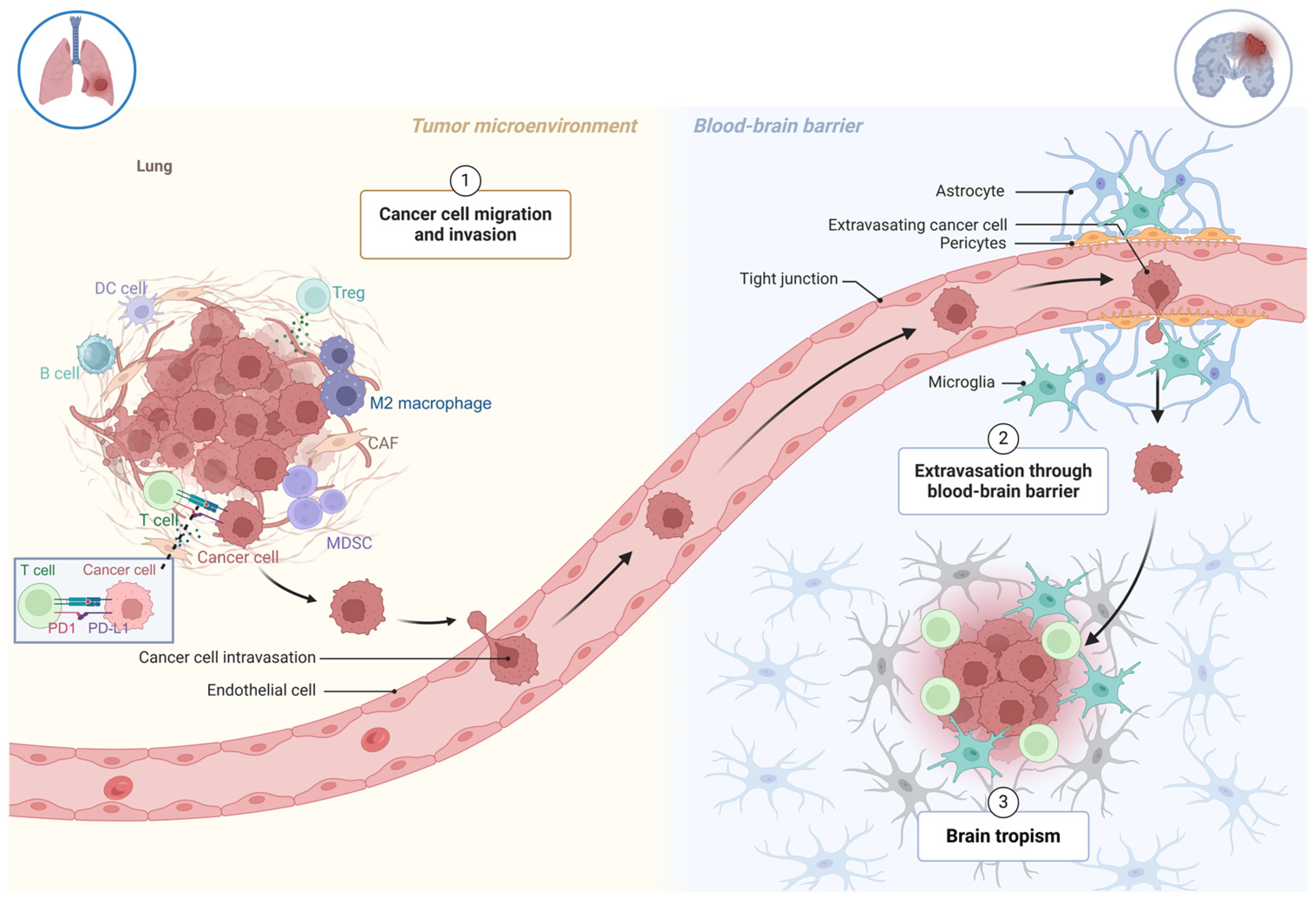
2. The Formation of Brain Metastases in NSCLC

Cancer metastasis, a hallmark of malignant tumors, involves a complex, multistep process known as the invasion–metastasis cascade. During this progression, tumor cells detach from their primary site, enter the vasculature, survive in the bloodstream, invade distant organs, and adapt to new microenvironments, enabling their survival and proliferation in metastatic sites [5]. The nervous system, especially the brain, is a common metastatic destination for non-small cell lung cancer (NSCLC) [6]. Understanding brain metastases (BMs) necessitates a comprehensive perspective integrating immunity and inflammation. T cell infiltration is evident in BM, with higher densities of CD8+ and CD45RO+ T cells correlating with improved prognoses. Conversely, the presence of immunosuppressive CD4+CD25+FOXP3+ regulatory T cells is linked to poorer outcomes. Notably, compared to primary extracranial tumors, T cells in BM exhibit reduced infiltration, clonal expansion, and diversity [7]. The formation of BM in NSCLC underscores the critical role of immune responses and tumor cell interactions within the brain microenvironment. Tumor cells can penetrate and colonize the central nervous system, facilitated by significant alterations to the blood–brain barrier, which compromise its protective integrity and promote metastatic progression (Figure 1).

3. Blood–Brain Barrier

The blood–brain barrier (BBB) is a highly selective, semipermeable boundary that protects the CNS from harmful substances circulating in the bloodstream [8]. This unique anatomical structure comprises a continuous layer of endothelial cells connected by tight junctions (TJs), pericytes, and astrocytic end-feet, collectively forming the neurovascular unit (NVU). The BBB meticulously regulates the bidirectional movement of molecules between the vascular system and the brain, maintaining CNS homeostasis. During tumor metastasis, the BBB undergoes significant modifications that compromise its protective functions and facilitate tumor cell invasion. These alterations involve coordinated disruptions in the barrier’s integrity and function, including changes in tight junction proteins, basement membrane composition, and interactions between endothelial cells and astrocytes. Such modifications enhance BBB permeability, creating a microenvironment conducive to tumor cell penetration and colonization.
TJs proteins play a pivotal role in maintaining the structural and functional integrity of the BBB. These proteins form a molecular seal between adjacent endothelial cells, preventing paracellular diffusion and maintaining brain homeostasis. The primary transmembrane components of these complexes include claudins (particularly claudin-5), occludin, and junctional adhesion molecules (JAMs), supported by cytoplasmic adapter proteins such as zonula occludens-1 (ZO-1), which anchor TJs to the actin cytoskeleton. This intricate protein network establishes the BBB as a highly selective barrier, protecting the CNS from harmful substances and maintaining a stable environment for neural function [9]. Sun. ZW et al. have confirmed that a dysregulated Cldn5 level and repression of histone methylation, both as promoters, contributed to BBB dysfunction in male C57BL/6 mice [10]. The disruption of TJ proteins can significantly increase BBB permeability, altering the brain microenvironment. This disruption is a critical factor in various pathological conditions, including metastasis, where individual variations in BBB properties influence the ability of circulating tumor cells (CTCs) to penetrate and colonize brain tissue [8].
The basement membrane and astrocytic end-feet are equally vital to the NVU, contributing to BBB integrity and overall brain homeostasis [11]. The basement membrane, predominantly composed of type IV collagen, laminin, nidogen, and perlecan, provides structural support and regulates molecular trafficking through specific binding sites for endothelial cells and astrocytes [12]. This specialized extracellular matrix ensures mechanical stability and mediates cell–matrix interactions, essential for maintaining BBB integrity. Astrocytic end-feet completely enclose cerebral vasculature and facilitate nutrient exchange and waste removal, further supporting brain homeostasis. However, during metastatic progression, the interplay between the basement membrane, astrocytes, and endothelial cells undergoes significant disruption. Tumor cells orchestrate basement membrane degradation by upregulating matrix metalloproteinases (MMPs), particularly MMP-2 and MMP-9, in astrocytes. This enzymatic activity enhances BBB permeability, facilitating tumor cell invasion into the brain parenchyma.

4. Astrocytes

Astrocytes, the most abundant glial cells in the CNS, are involved in a wide range of functions, including synapse formation, metabolic support for neurons, regulation of BBB integrity, and modulation of behavior [13]. They maintain CNS homeostasis, ensuring effective neuronal communication and overall brain function. Recent advancements in single-cell RNA sequencing (scRNA-seq) and spatial transcriptomics have unveiled the heterogeneity of astrocyte populations, highlighting their active involvement in neuroinflammatory processes. These studies have demonstrated that astrocytes can adopt diverse activation states, contributing to the pathogenesis of various neurological diseases by modulating immune responses and maintaining CNS equilibrium [14,15]. In the context of brain metastasis, astrocytes are integral components of both primary and metastatic tumors, exhibiting dual roles by either inhibiting or promoting tumor progression [16].
Astrocytes regulate the tumor immune microenvironment through multiple pathways. Notably, the Janus Kinase (JAK)-Signal Transducer and Activator of Transcription (STAT) pathway is pivotal in astrocyte–tumor interactions. Astrocytes expressing phosphorylated STAT3 (pSTAT3) have been observed in brain metastases, where they impede CD8+ cytotoxic T cells from infiltrating cancer cells. This immunosuppressive effect is mediated through the upregulation of programmed cell death-1 ligand 1 (PD-L1), vascular endothelial growth factor-A (VEGF-A), lipocalin-2, and tissue inhibitor of metalloproteinases-1 (TIMP-1) [17,18]. Furthermore, STAT3+ reactive astrocytes in brain metastases exhibit increased expression of the CD74 ligand macrophage migration inhibitory factor (MIF), enhancing interactions with CD74+ microglia [19]. Cancer cells can reprogram astrocytes via the cyclic GMP-AMP synthase (cGAS)-Stimulator of Interferon Genes (STING) pathway by transferring 2′,3′-cyclic GMP-AMP (cGAMP), leading to the production of inflammatory cytokines such as interferon-α (IFN-α) and tumor necrosis factor-α (TNF-α) [20]. These cytokines activate STAT1 and the nuclear factor κB (NF-κB) signaling pathways in cancer cells, thereby supporting tumor metastasis. Understanding the multifaceted roles of astrocytes in tumor progression and the tumor immune microenvironment is crucial. Future research is warranted to unravel these intricate interactions and develop targeted therapies that modulate astrocyte activity, aiming to inhibit tumor growth and improve outcomes for patients with brain metastases.

5. Microglia

Microglia, the resident macrophages and principal immune cells of the CNS, fulfill a diverse array of functions beyond their conventional immune roles [21]. Together with astrocytes and other glial cells, such as oligodendrocytes, microglia contribute to establishing and maintaining a tightly regulated microenvironment crucial for neuronal function and overall brain homeostasis [22]. These cells play a vital role in continuously monitoring the brain and engaging in three primary functions. First, microglia detect changes in the microenvironment by expressing a variety of sensory genes. Second, they perform physiological housekeeping tasks, such as migrating to injury sites. This migration is regulated by localized cyclic adenosine monophosphate (cAMP), enabling microglia to sense environmental alterations and swiftly respond to molecular signals [23]. Third, microglia act as protectors against harmful stimuli, including pathogen-associated molecular patterns (PAMPs) and damage-associated molecular patterns (DAMPs). In this protective role, microglia release pro-inflammatory cytokines, such as tumor necrosis factor (TNF)-α, interleukin (IL)-1β, IL-16, and chemokines, to combat injurious conditions [24].
Microglia and macrophages are also pivotal in modulating CNS repair and regeneration. However, shifts in their phenotypes can be triggered by changes in the surrounding microenvironment [25]. Microglia typically adopt an activated state during disturbances. This activation can lead to increased levels of inflammatory cytokines and enhanced ability to stimulate a T cell-mediated antitumor response [26,27]. This pro-inflammatory activation, termed M1 polarization, is characterized by microglia’s defensive role in detecting and addressing environmental disturbances. In contrast, M2 polarization represents an anti-inflammatory phenotype that supports angiogenesis, tumor growth, and immunosuppression. M2-polarized microglia promote these effects by recruiting immune checkpoints, such as PD-L1 and V-domain Ig suppressor of T cell activation (VISTA) [28]. When co-culturing with NSCLC A549 cell lines to study biological behavior, M2 macrophages can stimulate A549 cell proliferation and tumor growth. In contrast, the M1 subtype inhibits the cells, triggering apoptosis and senescence through increased expression of DNA damage-induced proteins [29]. During chronic inflammation, microglia and astrocytes in the brain metastasis microenvironment may shift towards an anti-inflammatory phenotype, thereby facilitating tumor progression.
A significant driver of microglial polarization from the M1 to the M2 phenotype is the STAT3 signaling pathway. In the context of brain metastasis, microglia influence key processes, including BM cell migration across the endothelium and the modulation of gene expression. This pathway reduces ERK activity, a tumor growth inhibitor, while enhancing STAT3 phosphorylation, which promotes tumor proliferation [30]. Recent research highlighted the potential of targeting the IL-6R-JAK2-STAT3 signaling pathway in activated microglia as a novel strategy to inhibit brain metastasis in NSCLC [31].
In addition to STAT3, microglia/macrophages are involved in several other mechanisms during brain metastasis, notably the PI3K/Akt pathway. Evidence suggests that inhibiting this pathway in lipopolysaccharide (LPS)-activated microglia reduces the expression of pro-inflammatory factors [32]. In addition, the PI3K/Akt pathway study reported that it can be activated and contribute to the attenuation of brain damage. It can also upregulate the expression of immune checkpoints VISTA and PD-L1, inhibiting the T-cell immune response [33].
Together, microglia exhibit dual roles in brain metastasis by transitioning between pro-inflammatory (M1) and anti-inflammatory (M2) phenotypes. Critical signaling pathways, such as STAT3 and PI3K/Akt, play central roles in this polarization process, influencing both tumor growth and immune modulation. Understanding these mechanisms can provide valuable insights into the dynamic roles of microglia in brain metastasis and potential therapeutic targets.

6. NSCLC Tumor Microenvironment

NSCLC is characterized by a high tumor mutation burden (TMB), with metastatic lesions often harboring distinct oncogenic driver mutations compared to primary lung tumors [34]. These mutations can lead to the generation of neoantigens, which have the potential to elicit an antitumor immune response. The tumor microenvironment (TME) in NSCLC includes tumor-infiltrating lymphocytes (TILs) and tertiary lymphoid structures (TLSs), both of which are linked to a T helper 1 (Th1) and cytotoxic immune signature. These immune components have been associated with patient survival and therapeutic responses [35]. However, NSCLC also presents with an immunosuppressive TME maintained by regulatory T cells (Tregs). The high density of Tregs and a low CD8+ T cell-to-Treg ratio within the tumor tissue are strongly correlated with poor prognosis [36]. Recent research by Peng et al. highlighted that CD8+PD-L1+ TILs were associated with increased TMB but were embedded within an immunosuppressive TME. This underscores the complexity of immune interactions within NSCLC [37].
The immune response in NSCLC is often mediated by the PD-1/PD-L1 regulatory axis, where the interaction of PD-1 on effector T cells with PD-L1 expressed by tumor cells acts as an inhibitory signal [38]. This interaction promotes T cell exhaustion, limiting their antitumor activity. PD-L1 has emerged as a predictive biomarker for response to anti-PD-1/PD-L1 therapies in NSCLC [39]. Research by Liu ZC et al. has shown that circIGF2BP3 is CD8+ T cell-mediated and causes immune escape through decreased PD-L1 ubiquitination. It has the potential mechanism of PD-L1 regulation in NSCLC treatment [40]. Intriguingly, a recent study found that knocking down PD-L1 can reduce the expression of hexokinase-2, an enzyme critical for glycolysis, and inhibit the PI3K/AKT/mTOR and ERK pathways, which are key drivers of tumor growth and survival [41].
Immune checkpoint molecules such as CTLA-4 and PD-1, both members of the CD28 family, play central roles in tumor immunity [42]. Tumor-infiltrating Treg cells exhibit increased expression of surface molecules like CTLA-4, which are associated with T cell activation suppression. Studies suggest that anti-CTLA-4 monoclonal antibodies (mAbs) can enhance immune responses by increasing the activity of CD8+ and CD4+ T cells, further supporting their potential in immunotherapy for NSCLC [43].
Within the NSCLC TME, macrophages known as tumor-associated macrophages (TAMs) contribute significantly to immune evasion, cancer cell proliferation, invasion, and metastasis [44]. Similar to microglia in brain metastases, TAMs exhibit two main phenotypes with distinct functions. M1 macrophages are activated by pro-inflammatory stimuli, including Th1 cytokines such as interferon-gamma (IFN-γ), toll-like receptor (TLR) agonists like lipopolysaccharide (LPS), and the granulocyte–macrophage colony-stimulating factor (GM-CSF). These macrophages secrete pro-inflammatory cytokines such as tumor necrosis factor-alpha (TNF-α), IL-1, IL-12, and nitric oxide (NO), promoting an antitumor immune response [45]. In contrast, M2 macrophages, associated with type 2 helper T cells (Th2), secrete anti-inflammatory mediators such as IL-10 and transforming growth factor-beta (TGF-β) and release matrix metalloproteinases (MMPs) that contribute to tissue remodeling, angiogenesis, and tumor progression. The M2 phenotype facilitates tumor immune evasion, highlighting the dual role of TAMs in the NSCLC TME [46]. Overall, NSCLC is defined by a highly complex tumor microenvironment shaped by diverse cellular and immune interactions. While substantial progress has been made in understanding these dynamics, further research is essential to fully elucidate the mechanisms driving TME composition and to advance drug development targeting NSCLC.

7. Immunotherapy Targeting the BM

For advanced NSCLC without actionable genomic alterations (AGAs), first-line immunotherapy monotherapy in PD-L1 high NSCLC or combination with platinum-based chemotherapy, regardless of PD-L1 status, has become the standard of care (SOC). Here, we discuss the efficacy of immunotherapy, alone or in combination with chemotherapy, in NSCLC with BM (Table 1).

7.1. Immunotherapy Alone

A pooled analysis of KEYNOTE-001 (phase 1), 010 (phase 2/3), 024 (phase 3), and 042 (phase 3) studies evaluated 3170 patients with stage IV NSCLC and PD-L1 positive status, of whom 293 (9.2%) had BM. Patients received pembrolizumab monotherapy and were compared to chemotherapy except for KEYNOTE-001. These studies excluded patients with active BM or carcinomatous meningitis. Among patients with BM and PD-L1 ≥ 50%, pembrolizumab revealed a numerically longer median OS compared to chemotherapy (19.7 m vs. 9.7 m, HR 0.67, 95% CI 0.44–1.02). Similarly, in patients with BM and PD-L1 ≥ 1%, pembrolizumab showed a median OS of 13.4 months versus 10.3 months with chemotherapy (HR 0.83, 95% CI 0.62–1.10). However, the survival benefit in both PD-L1 groups was not statistically significant [47].
The IMPOWER-LUNG 1, a phase 3 randomized controlled trial, evaluated patients with advanced NSCLC without EFGR/ALK/ROS1 alterations and with PD-L1 ≥ 50%. Patients were randomized to receive first-line cemiplimab versus chemotherapy. The study enrolled 12.1% treated and clinically stable BM patients. Among this subgroup, the PFS and OS outcomes favored cemiplimab over chemotherapy [48].
In the CheckMate 227 trial, a phase 3 open-label, randomized controlled study, patients with stage IV or recurrent NSCLC without EGFR/ALK/ROS1 alterations were randomized in a 1:1:1 ratio to receive ipilimumab and nivolumab, nivolumab alone (for PD-L1 ≥ 1%), nivolumab plus chemotherapy (for PD-L1 < 1%), or platinum-based chemotherapy. A subgroup analysis of patients with and without BM at a 5-year follow-up demonstrated that the combination of ipilimumab and nivolumab significantly improved the median OS with 17.4 months compared to chemotherapy with 13.7 months (HR 0.63, 95% CI 0.42–0.92). Although the median intracranial PFS was not significantly different, the 5-year intracranial PFS rate was higher with dual immunotherapy (16%) compared to chemotherapy (6%). Notably, the PD-L1 ≥ 1% patients with BM showed a higher 5-year OS rate (27%, 95% CI 15–40) compared to chemotherapy (8%, 95% CI 3–18). And this led to FDA approval of the combination of ipilimumab and nivolumab as a first-line treatment for PD-L1 positive metastatic NSCLC [50].
Goldberg et al. provided additional insights into intracranial responses through a phase 2 trial that involved 42 advanced NSCLC patients with untreated or progressing BMs following radiation therapy and treated with pembrolizumab. Among patients with PD-L1-positive tumors, 29.7% (95% CI 15.9–47.0) achieved intracranial responses, while no IC responses were observed in the PD-L1-negative group [51].
A subgroup analysis of the phase 3 OAK trial assessed the efficacy of atezolizumab compared to docetaxel in advanced NSCLC patients with and without BM in the second-line setting. Among the 14% of patients with baseline BM, atezolizumab showed a numerically longer median OS compared to docetaxel (16.0 months vs. 11.9 months, HR 0.74, 95% CI 0.49–1.13) [52].

7.2. Immunotherapy Combined with Chemotherapy

A pooled subgroup analysis from the KEYNOTE-021 (nonsquamous), KEYNOTE-189 (nonsquamous), and KEYNOTE-407 (squamous) trials assessed the efficacy of pembrolizumab combined with chemotherapy versus chemotherapy alone in advanced chemotherapy-naive NSCLC patients with or without BM. Among the 1298 patients enrolled, 171 (13.2%) had baseline BM. In patients with BM, pembrolizumab plus chemotherapy demonstrated improved systemic ORR (39.0%) versus chemotherapy (19.7%). Significantly longer median OS was also observed in the pembrolizumab plus chemotherapy arm compared to chemotherapy alone (18.8 months vs. 7.6 months, HR 0.48, 95% CI 0.32–0.70). PFS was also significantly improved in the combination therapy with 6.9 months compared to chemotherapy with 4.1 months (HR 0.44; 95% CI 0.31–0.62). Notably, the PFS benefits of pembrolizumab plus chemotherapy were consistent across all PD-L1 expression subgroups [54].
CheckMate 9LA is a phase 3 randomized controlled trial evaluating the efficacy of the combination of ipilimumab plus nivolumab plus chemotherapy compared to chemotherapy alone as first-line treatment in metastatic or recurrent NSCLC without EGFR/ALK alterations. The study demonstrated significant OS benefits that favor the combination therapy which led to FDA approval in 2020. A subgroup analysis revealed that among the 14% of patients with baseline BM, the combination therapy significantly improved median OS compared to chemotherapy alone (19.3 months vs. 6.8 months, HR 0.45, 95% CI 0.29–0.70). PFS also favored the combination therapy arm over the chemotherapy arm. Intracranial activity was assessed, showing a higher intracranial ORR in the combination therapy arm (39.2% vs. 20.0%). Additionally, intracranial median PFS (11.4 months vs. 4.6 months, HR 0.42, 95% CI 0.26–0.68) was significantly longer in the combination therapy arm compared to chemotherapy alone. Furthermore, for patients with baseline BMs, fewer patients in the combination group developed new BMs compared to the chemotherapy group (20% vs. 30%, respectively). And similarly, fewer new BMs developed in the combination arm in those without baseline BM (3.2% vs. 3.6%) [55].
The phase II Atezo-Brain trial, a single-arm study, evaluated the combination of atezolizumab, carboplatin, and pemetrexed in patients with advanced non-squamous NSCLC without EGFR/ALK alterations and with untreated, asymptomatic BMs. Among the 40 enrolled patients, the systemic and intracranial response rates were 45% and 42.7%, respectively, with no significant differences in ORR observed across different PD-L1 expression groups. The median PFS was 8.9 months (95% CI 6.7–13.8), while the median OS was 11.8 months (95% CI 7.6–16.9). The intracranial PFS was 6.9 months (95% CI 4.7–11.9) [56].
Notably, no new safety concerns were identified in NSCLC patients with BM treated with ICI alone or in combination with chemotherapy. Several studies, including KEYNOTE 189, KEYNOTE 407, Goldberg et al., and Atezo-Brain, have investigated ICIs in NSCLC patients with untreated BMs. These trials demonstrated intracranial responses, suggesting that upfront systemic therapy incorporating ICIs could be a viable treatment option for this vulnerable population [51,54,56].

7.3. Limitation of ICI and Future Strategies

Although ICI has demonstrated some efficacy in managing NSCLC with BMs in several pivotal studies, this population remains significantly underrepresented. This is largely due to the exclusion of active or untreated BMs in many trials and the lack of prioritization of BMs as evaluable lesions in trial design. Notably, several studies evaluating patients with untreated BM [51,56], showed comparable IC efficacy to those with treated BM, demonstrating a promising frontline strategy in this challenging population. Moreover, in trials that included and evaluated patients with BMs, the efficacy of IC ORR remained below 50%, underscoring an ongoing unmet need. The combination of ICI with brain radiotherapy or other targeted therapies has evolved as a new strategy to overcome the low IC efficacy of ICI or ICI resistance. In a meta-analysis, ICI+ brain radiotherapy demonstrated superior efficacy [57]. Although the sequence is to be determined, some studies suggested the concurrent model might be the optimal approach [57,58]. ICI plus the anti-VEGF antibody has evolved as a new frontline method. It was previously evaluated in some trials including IMPOWER 150 [59] and the TASUKI-52 trial [60], demonstrating superior PFS (both) and OS (IMPOWER 150) when adding ICI (atezolizumab in IMPOWER 150 and nivolumab in TASUKI-52, respectively) to bevacizumab, carboplatin, and paclitaxel. More recently, the phase III HARMONI-2 trial showed that first-line ivonescimab, a bispecific antibody targeting PD-1 and VEGF, significantly improved PFS over pembrolizumab (11.14 m vs. 5.82 m; HR 0.51; 95% CI0.38–0.69; p < 0.0001) in advanced NSCLC with a positive PD-L1 score. This benefit was consistent in patients with and without BMs [61]. The IC activity has been evaluated in the phase II trials AK112-201 and AK112-202, and showed a combined IC ORR of 34% [62]. Novel target agents are undergoing exploration in clinical trials including LAG-3, TIM-3, TIGIT [63], and STING (NCT02675439, NCT03172936).

8. Targeted Therapies in NSCLC with BM

Targeted therapies have become the SOC for advanced NSCLC with AGAs, particularly for patients with BMs, which are commonly observed across molecular subtypes, with incidence rates ranging from 20% to 50% [64]. Given the high BM involvement, the development of TKIs with strong IC activity has been critical in improving outcomes.

8.1. EGFR

In EGFR-mutated NSCLC, osimertinib, a third-generation EGFR TKI, has remained the standard first-line treatment since 2018, and demonstrated a robust IC ORR of 91% in the FLAURA trial [65]. The FLAURA 2 trial built on these findings by adding platinum-based chemotherapy to osimertinib, showing a significant improvement in (PFS for patients with baseline BM (24.9 vs. 13.8 m) [66]. Meanwhile, the MARIPOSA trial evaluated a novel combination of amivantamab (an EGFR-MET bispecific antibody) and lazertinib (third-generation EGFR TKI), which demonstrated superior PFS over osimertinib alone in patients with BM (18.3 vs. 13.0 m) [67]. Currently, all of the three regimens have been approved by the FDA for first-line management in EGFR-mutated advanced NSCLC.

8.2. ALK

For ALK-positive NSCLC, second- and third-generation ALK TKIs have dramatically improved IC control compared to the first-generation TKI crizotinib, which has limited CNS penetration. Alectinib, brigatinib, and ensartinib are currently FDA-approved second-generation ALK TKIs with IC ORR 63–78% [68,69,70]. The third-generation ALK TKI lorlatinib has shown exceptional CNS efficacy. Lorlatinib achieved an IC ORR of 60%, with a complete response (CR) rate of 49% and a median PFS that was not reached in patients with BM. Remarkably, the 5-year IC PFS rate with lorlatinib was 92%, making it the leading option for ALK-rearranged NSCLC with BM [71].

8.3. ROS1

Crizotinib was the first approved ROS1 TKI but has limited CNS activity [72]. Entrectinib has emerged as a more effective option for CNS disease, demonstrating a 52.2% IC ORR and median IC PFS of 8.3 months from pooled phase I/II studies [73]. More recently, repotrectinib, a next-generation ROS1 TKI, has shown promising results, achieving an 89% IC ORR in ROS1 TKI-naïve patients and 38% in patients previously treated with an ROS1 TKI, according to the TRIDENT-1 trial [74].

8.4. Other Targeted Therapies

Beyond EGFR, ALK, and ROS1, several other targeted therapies have shown IC activity in NSCLC. RET fusion-positive patients benefit from selpercatinib [75] and pralsetinib [76], with IC ORRs of 82% and 70%, respectively. For KRAS G12C mutations, sotorasib and adagrasib have modest IC efficacy, with adagrasib achieving around 33–42% IC ORR [77,78,79]. MET exon 14 skipping patients respond to capmatinib [80] and tepotinib [81], with IC ORRs of 57% and 66.7%. For NTRK fusions, larotrectinib [82] and entrectinib [83] show promising IC responses. These newer therapies expand options for NSCLC patients with BM, but further studies are needed to optimize BM-specific management.

9. Conclusions and Perspectives

NSCLC remains one of the most prevalent and challenging cancers globally. NSCLC is also the leading cause of brain metastases (BMs), which present a distinct and complex tumor microenvironment. When BMs occur, unique features such as the blood–brain barrier, specialized extracellular matrix (ECM) composition, and tissue-resident cells interact to facilitate blood-derived immune cell infiltration and ECM remodeling, establishing a TME distinct from that of primary NSCLC. This specialized microenvironment poses significant challenges for treatment but also offers opportunities for targeted therapeutic interventions. The integration of conventional therapies with immunotherapy has already demonstrated promise, highlighting the critical role of the TME in modulating responses to immunotherapy. Future research into the molecular and cellular mechanisms underlying BM formation and progression is essential for advancing more effective treatment strategies and shaping a more personalized and impactful therapeutic landscape.

Author Contributions

Conceptualization, H.C.; writing—original draft preparation, M.W., J.Y., and S.W.; writing—review and editing, J.Y., M.W., S.W., and H.G.; visualization, M.W.; supervision, H.C.; project administration, H.C. All authors have read and agreed to the published version of the manuscript.

Funding

This research received no external funding.

Data Availability Statement

The supporting data are not publicly available due to research participant privacy restrictions.

Conflicts of Interest

The authors declare no conflicts of interest.

References

  1. Nguyen, D.X.; Bos, P.D.; Massague, J. Metastasis: From dissemination to organ-specific colonization. Nat. Rev. Cancer 2009, 9, 274–284. [Google Scholar] [CrossRef]
  2. Eichler, A.F.; Chung, E.; Kodack, D.P.; Loeffler, J.S.; Fukumura, D.; Jain, R.K. The biology of brain metastases-translation to new therapies. Nat. Rev. Clin. Oncol. 2011, 8, 344–356. [Google Scholar] [CrossRef] [PubMed]
  3. Amin, S.B.M.; Meza, J.L.; Lin, C. Association of Immunotherapy with Survival Among Patients with Brain Metastases Whose Cancer Was Managed with Definitive Surgery of the Primary Tumor. JAMA Netw. Open 2020, 3, e2015444. [Google Scholar] [CrossRef] [PubMed]
  4. Cheng, H.; Perez-Soler, R. Leptomeningeal metastases in non-small-cell lung cancer. Lancet Oncol. 2018, 19, e43–e55. [Google Scholar] [CrossRef] [PubMed]
  5. Valastyan, S.; Weinberg, R.A. Tumor metastasis: Molecular insights and evolving paradigms. Cell 2011, 147, 275–292. [Google Scholar] [CrossRef]
  6. Riihimaki, M.; Hemminki, A.; Fallah, M.; Thomsen, H.; Sundquist, K.; Sundquist, J.; Hemminki, K. Metastatic sites and survival in lung cancer. Lung Cancer 2014, 86, 78–84. [Google Scholar] [CrossRef]
  7. Strickland, M.R.; Alvarez-Breckenridge, C.; Gainor, J.F.; Brastianos, P.K. Tumor Immune Microenvironment of Brain Metastases: Toward Unlocking Antitumor Immunity. Cancer Discov. 2022, 12, 1199–1216. [Google Scholar] [CrossRef]
  8. Arvanitis, C.D.; Ferraro, G.B.; Jain, R.K. The blood-brain barrier and blood-tumour barrier in brain tumours and metastases. Nat. Rev. Cancer 2020, 20, 26–41. [Google Scholar] [CrossRef]
  9. Stamatovic, S.M.; Keep, R.F.; Andjelkovic, A.V. Brain endothelial cell-cell junctions: How to “open” the blood brain barrier. Curr. Neuropharmacol. 2008, 6, 179–192. [Google Scholar] [CrossRef]
  10. Sun, Z.W.; Wang, X.; Zhao, Y.; Sun, Z.X.; Wu, Y.H.; Hu, H.; Zhang, L.; Wang, S.D.; Li, F.; Wei, A.J.; et al. Blood-brain barrier dysfunction mediated by the EZH2-Claudin-5 axis drives stress-induced TNF-alpha infiltration and depression-like behaviors. Brain Behav. Immun. 2024, 115, 143–156. [Google Scholar] [CrossRef]
  11. Nduom, E.K.; Yang, C.; Merrill, M.J.; Zhuang, Z.; Lonser, R.R. Characterization of the blood-brain barrier of metastatic and primary malignant neoplasms. J. Neurosurg. 2013, 119, 427–433. [Google Scholar] [CrossRef] [PubMed]
  12. Kim, S.H.; Turnbull, J.; Guimond, S. Extracellular matrix and cell signalling: The dynamic cooperation of integrin, proteoglycan and growth factor receptor. J. Endocrinol. 2011, 209, 139–151. [Google Scholar] [CrossRef]
  13. Linnerbauer, M.; Wheeler, M.A.; Quintana, F.J. Astrocyte Crosstalk in CNS Inflammation. Neuron 2020, 108, 608–622. [Google Scholar] [CrossRef] [PubMed]
  14. Sofroniew, M.V. Astrocyte barriers to neurotoxic inflammation. Nat. Rev. Neurosci. 2015, 16, 249–263. [Google Scholar] [CrossRef] [PubMed]
  15. Wasilewski, D.; Priego, N.; Fustero-Torre, C.; Valiente, M. Reactive Astrocytes in Brain Metastasis. Front. Oncol. 2017, 7, 298. [Google Scholar] [CrossRef]
  16. Valiente, M.; Obenauf, A.C.; Jin, X.; Chen, Q.; Zhang, X.H.; Lee, D.J.; Chaft, J.E.; Kris, M.G.; Huse, J.T.; Brogi, E.; et al. Serpins promote cancer cell survival and vascular co-option in brain metastasis. Cell 2014, 156, 1002–1016. [Google Scholar] [CrossRef]
  17. Priego, N.; Zhu, L.; Monteiro, C.; Mulders, M.; Wasilewski, D.; Bindeman, W.; Doglio, L.; Martinez, L.; Martinez-Saez, E.; Ramon, Y.C.S.; et al. STAT3 labels a subpopulation of reactive astrocytes required for brain metastasis. Nat. Med. 2018, 24, 1024–1035. [Google Scholar] [CrossRef]
  18. Ghoochani, A.; Schwarz, M.A.; Yakubov, E.; Engelhorn, T.; Doerfler, A.; Buchfelder, M.; Bucala, R.; Savaskan, N.E.; Eyupoglu, I.Y. MIF-CD74 signaling impedes microglial M1 polarization and facilitates brain tumorigenesis. Oncogene 2016, 35, 6246–6261. [Google Scholar] [CrossRef]
  19. Chen, Q.; Boire, A.; Jin, X.; Valiente, M.; Er, E.E.; Lopez-Soto, A.; Jacob, L.; Patwa, R.; Shah, H.; Xu, K.; et al. Carcinoma-astrocyte gap junctions promote brain metastasis by cGAMP transfer. Nature 2016, 533, 493–498. [Google Scholar] [CrossRef]
  20. Allen, N.J.; Lyons, D.A. Glia as architects of central nervous system formation and function. Science 2018, 362, 181–185. [Google Scholar] [CrossRef]
  21. Mayer, M.G.; Fischer, T. Microglia at the blood brain barrier in health and disease. Front. Cell. Neurosci. 2024, 18, 1360195. [Google Scholar] [CrossRef] [PubMed]
  22. Nishikawa, T.; Edelstein, D.; Du, X.L.; Yamagishi, S.; Matsumura, T.; Kaneda, Y.; Yorek, M.A.; Beebe, D.; Oates, P.J.; Hammes, H.P.; et al. Normalizing mitochondrial superoxide production blocks three pathways of hyperglycaemic damage. Nature 2000, 404, 787–790. [Google Scholar] [CrossRef] [PubMed]
  23. Izraely, S.; Ben-Menachem, S.; Sagi-Assif, O.; Telerman, A.; Zubrilov, I.; Ashkenazi, O.; Meshel, T.; Maman, S.; Orozco, J.I.J.; Salomon, M.P.; et al. The metastatic microenvironment: Melanoma-microglia cross-talk promotes the malignant phenotype of melanoma cells. Int. J. Cancer 2019, 144, 802–817. [Google Scholar] [CrossRef] [PubMed]
  24. Glass, C.K.; Saijo, K.; Winner, B.; Marchetto, M.C.; Gage, F.H. Mechanisms Underlying Inflammation in Neurodegeneration. Cell 2010, 140, 918–934. [Google Scholar] [CrossRef]
  25. Hanisch, U.K.; Kettenmann, H. Microglia: Active sensor and versatile effector cells in the normal and pathologic brain. Nat. Neurosci. 2007, 10, 1387–1394. [Google Scholar] [CrossRef]
  26. Lynch, M.A. The Multifaceted Profile of Activated Microglia. Mol. Neurobiol. 2009, 40, 139–156. [Google Scholar] [CrossRef]
  27. Thored, P.; Heldmann, U.; Gomes-Leal, W.; Gisler, R.; Darsalia, V.; Taneera, J.; Nygren, J.M.; Jacobsen, S.E.W.; Ekdahl, C.T.; Kokaia, Z.; et al. Long-Term Accumulation of Microglia with Proneurogenic Phenotype Concomitant with Persistent Neurogenesis in Adult Subventricular Zone After Stroke. Glia 2009, 57, 835–849. [Google Scholar] [CrossRef]
  28. Andreou, K.E.; Soto, M.S.; Allen, D.; Economopoulos, V.; de Bernardi, A.; Larkin, J.R.; Sibson, N.R. Anti-inflammatory Microglia/Macrophages As a Potential Therapeutic Target in Brain Metastasis. Front. Oncol. 2017, 7, 251. [Google Scholar] [CrossRef]
  29. Yuan, A.; Hsiao, Y.J.; Chen, H.Y.; Chen, H.W.; Ho, C.C.; Chen, Y.Y.; Liu, Y.C.; Hong, T.H.; Yu, S.L.; Chen, J.J.W.; et al. Opposite Effects of M1 and M2 Macrophage Subtypes on Lung Cancer Progression. Sci. Rep. 2015, 5, 14273. [Google Scholar] [CrossRef]
  30. Doron, H.; Pukrop, T.; Erez, N. A Blazing Landscape: Neuroinflammation Shapes Brain Metastasis. Cancer Res. 2019, 79, 423–436. [Google Scholar] [CrossRef]
  31. Jin, Y.; Kang, Y.; Wang, M.; Wu, B.; Su, B.; Yin, H.; Tang, Y.; Li, Q.; Wei, W.; Mei, Q.; et al. Targeting polarized phenotype of microglia via IL6/JAK2/STAT3 signaling to reduce NSCLC brain metastasis. Signal Transduct. Target. Ther. 2022, 7, 52. [Google Scholar] [CrossRef] [PubMed]
  32. Saponaro, C.; Cianciulli, A.; Calvello, R.; Dragone, T.; Iacobazzi, F.; Panaro, M.A. The PI3K/Akt pathway is required for LPS activation of microglial cells. Immunopharmacol. Immunotoxicol. 2012, 34, 858–865. [Google Scholar] [CrossRef] [PubMed]
  33. Cianciulli, A.; Porro, C.; Calvello, R.; Trotta, T.; Lofrumento, D.D.; Panaro, M.A. Microglia Mediated Neuroinflammation: Focus on PI3K Modulation. Biomolecules 2020, 10, 137. [Google Scholar] [CrossRef] [PubMed]
  34. Murciano-Goroff, Y.R.; Warner, A.B.; Wolchok, J.D. The future of cancer immunotherapy: Microenvironment-targeting combinations. Cell Res. 2020, 30, 507–519. [Google Scholar] [CrossRef]
  35. Goc, J.; Germain, C.; Vo-Bourgais, T.K.; Lupo, A.; Klein, C.; Knockaert, S.; de Chaisemartin, L.; Ouakrim, H.; Becht, E.; Alifano, M.; et al. Dendritic cells in tumor-associated tertiary lymphoid structures signal a Th1 cytotoxic immune contexture and license the positive prognostic value of infiltrating CD8+ T cells. Cancer Res. 2014, 74, 705–715. [Google Scholar] [CrossRef]
  36. Bravaccini, S.; Bronte, G.; Ulivi, P. TMB in NSCLC: A Broken Dream? Int. J. Mol. Sci. 2021, 22, 6536. [Google Scholar] [CrossRef]
  37. Zhang, L.; Chen, Y.; Wang, H.; Xu, Z.; Wang, Y.; Li, S.; Liu, J.; Chen, Y.; Luo, H.; Wu, L.; et al. Massive PD-L1 and CD8 double positive TILs characterize an immunosuppressive microenvironment with high mutational burden in lung cancer. J. Immunother. Cancer 2021, 9, e002356. [Google Scholar] [CrossRef]
  38. Lahiri, A.; Maji, A.; Potdar, P.D.; Singh, N.; Parikh, P.; Bisht, B.; Mukherjee, A.; Paul, M.K. Lung cancer immunotherapy: Progress, pitfalls, and promises. Mol. Cancer 2023, 22, 40. [Google Scholar] [CrossRef]
  39. Brody, R.; Zhang, Y.; Ballas, M.; Siddiqui, M.K.; Gupta, P.; Barker, C.; Midha, A.; Walker, J. PD-L1 expression in advanced NSCLC: Insights into risk stratification and treatment selection from a systematic literature review. Lung Cancer 2017, 112, 200–215. [Google Scholar] [CrossRef]
  40. Liu, Z.; Wang, T.; She, Y.; Wu, K.; Gu, S.; Li, L.; Dong, C.; Chen, C.; Zhou, Y. N(6)-methyladenosine-modified circIGF2BP3 inhibits CD8(+) T-cell responses to facilitate tumor immune evasion by promoting the deubiquitination of PD-L1 in non-small cell lung cancer. Mol. Cancer 2021, 20, 105. [Google Scholar] [CrossRef]
  41. Kim, S.; Jang, J.Y.; Koh, J.; Kwon, D.; Kim, Y.A.; Paeng, J.C.; Ock, C.Y.; Keam, B.; Kim, M.; Kim, T.M.; et al. Programmed cell death ligand-1-mediated enhancement of hexokinase 2 expression is inversely related to T-cell effector gene expression in non-small-cell lung cancer. J. Exp. Clin. Cancer Res. 2019, 38, 462. [Google Scholar] [CrossRef] [PubMed]
  42. Pulanco, M.C.; Madsen, A.T.; Tanwar, A.; Corrigan, D.T.; Zang, X. Recent advancements in the B7/CD28 immune checkpoint families: New biology and clinical therapeutic strategies. Cell Mol. Immunol. 2023, 20, 694–713. [Google Scholar] [CrossRef] [PubMed]
  43. Selby, M.J.; Engelhardt, J.J.; Quigley, M.; Henning, K.A.; Chen, T.; Srinivasan, M.; Korman, A.J. Anti-CTLA-4 antibodies of IgG2a isotype enhance antitumor activity through reduction of intratumoral regulatory T cells. Cancer Immunol. Res. 2013, 1, 32–42. [Google Scholar] [CrossRef]
  44. Conway, E.M.; Pikor, L.A.; Kung, S.H.; Hamilton, M.J.; Lam, S.; Lam, W.L.; Bennewith, K.L. Macrophages, Inflammation, and Lung Cancer. Am. J. Respir. Crit. Care Med. 2016, 193, 116–130. [Google Scholar] [CrossRef]
  45. Quatromoni, J.G.; Eruslanov, E. Tumor-associated macrophages: Function, phenotype, and link to prognosis in human lung cancer. Am. J. Transl. Res. 2012, 4, 376–389. [Google Scholar] [PubMed]
  46. Wang, L.X.; Zhang, S.X.; Wu, H.J.; Rong, X.L.; Guo, J. M2b macrophage polarization and its roles in diseases. J. Leukoc. Biol. 2019, 106, 345–358. [Google Scholar] [CrossRef]
  47. Mansfield, A.S.; Herbst, R.S.; de Castro, G.; Jr Hui, R.; Peled, N.; Kim, D.W.; Novello, S.; Satouchi, M.; Wu, Y.L.; Garon, E.B.; et al. Outcomes with Pembrolizumab Monotherapy in Patients with Programmed Death-Ligand 1-Positive NSCLC with Brain Metastases: Pooled Analysis of KEYNOTE-001, 010, 024, and 042. JTO Clin. Res. Rep. 2021, 2, 100205. [Google Scholar] [CrossRef]
  48. Sezer, A.; Kilickap, S.; Gumus, M.; Bondarenko, I.; Ozguroglu, M.; Gogishvili, M.; Turk, H.M.; Cicin, I.; Bentsion, D.; Gladkov, O.; et al. Cemiplimab monotherapy for first-line treatment of advanced non-small-cell lung cancer with PD-L1 of at least 50%: A multicentre, open-label, global, phase 3, randomised, controlled trial. Lancet 2021, 397, 592–604. [Google Scholar] [CrossRef]
  49. Gumus, M.; Chen, C.I.; Ivanescu, C.; Kilickap, S.; Bondarenko, I.; Ozguroglu, M.; Gogishvili, M.; Turk, H.M.; Cicin, I.; Harnett, J.; et al. Patient-reported outcomes with cemiplimab monotherapy for first-line treatment of advanced non-small cell lung cancer with PD-L1 of >/=50%: The EMPOWER-Lung 1 study. Cancer 2023, 129, 118–129. [Google Scholar] [CrossRef]
  50. Brahmer, J.R.; Lee, J.S.; Ciuleanu, T.E.; Bernabe Caro, R.; Nishio, M.; Urban, L.; Audigier-Valette, C.; Lupinacci, L.; Sangha, R.; Pluzanski, A.; et al. Five-year survival outcomes with nivolumab plus ipilimumab versus chemotherapy as first-line treatment for metastatic non-small-cell lung cancer in CheckMate 227. J. Clin. Oncol. 2023, 41, 1200–1212. [Google Scholar] [CrossRef]
  51. Goldberg, S.B.; Schalper, K.A.; Gettinger, S.N.; Mahajan, A.; Herbst, R.S.; Chiang, A.C.; Lilenbaum, R.; Wilson, F.H.; Omay, S.B.; Yu, J.B.; et al. Pembrolizumab for management of patients with NSCLC and brain metastases: Long-term results and biomarker analysis from a non-randomised, open-label, phase 2 trial. Lancet Oncol. 2020, 21, 655–663. [Google Scholar] [CrossRef]
  52. Gadgeel, S.M.; Lukas, R.V.; Goldschmidt, J.; Conkling, P.; Park, K.; Cortinovis, D.; de Marinis, F.; Rittmeyer, A.; Patel, J.D.; von Pawel, J.; et al. Atezolizumab in patients with advanced non-small cell lung cancer and history of asymptomatic, treated brain metastases: Exploratory analyses of the phase III OAK study. Lung Cancer 2019, 128, 105–112. [Google Scholar] [CrossRef] [PubMed]
  53. Rittmeyer, A.; Barlesi, F.; Waterkamp, D.; Park, K.; Ciardiello, F.; von Pawel, J.; Gadgeel, S.M.; Hida, T.; Kowalski, D.M.; Dols, M.C.; et al. Atezolizumab versus docetaxel in patients with previously treated non-small-cell lung cancer (OAK): A phase 3, open-label, multicentre randomised controlled trial. Lancet 2017, 389, 255–265. [Google Scholar] [CrossRef] [PubMed]
  54. Powell, S.F.; Rodriguez-Abreu, D.; Langer, C.J.; Tafreshi, A.; Paz-Ares, L.; Kopp, H.G.; Rodriguez-Cid, J.; Kowalski, D.M.; Cheng, Y.; Kurata, T.; et al. Outcomes with Pembrolizumab Plus Platinum-Based Chemotherapy for Patients with NSCLC and Stable Brain Metastases: Pooled Analysis of KEYNOTE-021, -189, and -407. J. Thorac. Oncol. 2021, 16, 1883–1892. [Google Scholar] [CrossRef]
  55. Paz-Ares, L.G.; Carbone, D.P. Response to the Letter to the Editor Titled “First-Line Nivolumab Plus Ipilimumab with Chemotherapy for Metastatic NSCLC: The Updated Outcomes From CheckMate 9LA”. J. Thorac. Oncol. 2023, 18, e102–e103. [Google Scholar] [CrossRef] [PubMed]
  56. Nadal, E.; Rodriguez-Abreu, D.; Simo, M.; Massuti, B.; Juan, O.; Huidobro, G.; Lopez, R.; De Castro, J.; Estival, A.; Mosquera, J.; et al. Phase II Trial of Atezolizumab Combined with Carboplatin and Pemetrexed for Patients with Advanced Nonsquamous Non-Small-Cell Lung Cancer with Untreated Brain Metastases (Atezo-Brain, GECP17/05). J. Clin. Oncol. 2023, 41, 4478–4485. [Google Scholar] [CrossRef]
  57. Yang, Y.; Deng, L.; Yang, Y.; Zhang, T.; Wu, Y.; Wang, L.; Bi, N. Efficacy and Safety of Combined Brain Radiotherapy and Immunotherapy in Non-Small-Cell Lung Cancer with Brain Metastases: A Systematic Review and Meta-Analysis. Clin. Lung Cancer 2022, 23, 95–107. [Google Scholar] [CrossRef]
  58. Kotecha, R.; Kim, J.M.; Miller, J.A.; Juloori, A.; Chao, S.T.; Murphy, E.S.; Peereboom, D.M.; Mohammadi, A.M.; Barnett, G.H.; Vogelbaum, M.A.; et al. The impact of sequencing PD-1/PD-L1 inhibitors and stereotactic radiosurgery for patients with brain metastasis. Neuro Oncol. 2019, 21, 1060–1068. [Google Scholar] [CrossRef]
  59. Socinski, M.A.; Jotte, R.M.; Cappuzzo, F.; Orlandi, F.; Stroyakovskiy, D.; Nogami, N.; Rodriguez-Abreu, D.; Moro-Sibilot, D.; Thomas, C.A.; Barlesi, F.; et al. Atezolizumab for First-Line Treatment of Metastatic Nonsquamous NSCLC. N. Engl. J. Med. 2018, 378, 2288–2301. [Google Scholar] [CrossRef]
  60. Sugawara, S.; Lee, J.S.; Kang, J.H.; Kim, H.R.; Inui, N.; Hida, T.; Lee, K.H.; Yoshida, T.; Tanaka, H.; Yang, C.T.; et al. Nivolumab with carboplatin, paclitaxel, and bevacizumab for first-line treatment of advanced nonsquamous non-small-cell lung cancer. Ann. Oncol. 2021, 32, 1137–1147. [Google Scholar] [CrossRef]
  61. Zhou, C.; Chen, J.; Wu, L.; Wang, L.; Liu, B.; Yao, J.; Zhong, H.; Li, J.; Cheng, Y.; Sun, Y.; et al. PL02.04 Phase 3 Study of Ivonescimab (AK112) vs. Pembrolizumab as First-line Treatment for PD-L1-positive Advanced NSCLC: Primary Analysis of HARMONi-2. J. Thorac. Oncol. 2024, 19, S1. [Google Scholar] [CrossRef]
  62. Zhang, L.; Zhou, C.; Fang, W.F.; Du, Y.; Zhao, Y.; Chen, J.; Luo, Y.; Yang, Y.; Xiong, A.; Zhao, H.; et al. 174P Intracranial (IC) activity of ivonescimab (ivo) alone or in combination with platinum doublet chemotherapy (PC) in patients (Pts) with advanced non-small cell lung cancer (aNSCLC) and brain metastases (BMs). ESMO Open 2024, 9, 102749. [Google Scholar] [CrossRef]
  63. Lu, C.; Tan, Y. Promising immunotherapy targets: TIM3, LAG3, and TIGIT joined the party. Mol. Ther. Oncol. 2024, 32, 200773. [Google Scholar] [CrossRef] [PubMed]
  64. Boldig, C.; Boldig, K.; Mokhtari, S.; Etame, A.B. A Review of the Molecular Determinants of Therapeutic Response in Non-Small Cell Lung Cancer Brain Metastases. Int. J. Mol. Sci. 2024, 25, 6961. [Google Scholar] [CrossRef]
  65. Reungwetwattana, T.; Nakagawa, K.; Cho, B.C.; Cobo, M.; Cho, E.K.; Bertolini, A.; Bohnet, S.; Zhou, C.; Lee, K.H.; Nogami, N.; et al. CNS Response to Osimertinib Versus Standard Epidermal Growth Factor Receptor Tyrosine Kinase Inhibitors in Patients with Untreated EGFR-Mutated Advanced Non-Small-Cell Lung Cancer. J. Clin. Oncol. 2018, 36, JCO2018783118. [Google Scholar] [CrossRef]
  66. Planchard, D.; Janne, P.A.; Cheng, Y.; Yang, J.C.; Yanagitani, N.; Kim, S.W.; Sugawara, S.; Yu, Y.; Fan, Y.; Geater, S.L.; et al. Osimertinib with or without Chemotherapy in EGFR-Mutated Advanced NSCLC. N. Engl. J. Med. 2023, 389, 1935–1948. [Google Scholar] [CrossRef]
  67. Cho, B.C.; Lu, S.; Felip, E.; Spira, A.I.; Girard, N.; Lee, J.S.; Lee, S.H.; Ostapenko, Y.; Danchaivijitr, P.; Liu, B.; et al. Amivantamab plus Lazertinib in Previously Untreated EGFR-Mutated Advanced NSCLC. N. Engl. J. Med. 2024, 391, 1486–1498. [Google Scholar] [CrossRef]
  68. Mok, T.; Camidge, D.R.; Gadgeel, S.M.; Rosell, R.; Dziadziuszko, R.; Kim, D.W.; Perol, M.; Ou, S.I.; Ahn, J.S.; Shaw, A.T.; et al. Updated overall survival and final progression-free survival data for patients with treatment-naive advanced ALK-positive non-small-cell lung cancer in the ALEX study. Ann. Oncol. 2020, 31, 1056–1064. [Google Scholar] [CrossRef]
  69. Camidge, D.R.; Kim, H.R.; Ahn, M.J.; Yang, J.C.H.; Han, J.Y.; Hochmair, M.J.; Lee, K.H.; Delmonte, A.; Garcia Campelo, M.R.; Kim, D.W.; et al. Brigatinib Versus Crizotinib in ALK Inhibitor-Naive Advanced ALK-Positive NSCLC: Final Results of Phase 3 ALTA-1L Trial. J. Thorac. Oncol. 2021, 16, 2091–2108. [Google Scholar] [CrossRef]
  70. Horn, L.; Wang, Z.; Wu, G.; Poddubskaya, E.; Mok, T.; Reck, M.; Wakelee, H.; Chiappori, A.A.; Lee, D.H.; Breder, V.; et al. Ensartinib vs Crizotinib for Patients with Anaplastic Lymphoma Kinase-Positive Non-Small Cell Lung Cancer: A Randomized Clinical Trial. JAMA Oncol. 2021, 7, 1617–1625. [Google Scholar] [CrossRef]
  71. Solomon, B.J.; Liu, G.; Felip, E.; Mok, T.S.K.; Soo, R.A.; Mazieres, J.; Shaw, A.T.; de Marinis, F.; Goto, Y.; Wu, Y.L.; et al. Lorlatinib Versus Crizotinib in Patients with Advanced ALK-Positive Non-Small Cell Lung Cancer: 5-Year Outcomes From the Phase III CROWN Study. J. Clin. Oncol. 2024, 42, 3400–3409. [Google Scholar] [CrossRef] [PubMed]
  72. Shaw, A.T.; Ou, S.H.; Bang, Y.J.; Camidge, D.R.; Solomon, B.J.; Salgia, R.; Riely, G.J.; Varella-Garcia, M.; Shapiro, G.I.; Costa, D.B.; et al. Crizotinib in ROS1-rearranged non-small-cell lung cancer. N. Engl. J. Med. 2014, 371, 1963–1971. [Google Scholar] [CrossRef] [PubMed]
  73. Dziadziuszko, R.; Krebs, M.G.; De Braud, F.; Siena, S.; Drilon, A.; Doebele, R.C.; Patel, M.R.; Cho, B.C.; Liu, S.V.; Ahn, M.J.; et al. Updated Integrated Analysis of the Efficacy and Safety of Entrectinib in Locally Advanced or Metastatic ROS1 Fusion-Positive Non-Small-Cell Lung Cancer. J. Clin. Oncol. 2021, 39, 1253–1263. [Google Scholar] [CrossRef] [PubMed]
  74. Drilon, A.; Camidge, D.R.; Lin, J.J.; Kim, S.W.; Solomon, B.J.; Dziadziuszko, R.; Besse, B.; Goto, K.; de Langen, A.J.; Wolf, J.; et al. Repotrectinib in ROS1 Fusion-Positive Non-Small-Cell Lung Cancer. N. Engl. J. Med. 2024, 390, 118–131. [Google Scholar] [CrossRef]
  75. Drilon, A.; Subbiah, V.; Gautschi, O.; Tomasini, P.; de Braud, F.; Solomon, B.J.; Shao-Weng Tan, D.; Alonso, G.; Wolf, J.; Park, K.; et al. Selpercatinib in Patients with RET Fusion-Positive Non-Small-Cell Lung Cancer: Updated Safety and Efficacy From the Registrational LIBRETTO-001 Phase I/II Trial. J. Clin. Oncol. 2023, 41, 385–394. [Google Scholar] [CrossRef]
  76. Griesinger, F.; Curigliano, G.; Thomas, M.; Subbiah, V.; Baik, C.S.; Tan, D.S.W.; Lee, D.H.; Misch, D.; Garralda, E.; Kim, D.W.; et al. Safety and efficacy of pralsetinib in RET fusion-positive non-small-cell lung cancer including as first-line therapy: Update from the ARROW trial. Ann. Oncol. 2022, 33, 1168–1178. [Google Scholar] [CrossRef]
  77. de Langen, A.J.; Johnson, M.L.; Mazieres, J.; Dingemans, A.C.; Mountzios, G.; Pless, M.; Wolf, J.; Schuler, M.; Lena, H.; Skoulidis, F.; et al. Sotorasib versus docetaxel for previously treated non-small-cell lung cancer with KRAS(G12C) mutation: A randomised, open-label, phase 3 trial. Lancet 2023, 401, 733–746. [Google Scholar] [CrossRef]
  78. Barlesi, F.; Yao, W.; Duruisseaux, M.; Doucet, L.; Shi, J.; Juan Vidal, O.J.; Kim, Y.-C.; García Campelo, M.R.; Azkárate Martínez, A.; Lu, S.; et al. LBA57 Adagrasib (ADA) vs docetaxel (DOCE) in patients (pts) with KRASG12C-mutated advanced NSCLC and baseline brain metastases (BM): Results from KRYSTAL-12. Ann. Oncol. 2024, 35, S1247–S1248. [Google Scholar] [CrossRef]
  79. Mok, T.S.K.; Yao, W.; Duruisseaux, M.; Doucet, L.; Martínez, A.A.; Gregorc, V.; Juan-Vidal, O.; Lu, S.; Bondt, C.D.; Marinis, F.D.; et al. KRYSTAL-12, Phase 3 study of adagrasib versus docetaxel in patients with previously treated advanced/metastatic non-small cell lung cancer (NSCLC) harboring a KRASG12C mutation. J. Clin. Oncol. 2024, 42, LBA8509. [Google Scholar] [CrossRef]
  80. Wolf, J.; Hochmair, M.; Han, J.Y.; Reguart, N.; Souquet, P.J.; Smit, E.F.; Orlov, S.V.; Vansteenkiste, J.; Nishio, M.; de Jonge, M.; et al. Capmatinib in MET exon 14-mutated non-small-cell lung cancer: Final results from the open-label, phase 2 GEOMETRY mono-1 trial. Lancet Oncol. 2024, 25, 1357–1370. [Google Scholar] [CrossRef]
  81. Thomas, M.; Garassino, M.; Felip, E.; Sakai, H.; Le, X.; Veillon, R.; Smit, E.; Mazieres, J.; Cortot, A.; Raskin, J.; et al. OA03.05 Tepotinib in Patients with MET Exon 14 (METex14) Skipping NSCLC: Primary Analysis of the Confirmatory VISION Cohort C. J. Thorac. Oncol. 2022, 17, S9–S10. [Google Scholar] [CrossRef]
  82. Drilon, A.; Tan, D.S.W.; Lassen, U.N.; Leyvraz, S.; Liu, Y.; Patel, J.D.; Rosen, L.; Solomon, B.; Norenberg, R.; Dima, L.; et al. Efficacy and Safety of Larotrectinib in Patients with Tropomyosin Receptor Kinase Fusion-Positive Lung Cancers. JCO Precis. Oncol. 2022, 6, e2100418. [Google Scholar] [CrossRef]
  83. Cho, B.C.; Chiu, C.H.; Massarelli, E.; Buchschacher, G.L.; Goto, K.; Overbeck, T.R.; Loong, H.H.F.; Chee, C.E.; Garrido, P.; Dong, X.; et al. Updated efficacy and safety of entrectinib in NTRK fusion-positive non-small cell lung cancer. Lung Cancer 2024, 188, 107442. [Google Scholar] [CrossRef]
Figure 1. A summary of the non-small cell lung cancer brain metastasis. Created in BioRender. Wang, M. (2025) https://BioRender.com/e30a983 (accessed on 8 March 2025).
Curroncol 32 00171 g001
Table 1. Clinical trials evaluating ICI with or without chemotherapy in advanced NSCLC with BM.
TrialTypeTreatmentBM EligibilityPatient EligibilityPatient Number/BM NumberSystemic Outcome in Patients with BMIC Outcome
Pooled analysis of KEYNOTE-001, -010, -024, -042 [47]KEYNOTE-001 phase 1;
KEYNOTE-010 phase 2/3; KEYNOTE -024 and -042 phase 3
Pembro vs. ChemoNo active BM, no carcinomatous meningitisPreviously treated and treatment naïve, PD-L1 positive. No EGFR/ALK alteration, or failed EGFR or ALK TKI (KEYNOTE 001 and KEYNOTE 010)3170/293PD-L1 ≥ 50% and BM:
ORR: 33.9% vs. 14.6%
mPFS 4.1 m vs. 4.6 m (HR 0.70, 95% CI 0.47–1.03)
mOS 19.7 m vs. 9.7 m (HR 0.67, 95% CI 0.44–1.02)
PD-L1 ≥ 1% and BM:
ORR: 26.1% vs. 18.1%
mPFS 2.3 m vs. 5.2 m (HR 0.96, 95% CI 0.73–1.25)
mOS 13.4 m vs. 10.3 m (HR 0.83, 95% CI 0.62–1.10)
/
IMPOWER-Lung 1 [48,49]Phase 3, randomized, controlled studyCEMI vs. ChemoTreated and clinically stable BMAdvanced NSCLC with PD-L1 ≥ 50%, no EGFR/ALK/ROS1 alterations710 enrolled, 563 PD-L1 ≥ 50%/68mPFS 10.4 m vs. 5.3 m (HR 0.45, 95% CI 022–0.92)
mOS 18.7 m vs. 11.7 m (HR 0.17, 95% CI 0.04–0.76)
/
CheckMate-227 part 1 [50]Phase 3, open label, randomized controlled studyIpi + Nivo vs. ChemoTreated and asymptomatic BMStage IV or recurrent NSCLC, treatment-naïve, no EGFR/ALK alterations1739/202ORR 32% vs. 26%
mPFS 5.4 m vs. 5.8 m (HR 0.77, 95% CI 0.51–1.15)
mOS 17.4 m vs. 13.7 m (HR 0.63, 95% CI 0.42–0.92)
IC PFS 8.6 m vs. 8.7 m (HR 0.82, 95% CI 0.52–1.30)
5-year IC PFS 16% vs. 6%
New BM: 4% vs. 20%
Goldberg et al. [51]Phase 2, open label, single armPembroUntreated or progressing after RT; no neurologic symptoms or steroid requirementStage IV NSCLC42/42
(cohort 1 PD-L1 ≥ 1%: 37; cohort 2 PD-L1 < 1% or unevaluable: 5)
Cohort 1:
mPFS 2.3 m (95% CI 1.9-NE)
mOS 9.9 m (95% CI 7.5–29.8)
Cohort 1:
IC ORR
29.7% (95% CI 15.9–47.0)
Cohort 2:
IC ORR 0
OAK study [52,53]Phase 3, open label, randomized controlled studyAtezo vs. docetaxelTreated, asymptomatic, supratentorial BMAdvanced NSCLC previously treated with platinum-based Chemo850/123mOS 20.1 m vs. 11.9 m (HR 0.54, 95% CI 0.31–0.94)IC PFS NR vs. 9.5 m (HR 0.38, 95% CI 0.16–0.91, p = 0.024)
IC OS 16.0 m vs. 11.9 m (HR 0.74, 95% CI 0.49–1.13, p = 0.16)
Pooled analysis of KEYNOTE-021, -189, -407 [54]KEYNOTE-021 phase 2; KEYNOTE-189 and -407 phase 3Pembro +
Chemo vs. Chemo
Treated or untreated (KEYNOTE-189 and KEYNOTE-407 only) stable BMStage IIIB or IV (KEYNOTE-021 cohort G), Stage IV (KEYNOTE-189 and -407) nonsquamous without EGFR/ALK
alteration (KEYNOTE-021 cohort G and KEYNOTE-189) or squamous (KEYNOTE-407), chemotherapy naïve NSCLC
1299/171ORR 39.0% vs. 19.7%
mPFS 6.9 m vs. 4.1 m (HR 0.44, 95% CI 0.31–0.62)
mOS 18.8 m vs. 7.6 m (HR 0.48, 95% CI 0.32–0.70)
/
CheckMate 9LA [55]Phase 3, open label, randomized controlled studyIpi + Nivo + Chemo vs. ChemoTreated and asymptomatic BMStage IV or recurrent NSCLC without EGFR/ALK alterations719/101ORR 43% vs. 24%
mPFS 9.7 m vs. 4.1 m (HR 0.44, 95% CI 0.28–0.69)
mOS 19.3 m vs. 6.8 m (HR 0.45, 95% CI 0.29–0.70)
IC ORR 39% vs. 20%
IC PFS 11.4 m vs. 4.6 m (HR 0.42, 95% CI 0.26–0.68)
New BM in pt with baseline BM: 20% vs. 30%
New BM in pt without baseline BM: 3.2% vs. 3.6%
Atezo-Brain [56]Phase 2, single armAtezo + c arboplatin + pemetrexedUntreated and asymptomatic BMAdvanced nonsquamous NSCLC with BM, no EGFR/ALK alterations40/40ORR 45% (95% credibility interval Crl 28.1–57.9)
mPFS 8.9 m (95% CI 6.7–13.8)
mOS 11.8 m (95% CI 7.6–16.9)
IC ORR 42.7% (95% Crl 28.1–57.9)
IC PFS 6.9 m (95% CI 4.7–11.9)
NSCLC: non-small cell lung cancer; BMs: brain metastases; Pembro: pembrolizumab; CEMI: Cemiplimab; Atezo: atezolizumab; Ipi: ipilimumab; Nivo: nivolumab; Chemo: chemotherapy; IC: intracranial; mPFS: median progression-free survival; mOS: median overall survival; ORR: objective response rate; NR: not reached; RT: radiotherapy.
Disclaimer/Publisher’s Note: The statements, opinions and data contained in all publications are solely those of the individual author(s) and contributor(s) and not of MDPI and/or the editor(s). MDPI and/or the editor(s) disclaim responsibility for any injury to people or property resulting from any ideas, methods, instructions or products referred to in the content.

