Article

Breast Volume Is a Predictor of Higher Heart Dose in Whole-Breast Supine Free-Breathing Volumetric-Modulated Arc Therapy Planning

1
Radiation Oncology, Fondazione Policlinico Universitario Campus Bio-Medico, Via Alvaro del Portillo, 200, 00128 Rome, Italy
2
Department of Radiation Oncology (Medicine and Surgery), Università Campus Bio-Medico di Roma, 00128 Rome, Italy
3
Medical Physics Unit, S. Filippo Neri Hospital, ASL Roma 1, 00135 Rome, Italy
4
Elekta AB, 113 57 Stockholm, Sweden
5
Radiation Oncology, Tor Vergata University, 00133 Rome, Italy
*
Author to whom correspondence should be addressed.
Curr. Oncol. 2023, 30(12), 10530-10538; https://doi.org/10.3390/curroncol30120768
Submission received: 23 November 2023 / Accepted: 11 December 2023 / Published: 18 December 2023
(This article belongs to the Topic Innovative Radiation Therapies)

Abstract

:
In breast cancer volumetric-modulated arc therapy (VMAT) planning, the rotation of the gantry around the target implies a greater dose spreading to the whole heart, compared to tangential-field standard treatment. A consecutive cohort of 121 breast cancer patients treated with the VMAT technique was investigated. The correlation of breast volume, heart volume and lung volume with mean heart dose (mHD) and mean and maximum LAD dose (mLAD dose, MLAD dose) was tested, and a subsequent a linear regression analysis was carried out. VMAT treatment plans from 56 left breast cancer and 65 right breast cancer patients were analyzed. For right-sided patients, breast volume was significantly correlated with mHD, mLAD and MLAD dose, while for left-sided patients, breast volume was significantly correlated with mHD and mLAD, while heart volume and lung volume were correlated with mHD, mLAD and MLAD dose. Breast volume was the only predictor of increased heart and LAD dose (p ≤ 0.001) for right-sided patients. In left-sided patients, heart and lung were also predictors of increased mHD (p = 0.005, p ≤ 0.001) and mean LAD dose (p = 0.009, p ≤ 0.001). In this study, we observed an increase in heart and LAD doses in larger-breasted patients treated with VMAT planning. In right-sided patients, breast volume was shown to be the only predictor of increased heart dose and LAD dose.

1. Introduction

Radiotherapy (RT) is an established part of the multidisciplinary treatment of breast cancer. After breast-conserving surgery, adjuvant RT reduces the risk of loco-regional recurrence and is widely used as the standard of care [1]. Several fractionation schemes and treatment modalities have been applied and investigated. However, adjuvant treatments may have some detrimental late effects on nearby healthy organs.
In particular, there are data regarding patients with left-side breast cancer treated with older techniques, where radiotherapy increases the risk of heart disease [2,3]. A meta-analysis recorded a link between cardiac deaths following breast radiotherapy and the volume of the heart receiving 5 Gy [4].
Several predictors of heart exposure during RT for breast cancer have been identified, including clinical, anatomical and planning factors [5,6,7,8,9,10]. Among these, larger PTV, requiring a longer distance between the medial and lateral entry points and a larger portion of the heart in the radiation field, was found to worsen heart dosimetry [9]. However, most studies included only patients treated with three-dimensional conformal RT (3D-CRT).
Since the introduction of volumetric-modulated arc therapy (VMAT) after the publication of the seminal work of Otto [11] and the subsequent implementation of optimization algorithms in treatment planning systems, VMAT has been applied to almost all type of cancers. When this technique is used for breast cancer postoperative treatment, the rotation of the gantry around the target implies a greater dose spreading to the whole heart compared to tangential-field treatment [12]. This can be even greater in cases of treatment of large-breasted patients. For this reason, we wanted to evaluate how breast volume affects heart dose in supine free-breathing VMAT radiotherapy planning for breast cancer patients.

2. Materials and Methods

2.1. Patient Selection

We retrospectively investigated a consecutive cohort of 56 left breast cancer and 65 right breast cancer patients treated with the VMAT technique. All patients were consecutively treated at the University Campus Bio-Medico of Rome, Italy, from June 2022 to December 2022. Patients enrolled signed a consent form for data collection according to the study design requirements and Fondazione Policlinico Universitario Campus Bio-Medico ethical committee.

2.2. Simulation and Target Definition

All patients were positioned supine on a C–Qual M™ Breastboard (angle 10–12 degrees) with both arms lifted up above the head. The planning CT was performed on a 16-slice Computed Tomography (CT) scanner (Somatom Sensation CT-scanner, Siemens Medical Systems, Erlangen, Germany) with a slice thickness of 3 mm. Clinical target volume (CTV), planning target volume (PTV) and organs at risk (OARs) were delineated according to ESTRO guidelines for breast cancer [13]. For heart contouring, the atlas by Feng et al. was used [14]. The breast CTV included the breast volume, after a reduction of 5 mm from the surface edge, not taking into account the major pectoral muscle, the lung and the ribs. The breast PTV was defined with a 5 mm margin around the CTV.

2.3. Treatment Planning

For all patients, we employed the VMAT technique. The dose calculation was performed using the Montecarlo (MC) Algorithm provided by the Monaco 5.51.10 Treatment Planning System (Elekta A.B., Stockholm, Sweden). A grid calculation size of 3 mm was used with 1% statistical MC variance. The plan design consisted of two small tangential arcs (each partial arc, geometrically resembling the 3D-CRT tangential beams, consisted of four arcs spanning 40–60 degrees amplitude each: the first in the clockwise direction follow by another in the anti-clockwise direction, the third again covering 40–60 degrees amplitude in the clockwise direction and the last coming back to the start point) with 6 MV photons, aiming to conform the prescribed dose to the breast target, reducing cardiac and lung doses. Since breast cancer extends towards the patient’s surface, the target volume could move outside the treatment field. In order to take into account this issue, an Auto Flash margin (value from 1.5 cm up to 2.5 cm) was used, leading the multileaf collimator (MLC) leaves opening outside of the body contour. The prescribed dose was 40.05 Gy (2.67 Gy/d). The optimization objectives were as follows: 95% of prescription dose to 95% of the PTV volume; 105% of prescription dose to less than 5% of PTV volume. For the OARs, the following constraints were used: heart mean dose < 5 Gy (optimal < 3.5 Gy), left descending artery (LAD) Dmax < 20 Gy (optimal < 15 Gy), LAD Dmean < 8 Gy, volume of lung receiving 5 Gy- V5 < 60%, contralateral breast mean dose < 3 Gy. Treatments were delivered with two energy-matched ELEKTA VERSAHD Linac devices (Elekta, Crawley, UK). Patient positioning was performed with the AlignRT Advance (Vision RT, Ltd., London, UK) SGRT system, which provides a real-time motion monitoring of the surface. The real-time surface was compared with the CT planning surface that was set as reference. An additional position verification was performed with a daily CBTC in order to make sure the internal OARs’ location was correct. All patients were treated with free breathing.

2.4. Statistical Analysis

The treatment plans were evaluated using the ProKnow data analysis platform (Elekta). The dose–volume parameters for each OAR and anatomic volumes such as breast volume, heart volume and lung volume were recorded for each patient. Comparisons by groups according to breast volume (cut-off = mean breast volume, 892 cc) was performed using the paired t-test. The correlation of breast volume, heart volume and lung volume with mHD, mean dose and maximum dose of LAD (mLAD, MLAD, respectively) was tested using Pearson’s correlation coefficient “r”. The variance inflation factor (VIF) was used to judge whether there was collinearity among variables. The volumes that significantly correlated with mHD, mLAD and MLAD dose were examined using a linear regression model. A linear regression model was created using the variable with a significant value of the correlation coefficient. Linear regression requires that residuals conform to normal analysis and are independent of each other, so residuals of each regression model were calculated, and histograms were used to explore whether the residuals conformed to normal distribution. The Durbin–Watson test was used to test whether the residuals in the linear regression model were independent of each other. Variables with a p < 0.01 in the univariate regression model were evaluated in the multivariate regression model. Statistical analysis was performed using IBM SPSS (Statistical Package for the Social Sciences) v.26 (APA, MLA, Chicago, IL, USA).

3. Results

3.1. Patients’ Characteristics

In this study, 121 patients affected by breast cancer receiving adjuvant whole-breast RT were taken into account. The mean age of the whole patient population was 65.2 years (SD 11.12 years). Of these patients, 56 patients were treated for left breast cancer, and 65 patients were treated for right breast cancer. The mean breast volume for the whole population was 892.00 cc (SD 389.77), 892.64 cc (SD 435.56) in left-sided patients and 864.09 cc (SD = 348.45) in right-sided patients, respectively. Dosimetric parameters (mean value, SD) for right-sided and left-sided breast cancer patients are summarized in Table 1.
mHD was 1.4 Gy (SD 0.40) for right-sided patients and 2.5 Gy (SD 1.0) for left-sided patients. In right-sided patients, mHD (p = 0.03), mLAD (p = 0.015) and MLAD (p = 0.07) were significantly higher in patients with larger breast volume (cut-off value = 892 cc). Their values increased by 25%, 18% and 13%, respectively. In left-sided patients, mHD was increased by 30% (p = 0.01) in patients with a larger breast volume (see Figure 1). No significant differences were observed in mLAD and MLAD.
Breast volume, heart volume and lung volume did not exceed the maximum acceptance VIF level and therefore were included in the following statistical analysis.

3.2. Correlation Analysis

The results of correlation analysis with mHD, mLAD and MLAD dose are shown in Table 2. For right-sided patients, breast volume was significantly correlated with mHD, mLAD and MLAD dose, while lung volume was significantly inversely correlated to mHD only. For left-sided patients, breast volume was significantly correlated to mHD and mLAD, while heart volume and lung volume were correlated (lung inversely) with mHD, mLAD and MLAD dose.

3.3. Regression Analysis

The results for regression analysis are summarized in Table 3.

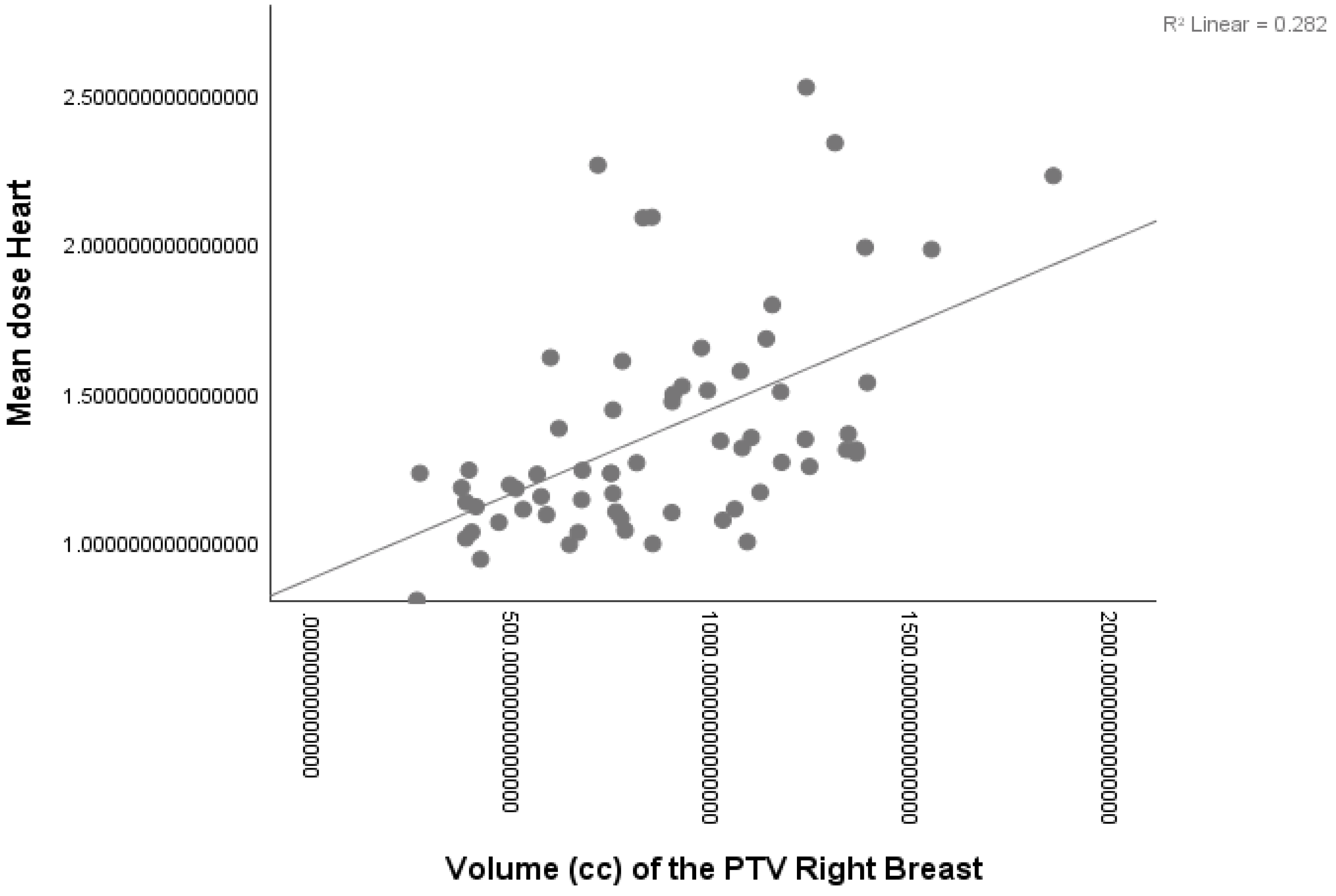
3.3.1. Right-Sided Patients

Larger breast volumes were associated with increased mHD, mLAD and MLAD. Lung volume did not show a strong relationship with the heart dose variable (p = 0.024) and therefore a multivariate analysis was not performed. The following formulas specify the univariate linear regression fitting model for mHD (a, see Figure 2), mLAD (b) and MLAD (c):
(a)
mHD (Gy) =0.881 + (0.531 × breast volume)
(b)
mLAD (Gy) = 0.860 + (0.443 × breast volume)
(c)
MLAD (Gy) =1.100 + (0.323 × breast volume)
The mean (SD) of absolute residuals was 0.0 (0.2) Gy for (a), 0.0 (0.3) Gy for (b) and 0.0 (0.5) Gy for (c). The value of the Durbin–Watson test was 2.0 for (a), 1.92 for (b) and 2.06 for (c), showing the independence of residuals.

3.3.2. Left-Sided Patients

The results for regression analysis are summarized in Table 3. Breast volume was only shown to be highly related to mHD in univariate analysis, not in multivariate analysis. Heart volume and lung volume were shown to be significantly related to mHD and mLAD dose in both univariate and multivariate analysis. Lung volume was only the best predictor of increased MLAD dose. The following specify the multiple linear regression fitting model of mHD (a) and mLAD dose (b):
(a)
mHD (Gy) = 1.705 + (0.208 × breast volume) + (0.312 × heart volume) − (0.411 × lung volume)
(b)
mLAD dose (Gy) = 6.528 + (0.281 × heart volume) − (0.565 × lung volume)
The mean (SD) of absolute residuals was 0.0 (0.7) Gy for (a) and 0.0 (1.6) Gy for (b). The value of the Durbin–Watson test was 2.0 for (a) and 2.1 for (b), showing independence of residuals.
The following equation specifies the linear regression fitting model of MLAD dose:
(a)
MLAD dose (Gy) = 22.812 − (0.639 × lung volume)
The mean (SD) of absolute residuals was 0.0 (3.0) Gy. The value of the Durbin–Watson test was 2.2, showing the independence of residuals.

4. Discussion

In this study, we analyzed heart radiation exposure using VMAT radiotherapy planning with regard to breast volume in a cohort of 121 breast cancer patients treated with whole-breast RT only (WBRT). In all patients, predefined mandatory heart constraints were respected in both left- and right-sided patients. However, recent guidelines recommend more stringent heart constraints [15] for whole-breast radiotherapy. Considering these latter constraints, we would have exceeded the recommended dose in 9% of right-sided and 45% of left-sided patients.
In the whole patient population, larger-breasted patients showed increased mHD by 25% and 30% in right and left patients, respectively. However, the contribution of breast volume in left-sided patients to heart and LAD doses was shown to be less relevant compared to the lung and heart volume. On the contrary, in right-sided patients, breast volume seems to be the main predictor of increased heart and LAD doses.
Several studies have investigated anatomical and clinical predictors of heart exposure in 3D breast radiotherapy and the relative benefit from DIBH in left breast cancer patients. Among anatomical factors, smaller lung volumes [5,6], as well as the cardiac contact distance measured on different planes and larger PTV [7,8,9], were considered predictors of increased heart exposure. Among clinical factors, higher BMI and vital capacity measured by spirometry were predictive of higher heart dose [10].
The introduction of intensity-modulated radiotherapy (IMRT) compared with the use of 3D-CRT was associated with greater mHD [16]. However, few studies investigated factors influencing heart exposure in patients treated with intensity-modulated techniques. Kang et al. evaluated cardiac junction and pulmonary junction (anatomic variable derived from 3D plans) to predict the benefit from the use of VMAT in patients with left-sided breast cancer [17].
Different heart-sparing techniques have been investigated [18]. Among them, several studies and consistent literature data support the use of the deep inspiration breath hold (DIBH) technique, which can reduce radiation-induced cardiac toxicity by increasing the distance between the breast and the heart [19,20,21,22].
Despite the widespread usage of DIBH for left-breast RT, few studies have investigated its role in right-breast cancer [23,24,25]. Some studies suggested that DIBH for right-sided breast cancer should be adopted to reduce ipsilateral lung and liver dose in loco-regional radiation therapy [26,27]. A significant benefit was detected in reducing the maximum dose to the heart and the right coronary artery in cases of regional nodal irradiation [23,24]. Interestingly, in a study evaluating the dosimetric benefit of DIBH for locoregional irradiation of R-BC with VMAT, Loap et al. concluded that adding DIBH to VMAT is not justified for all patient candidates for right-breast and regional nodal irradiation. Therefore, specific patient subpopulations who could benefit from additional DIBH combination with locoregional VMAT needed to be identified [25].
Also, prone radiotherapy has been investigated in both left- and right-sided patients in terms of dosimetric benefit over supine radiotherapy [28,29,30]. Median higher heart doses were observed in prone radiotherapy both in left-sided patients when compared to DIBH supine radiotherapy (mean dose 3.4 vs. 1.9 Gy) and in right-sided patients when compared with supine radiotherapy (mean dose 1.9 vs. 1.3 Gy) [29,30]. However, the overall dosimetric benefit, taking into account not only heart dose but also lung, PTV and extra-target dose favored prone positioning in 61% of left-sided patients and in 81.5% of right-sided patients, and in both cases, breast volume predicted the benefit from prone positioning [29,30].
This study has some limitations. First, it was retrospective and therefore is subject to selection biases. Second, there is a lack of comparison with alternative techniques, such as DIBH or alternative positioning (prone radiotherapy), to verify if these techniques would obviate the higher heart dose in larger-breasted patients. This would be of great interest and should be investigated in future analyses.

5. Conclusions

In conclusion, in this study, we observed an increase in heart and LAD doses in larger-breasted patients undergoing breast-only radiotherapy. In right-sided patients, breast volume was shown to be the only predictor of increased heart dose and LAD dose. Even if the absolute increase in doses was limited, given the low doses of heart dosimetry in these patients, the role of breast volume is certain and should also be further evaluated in patients undergoing regional nodal irradiation when VMAT planning is used. This is of particular relevance as patients are routinely treated with free breathing.

Author Contributions

Conceptualization, E.I. and R.A.; methodology, R.A.; software, R.P.; validation, S.R., M.F. and S.S.; formal analysis, R.A.; investigation, C.S. and A.C.; data curation, F.P.C. and R.F.; writing—original draft preparation, E.I. and C.G.; writing—review and editing, R.A.; visualization, M.F.; supervision, S.R. and R.M.D. All authors have read and agreed to the published version of the manuscript.

Funding

This research received no external funding.

Institutional Review Board Statement

All procedures performed in studies involving human participants conformed to the ethical standards of the institutional and national research committees and the Helsinki Declaration. No specific ethical approval is required for retrospective dose analysis studies in our institution.

Informed Consent Statement

All patients signed informed consent forms for treatment and use of their clinical data for research or educational purposes.

Data Availability Statement

The data that support the findings of this study are available from the corresponding author (RA) upon reasonable request.

Conflicts of Interest

The authors declare no conflict of interest.

References

  1. Early Breast Cancer Trialists’ Collaborative Group (EBCTCG); Darby, S.; McGale, P.; Correa, C.; Taylor, C.; Arriagada, R.; Clarke, M.; Cutter, D.; Davies, C.; Ewertz, M.; et al. Effect of radiotherapy after breast-conserving surgery on 10-year recurrence and 15-year breast cancer death: Meta-analysis of individual patient data for 10,801 women in 17 randomized trials. Lancet 2011, 378, 1707–1716. [Google Scholar] [CrossRef] [PubMed]
  2. Yeboa, D.N.; Evans, S.B. Contemporary Breast Radiotherapy and Cardiac Toxicity. Semin. Radiat. Oncol. 2016, 26, 71–78. [Google Scholar] [CrossRef] [PubMed]
  3. Darby, S.C.; Ewertz, M.; McGale, P.; Bennet, A.M.; Blom-Goldman, U.; Brønnum, D.; Correa, C.; Cutter, D.; Gagliardi, G.; Gigante, B.; et al. Risk of Ischemic Heart Disease in Women after Radiotherapy for Breast Cancer. N. Engl. J. Med. 2013, 368, 987–998. [Google Scholar] [CrossRef] [PubMed]
  4. Clarke, M.; Collins, R.; Darby, S.; Davies, C.; Elphinstone, P.; Evans, V.; Godwin, J.; Gray, R.; Hicks, C.; James, S.; et al. Effects of radiotherapy and of differences in the extent of surgery for early breast cancer on local recurrence and 15-year survival: An overview of the randomized trials. Lancet 2005, 366, 2087–2106. [Google Scholar] [CrossRef] [PubMed]
  5. Dell’Oro, M.; Giles, E.; Sharkey, A.; Borg, M.; Connell, C.; Bezak, E. A Retrospective Dosimetric Study of Radiotherapy Patients with Left-Sided Breast Cancer; Patient Selection Criteria for Deep Inspiration Breath Hold Technique. Cancers 2019, 11, 259. [Google Scholar] [CrossRef] [PubMed]
  6. Xin, X.; Li, J.; Zhao, Y.; Wang, P.; Tang, B.; Yao, X.; Liao, X.; Ma, J.; Orlandini, L.C. Retrospective Study on Left-Sided Breast Radiotherapy: Dosimetric Results and Correlation with Physical Factors for Free Breathing and Breath Hold Irradiation Techniques. Technol. Cancer Res. Treat. 2021, 20, 15330338211062429. [Google Scholar] [CrossRef]
  7. Cao, N.; Kalet, A.M.; Young, L.A.; Fang, L.C.; Kim, J.N.; Mayr, N.A.; Meyer, J. Predictors of cardiac and lung dose sparing in DIBH for left breast treatment. Phys. Med. 2019, 67, 27–33. [Google Scholar] [CrossRef]
  8. Rochet, N.; Drake, J.I.; Harrington, K.; Wolfgang, J.A.; Napolitano, B.; Sadek, B.T.; Shenouda, M.N.; Keruakous, A.R.; Niemierko, A.; Taghian, A.G. Deep inspiration breath-hold technique in left-sided breast cancer radiation therapy: Evaluating cardiac contact distance as a predictor of cardiac exposure for patient selection. Pract. Radiat. Oncol. 2015, 5, e127–e134. [Google Scholar] [CrossRef]
  9. Ferini, G.; Molino, L.; Tripoli, A.; Valenti, V.; Illari, S.I.; Marchese, V.A.; Cravagno, I.R.; Borzi, G.R. Anatomical Predictors of Dosimetric Advantages for Deep-inspiration-breath-hold 3D-conformal Radiotherapy Among Women with Left Breast Cancer. Anticancer Res. 2021, 41, 1529–1538. [Google Scholar] [CrossRef]
  10. Koide, Y.; Shimizu, H.; Aoyama, T.; Kitagawa, T.; Miyauchi, R.; Watanabe, Y.; Tachibana, H.; Kodaira, T. Preoperative spirometry and BMI in deep inspiration breath-hold radiotherapy: The early detection of cardiac and lung dose predictors without radiation exposure. Radiat. Oncol. 2022, 17, 35. [Google Scholar] [CrossRef]
  11. Otto, K. Volumetric modulated arc therapy: IMRT in a single gantry arc. Med. Phys. 2008, 35, 310–317. [Google Scholar] [CrossRef]
  12. Ma, C.; Zhang, W.; Lu, J.; Wu, L.; Wu, F.; Huang, B.; Lin, Y.; Li, D. Dosimetric comparison and evaluation of three radiotherapy techniques for use after modified radical mastectomy for locally advanced left-sided breast cancer. Sci. Rep. 2015, 5, 12274. [Google Scholar] [CrossRef] [PubMed]
  13. Offersen, B.V.; Boersma, L.J.; Kirkove, C.; Hol, S.; Aznar, M.C.; Biete Sola, A.; Kirova, Y.M.; Pignol, J.P.; Remouchamps, V.; Verhoeven, K.; et al. ESTRO consensus guideline on target volume delineation for elective radiation therapy of early stage breast cancer. Radiother. Oncol. 2015, 114, 3–10. [Google Scholar] [CrossRef]
  14. Feng, M.; Moran, J.M.; Koelling, T.; Chughtai, A.; Chan, J.L.; Freedman, L.; Hayman, J.A.; Jagsi, R.; Jolly, S.; Larouere, J.; et al. Development and validation of a heart atlas to study cardiac exposure to radiation following treatment for breast cancer. Int. J. Radiat. Oncol. Biol. Phys. 2011, 79, 10–18. [Google Scholar] [CrossRef] [PubMed]
  15. Piroth, M.D.; Baumann, R.; Budach, W.; Dunst, J.; Feyer, P.; Fietkau, R.; Haase, W.; Harms, W.; Hehr, T.; Krug, D.; et al. Heart toxicity from breast cancer radiotherapy: Current findings, assessment, and prevention. Strahlenther. Onkol. 2019, 195, 1–12. [Google Scholar] [CrossRef] [PubMed]
  16. Pierce, L.J.; Feng, M.; Griffith, K.A.; Jagsi, R.; Boike, T.; Dryden, D.; Gustafson, G.S.; Benedetti, L.; Matuszak, M.M.; Nurushev, T.S.; et al. Recent Time Trends and Predictors of Heart Dose From Breast Radiation Therapy in a Large Quality Consortium of Radiation Oncology Practices. Int. J. Radiat. Oncol. Biol. Phys. 2017, 99, 1154–1161. [Google Scholar] [CrossRef] [PubMed]
  17. Kang, Z.; Chen, S.; Shi, L.; He, Y.; Gao, X. Predictors of heart and lung dose in left-sided breast cancer treated with VMAT relative to 3D-CRT: A retrospective study. PLoS ONE 2021, 16, e0252552. [Google Scholar] [CrossRef]
  18. Duma, M.N.; Baumann, R.; Budach, W.; Dunst, J.; Feyer, P.; Fietkau, R.; Haase, W.; Harms, W.; Hehr, T.; Krug, D.; et al. Heart-sparing radiotherapy techniques in breast cancer patients: A recommendation of the breast cancer expert panel of the German society of radiation oncology (DEGRO). Strahlenther. Onkol. 2019, 195, 861–871. (In English) [Google Scholar] [CrossRef]
  19. Al-Hammadi, N.; Caparrotti, P.; Naim, C.; Hayes, J.; Benson, K.R.; Vasic, A.; Al-Abdulla, H.; Hammoud, R.; Divakar, S.; Petric, P. Voluntary Deep Inspiration Breath-hold Reduces the Heart Dose without Compromising the Target Volume Coverage during Radiotherapy for Left-Sided Breast Cancer. Radiol. Oncol. 2018, 52, 112–120. [Google Scholar] [CrossRef]
  20. Kügele, M.; Edvardsson, A.; Berg, L.; Alkner, S.; Ljus, C.A.; Ceberg, S. Dosimetric effects of intrafractional isocenter variation during deep inspiration breath-hold for breast cancer patients using surface-guided radiotherapy. J. Appl. Clin. Med. Phys. 2018, 19, 25–38. [Google Scholar] [CrossRef]
  21. Koivumäki, T.; Fogliata, A.; Zeverino, M.; Boman, E.; Sierpowska, J.; Moeckli, R.; Vallet, V.; Cozzi, L.; Heikkilä, J.; Väänänen, A.; et al. Dosimetric evaluation of modern radiation therapy techniques for left breast in deep-inspiration breath-hold. Phys. Med. 2018, 45, 82–87. [Google Scholar] [CrossRef] [PubMed]
  22. Simonetto, C.; Eidemüller, M.; Gaasch, A.; Pazos, M.; Schönecker, S.; Reitz, D.; Kääb, S.; Braun, M.; Harbeck, N.; Niyazi, M.; et al. Does deep inspiration breath-hold prolong life? Individual risk estimates of ischaemic heart disease after breast cancer radiotherapy. Radiother. Oncol. 2019, 131, 202–207. [Google Scholar] [CrossRef] [PubMed]
  23. Pandeli, C.; Smyth, L.M.L.; David, S.; See, A.W. Dose reduction to organs at risk with deep-inspiration breath-hold during right breast radiotherapy: A treatment planning study. Radiat. Oncol. 2019, 14, 223. [Google Scholar] [CrossRef] [PubMed]
  24. Lai, J.; Luo, Z.; Hu, H.; Jiang, L.; Wu, J.; Lei, L.; Qu, L.; Wu, Z. SGRT-based DIBH radiotherapy practice for right-sided breast cancer combined with RNI: A retrospective study on dosimetry and setup accuracy. J. Appl. Clin. Med. Phys. 2023, 24, e13998. [Google Scholar] [CrossRef] [PubMed]
  25. Loap, P.; Vu-Bezin, J.; Monceau, V.; Jacob, S.; Fourquet, A.; Kirova, Y. Dosimetric evaluation of the benefit of deep inspiration breath hold (DIBH) for locoregional irradiation of right breast cancer with volumetric modulated arctherapy (VMAT). Acta Oncol. 2023, 62, 150–158. [Google Scholar] [CrossRef] [PubMed]
  26. Conway, J.L.; Conroy, L.; Harper, L.; Scheifele, M.; Li, H.; Smith, W.L.; Graham, T.; Phan, T.; Olivotto, I.A. Deep inspiration breath-hold produces a clinically meaningful reduction in ipsilateral lung dose during locoregional radiation therapy for some women with right-sided breast cancer. Pract. Radiat. Oncol. 2017, 7, 147–153. [Google Scholar] [CrossRef] [PubMed]
  27. Rice, L.; Harris, S.; Green, M.M.L.; Price, P.M. Deep inspiration breath-hold (DIBH) technique applied in right breast radiotherapy to minimize liver radiation. BJR Case Rep. 2015, 1, 20150038. [Google Scholar] [CrossRef]
  28. Ramella, S.; Trodella, L.; Ippolito, E.; Fiore, M.; Cellini, F.; Stimato, G.; Gaudino, D.; Greco, C.; Ramponi, S.; Cammilluzzi, E.; et al. Whole-breast irradiation: A subgroup analysis of criteria to stratify for prone position treatment. Med. Dosim. 2012, 37, 186–191. [Google Scholar] [CrossRef]
  29. Wang, X.; Fargier-Bochaton, O.; Dipasquale, G.; Laouiti, M.; Kountouri, M.; Gorobets, O.; Nguyen, N.P.; Miralbell, R.; Vinh-Hung, V. Is prone free breathing better than supine deep inspiration breath-hold for left whole-breast radiotherapy? A dosimetric analysis. Strahlenther. Onkol. 2021, 197, 317–331. [Google Scholar] [CrossRef]
  30. Fargier-Bochaton, O.; Wang, X.; Dipasquale, G.; Laouiti, M.; Kountouri, M.; Gorobets, O.; Nguyen, N.P.; Miralbell, R.; Vinh-Hung, V. Prone versus supine free-breathing for right-sided whole breast radiotherapy. Sci. Rep. 2022, 12, 525. [Google Scholar] [CrossRef]
Figure 1. Dosimetric parameters (mean dose heart, max dose LAD, mean Dose LAD) for right-sided and left-sided (cut-off = mean breast volume, 892 cc).
Curroncol 30 00768 g001
Figure 2. Linear regression fitting model of mHD in right-sided patients.
Curroncol 30 00768 g002
Table 1. Dosimetric parameters (mean value, SD) for right-sided and left-sided breast cancer patients.
VariablesLeft-Sided PatientsRight-Sided Patients
N° Patients5665
MinimumMaximumMeanSDMinimumMaximumMeanSD
Breast Volume (cc)233.482189.57892.64435.56256.791852.58864.09348.45
Heart Volume (cc)436.781029.84643.77118.20419.531359.37637.85150.53
Ipsilateral Lung Volume (cc)668.873076.971203.13363.65828.872291.821502.13336.13
mHD0.965.082.550.100.802.521.370.37
MLAD3.7820.1514.294.030.974.531.590.62
mLAD1.8111.175.772.240.722.431.240.35
Abbreviations: mHD = mean heart dose; mLAD = mean LAD dose; MLAD = maximum LAD dose.
Table 2. Correlation analysis between CT volumes (breast, lung, heart) and heart dosimetry in right- and left-sided breast patients.
SideVolumemHD (r, p)mLAD (r, p)MLAD (r, p)
RightBreast0.531, <0.001 *0.443, <0.001 *0.323, <0.001 *
Heart0.243, 0.0510.156, 0.2150.125, 0.323
Ipsilateral Lung−0.280, 0.024 *−0.125, 0.320−0.130, 0.301
LeftBreast0.392, 0.003 *0.327, 0.014 *0.180, 0.183
Heart0.378, 0.004 *0.345, 0.0900.332, 0.012 *
Ipsilateral Lung−0.524, <0.001 *−0.597, <0.001 *−0.639, <0.001 *
Abbreviations: r: Pearson’s correlation coefficient; p: p value, * statistically significant, LAD = left descending artery; mHD = mean heart dose; mLAD = mean LAD dose; MLAD = maximum LAD dose.
Table 3. Linear regression coefficients for prediction of mHD, mean LAD dose and maximum LAD dose (univariate and multivariate).
UnivariateMultivariate
 
Side RIGHTmHDR2FpR2Fp
Breast Volume0.28224.790<0.001 *
Lung Volume0.0855.3590.024
mLAD
Breast Volume0.19615.395<0.001 *
MLAD
Breast Volume0.1047.3310.009 *
 
Side LEFTmHDR2FpR2Fp
Breast Volume0.1549.8000.003 *0.48316.1650.349
Heart Volume0.1439.0240.004 *0.005
Lung Volume0.27420.429<0.001 *<0.001
mLAD
Breast Volume0.1076.4620.014
Heart Volume0.1197.3010.009 *0.43420.3500.009
Lung Volume0.35629.893<0.001 *<0.001
MLAD
Heart Volume0.1106.7060.012
Lung Volume0.40837.213<0.001 *
Abbreviations: mHD = mean heart dose; LAD = left descending artery; mLAD = mean LAD dose; MLAD = maximum LAD dose. * statistically significant.
Disclaimer/Publisher’s Note: The statements, opinions and data contained in all publications are solely those of the individual author(s) and contributor(s) and not of MDPI and/or the editor(s). MDPI and/or the editor(s) disclaim responsibility for any injury to people or property resulting from any ideas, methods, instructions or products referred to in the content.

