Growing Teratoma Syndrome with Synchronous Gliomatosis Peritonei during Chemotherapy in Ovarian Immature Teratoma: A Case Report and Literature Review


Abstract
:1. Introduction
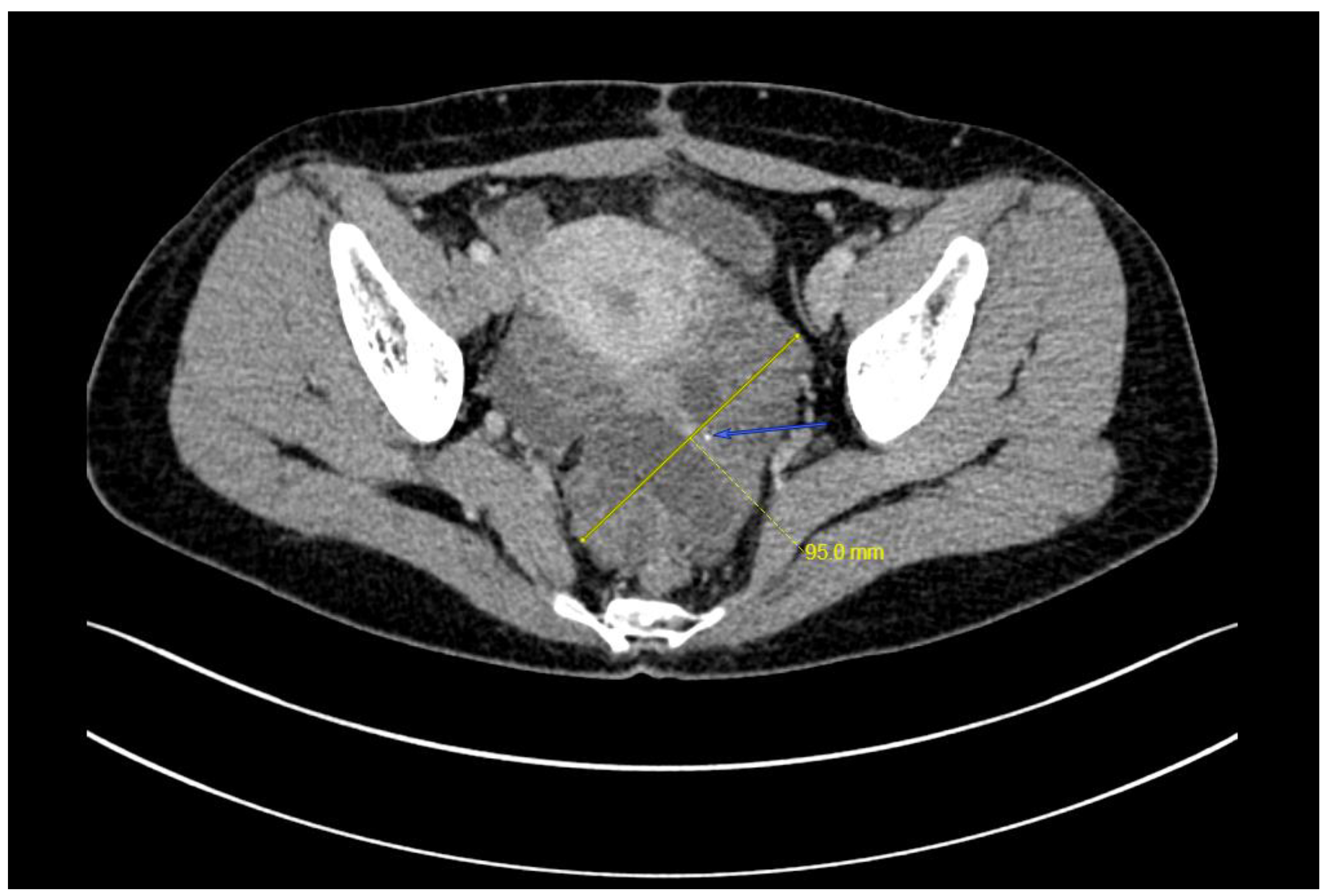
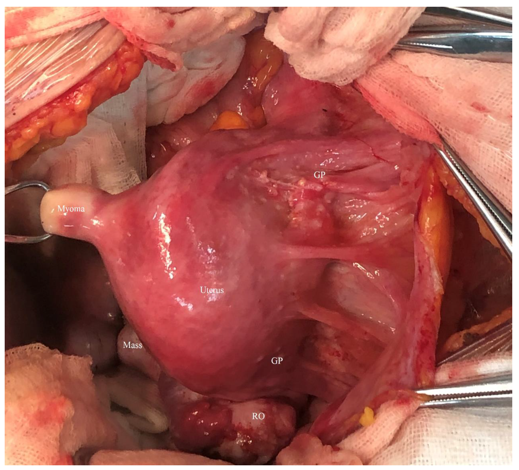
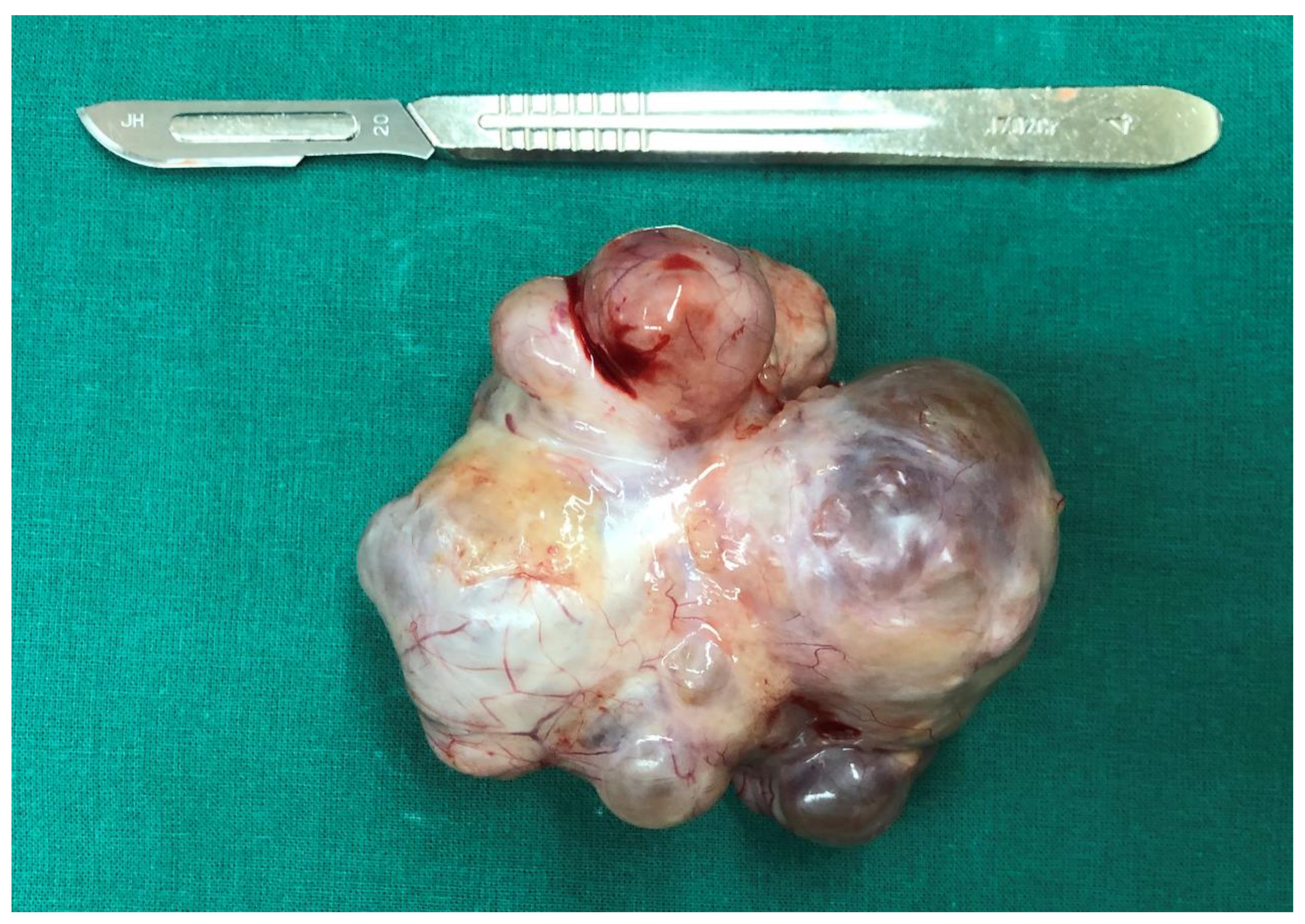
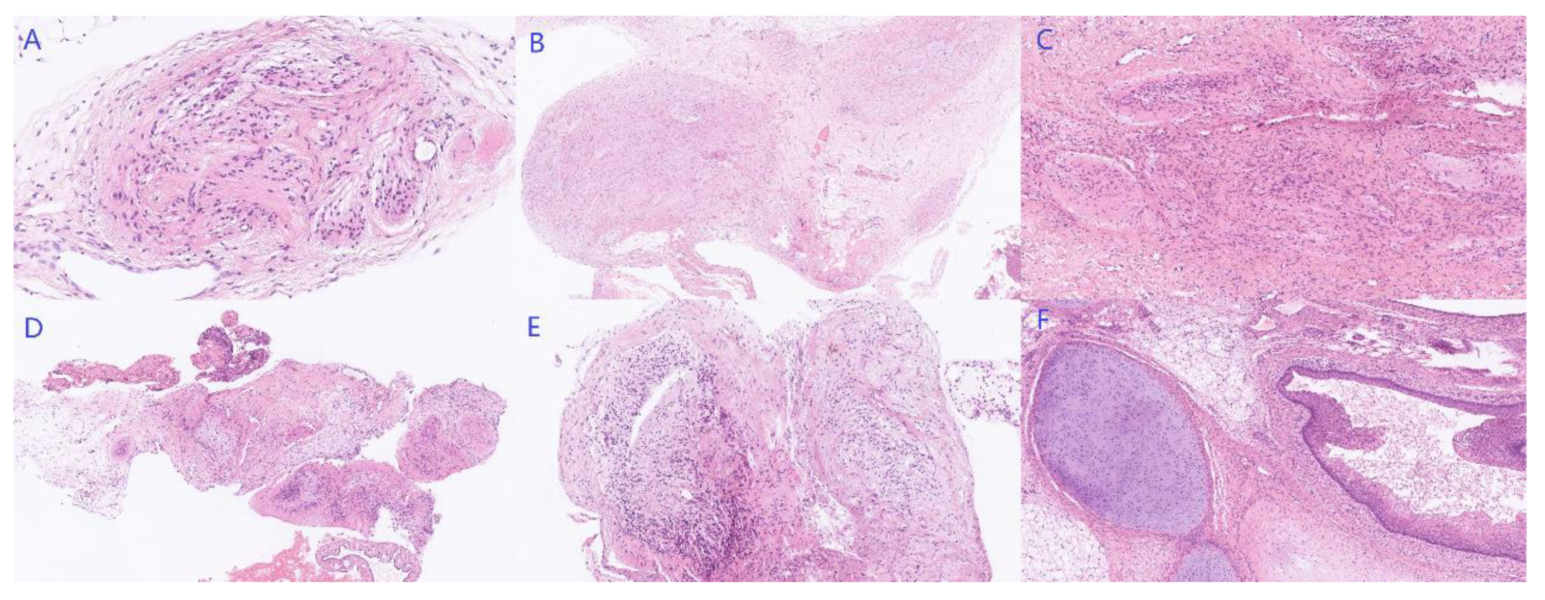
2. Case
3. Discussion
4. Conclusions
Supplementary Materials
Author Contributions
Funding
Institutional Review Board Statement
Informed Consent Statement
Data Availability Statement
Acknowledgments
Conflicts of Interest
References
- Smith, H.O.; Berwick, M.; Verschraegen, C.F.; Wiggins, C.; Lansing, L.; Muller, C.Y.; Qualls, C.R. Incidence and survival rates for female malignant germ cell tumors. Obstet. Gynecol. 2006, 107, 1075–1085. [Google Scholar] [CrossRef]
- Park, M.; Lim, J.; Lee, J.A.; Park, H.J.; Park, B.K.; Lim, M.C.; Park, S.-Y.; Won, Y.-J. Incidence and outcomes of malignant ovarian germ cell tumors in Korea, 1999-2017. Gynecol. Oncol. 2021, 163, 79–84. [Google Scholar] [CrossRef] [PubMed]
- Wang, D.; Zhu, S.; Jia, C.; Cao, D.; Wu, M.; Shen, K.; Yang, J.; Pan, L.; Cheng, N.; Xiang, Y. Diagnosis and management of growing teratoma syndrome after ovarian immature teratoma: A single center experience. Gynecol. Oncol. 2020, 157, 94–100. [Google Scholar] [CrossRef]
- Bentivegna, E.; Azaïs, H.; Uzan, C.; Leary, A.; Pautier, P.; Gonthier, C.; Genestie, C.; Balleyguier, C.; Lhommé, C.; Duvillard, P.; et al. Surgical Outcomes After Debulking Surgery for Intraabdominal Ovarian Growing Teratoma Syndrome: Analysis of 38 Cases. Ann. Surg. Oncol. 2015, 22 (Suppl. S3), S964–S970. [Google Scholar] [CrossRef]
- Bonazzi, C.; Peccatori, F.; Colombo, N.; Lucchini, V.; Cantù, M.G.; Mangioni, C. Pure ovarian immature teratoma, a unique and curable disease: 10 years’ experience of 32 prospectively treated patients. Obstet. Gynecol. 1994, 84, 598–604. [Google Scholar]
- Bentivegna, E.; Gonthier, C.; Uzan, C.; Genestie, C.; Duvillard, P.; Morice, P.; Gouy, S. Gliomatosis peritonei: A particular entity with specific outcomes within the growing teratoma syndrome. Int. J. Gynecol. Cancer 2015, 25, 244–249. [Google Scholar] [CrossRef]
- Wang, D.; Jia, C.W.; Feng, R.E.; Shi, H.H.; Sun, J. Gliomatosis peritonei: A series of eight cases and review of the literature. J. Ovarian Res. 2016, 9, 45. [Google Scholar] [CrossRef]
- Li, S.; Liu, Z.; Dong, C.; Long, F.; Liu, Q.; Sun, D.; Gao, Z.; Wang, L. Growing Teratoma Syndrome Secondary to Ovarian Giant Immature Teratoma in an Adolescent Girl: A Case Report and Literature Review. Medicine 2016, 95, e2647. [Google Scholar] [CrossRef]
- Gorbatiy, V.; Spiess, P.E.; Pisters, L.L. The growing teratoma syndrome: Current review of the literature. Indian J. Urol. 2009, 25, 186–189. [Google Scholar] [CrossRef]
- Favara, B.E.; Franciosi, R.A. Ovarian teratoma and neuroglial implants on the peritoneum. Cancer 1973, 31, 678–681. [Google Scholar] [CrossRef]
- Kim, N.R.; Lim, S.; Jeong, J.; Cho, H.Y. Peritoneal and nodal gliomatosis with endometriosis, accompanied with ovarian immature teratoma: A case study and literature review. Korean J. Pathol. 2013, 47, 587–591. [Google Scholar] [CrossRef] [PubMed]
- Liang, L.; Zhang, Y.; Malpica, A.; Ramalingam, P.; Euscher, E.D.; Fuller, G.N.; Liu, J. Gliomatosis peritonei: A clinicopathologic and immunohistochemical study of 21 cases. Mod. Pathol. 2015, 28, 1613–1620. [Google Scholar] [CrossRef] [PubMed]
- Robboy, S.J.; Scully, R.E. Ovarian teratoma with glial implants on the peritoneum. An analysis of 12 cases. Hum. Pathol. 1970, 1, 643–653. [Google Scholar] [CrossRef]
- Ferguson, A.W.; Katabuchi, H.; Ronnett, B.M.; Cho, K.R. Glial implants in gliomatosis peritonei arise from normal tissue, not from the associated teratoma. Am. J. Pathol. 2001, 159, 51–55. [Google Scholar] [CrossRef]
- Małgorzata, S.; Anna, K.G.; Reszeć, J.; Krawczuk-Rybak, M. Growing Teratoma Syndrome and Gliomatosis Peritonei in a 15-Year-Old Girl With Immature Ovarian Teratoma: Case Report and Review of the Literature. J. Pediatr. Adolesc. Gynecol. 2021, 34, 885–889. [Google Scholar] [CrossRef]
- Van Nguyen, J.M.; Bouchard-Fortier, G.; Ferguson, S.E.; Covens, A. How Common Is the Growing Teratoma Syndrome in Patients with Ovarian Immature Teratoma? Int. J. Gynecol. Cancer 2016, 26, 1201–1206. [Google Scholar] [CrossRef]
- Kikawa, S.; Todo, Y.; Minobe, S.; Yamashiro, K.; Kato, H.; Sakuragi, N. Growing teratoma syndrome of the ovary: A case report with FDG-PET findings. J. Obstet. Gynaecol. Res. 2011, 37, 926–932. [Google Scholar] [CrossRef]
- Frazer, J.L.; Hook, C.E.; Addley, H.C.; Jackson, C.R.; Latimer, J.A.; Nicholson, J.C.; Murray, M.J. Recurrent ovarian immature teratoma in a 12-year-old girl: Implications for management. Gynecol. Oncol. 2019, 154, 259–265. [Google Scholar] [CrossRef]
- André, F.; Fizazi, K.; Culine, S.; Droz, J.; Taupin, P.; Lhommé, C.; Terrier-Lacombe, M.-J.; Théodore, C. The growing teratoma syndrome: Results of therapy and long-term follow-up of 33 patients. Eur. J. Cancer 2000, 36, 1389–1394. [Google Scholar] [CrossRef]
- Zagamé, L.; Pautier, P.; Duvillard, P.; Castaigne, D.; Patte, C.; Lhommé, C. Growing teratoma syndrome after ovarian germ cell tumors. Obstet. Gynecol. 2006, 108 Pt 1, 509–514. [Google Scholar] [CrossRef]
- Ahlgren, A.D.; Simrell, C.R.; Triche, T.J.; Ozols, R.; Barsky, S.H. Sarcoma arising in a residual testicular teratoma after cytoreductive chemotherapy. Cancer 1984, 54, 2015–2018. [Google Scholar] [CrossRef]
- Dunne, R.F.; Sahasrabudhe, D.M.; Messing, E.M.; Jean-Gilles, J.; Fung, C. A case series of transformation of teratoma to primitive neuroectodermal tumor: Evolving management of a rare malignancy. Rare Tumors 2014, 6, 5268. [Google Scholar] [CrossRef] [PubMed]
- Ahmed, F.Y.; Gore, M.E.; Shepherd, J.; Fisher, C. Transformation of residual mature ovarian teratoma to adenocarcinoma 10 years after initial treatment. Clin. Oncol. 1996, 8, 125–126. [Google Scholar] [CrossRef]
- Comerci, J.T., Jr.; Licciardi, F.; Bergh, P.A.; Gregori, C.; Breen, J.L. Mature cystic teratoma: A clinicopathologic evaluation of 517 cases and review of the literature. Obstet. Gynecol. 1994, 84, 22–28. [Google Scholar]


Publisher’s Note: MDPI stays neutral with regard to jurisdictional claims in published maps and institutional affiliations. |
© 2022 by the authors. Licensee MDPI, Basel, Switzerland. This article is an open access article distributed under the terms and conditions of the Creative Commons Attribution (CC BY) license (https://creativecommons.org/licenses/by/4.0/).
Share and Cite
Li, S.; Su, N.; Jia, C.; Zhang, X.; Yin, M.; Yang, J. Growing Teratoma Syndrome with Synchronous Gliomatosis Peritonei during Chemotherapy in Ovarian Immature Teratoma: A Case Report and Literature Review. Curr. Oncol. 2022, 29, 6364-6372. https://doi.org/10.3390/curroncol29090501
Li S, Su N, Jia C, Zhang X, Yin M, Yang J. Growing Teratoma Syndrome with Synchronous Gliomatosis Peritonei during Chemotherapy in Ovarian Immature Teratoma: A Case Report and Literature Review. Current Oncology. 2022; 29(9):6364-6372. https://doi.org/10.3390/curroncol29090501
Chicago/Turabian StyleLi, Sijian, Na Su, Congwei Jia, Xinyue Zhang, Min Yin, and Jiaxin Yang. 2022. "Growing Teratoma Syndrome with Synchronous Gliomatosis Peritonei during Chemotherapy in Ovarian Immature Teratoma: A Case Report and Literature Review" Current Oncology 29, no. 9: 6364-6372. https://doi.org/10.3390/curroncol29090501
APA StyleLi, S., Su, N., Jia, C., Zhang, X., Yin, M., & Yang, J. (2022). Growing Teratoma Syndrome with Synchronous Gliomatosis Peritonei during Chemotherapy in Ovarian Immature Teratoma: A Case Report and Literature Review. Current Oncology, 29(9), 6364-6372. https://doi.org/10.3390/curroncol29090501