Article

Mutation Profiles of Ovarian Seromucinous Borderline Tumors in Japanese Patients

1
Department of Obstetrics and Gynecology, Shimane University School of Medicine, Izumo 6938501, Japan
2
Department of Obstetrics and Gynecology, Seirei Hamamatsu General Hospital, Hamamatsu 4308558, Japan
3
Department of Organ Pathology, Seirei Hamamatsu General Hospital, Hamamatsu 4308558, Japan
4
Department of Obstetrics and Gynecology, Matsue Red Cross Hospital, Matsue 6908506, Japan
5
Department of Pathology, Shonan Fujisawa Tokushukai Hospital, Fujisawa 2510041, Japan
*
Author to whom correspondence should be addressed.
Curr. Oncol. 2022, 29(5), 3658-3667; https://doi.org/10.3390/curroncol29050294
Submission received: 25 March 2022 / Revised: 22 April 2022 / Accepted: 29 April 2022 / Published: 18 May 2022

Abstract

:
Ovarian seromucinous tumors (SMBTs) are relatively rare, and their carcinogenesis is largely unknown. In this study, the molecular features of SMBTs in Japan are assessed. DNA was extracted from microdissected paraffin-embedded sections from 23 SMBT cases. Genetic mutations (KRAS, BRAF, PIK3CA, and ERBB2) were evaluated using Sanger sequencing. Immunohistochemistry for p53, ARID1A, and PTEN was also performed as a surrogate for the loss of functional mutations in these tumor suppressor genes. The prevalence of KRAS, BRAF, PIK3CA, and ERBB2 mutations was 4.3% (1/23), 8.6% (2/23), 8.6% (2/23), and 17.3% (4/23), respectively. Overexpression or loss of p53 expression occurred in 26% (6/23), loss of ARID1A expression in 4.3% (1/23), and none of the cases showed expression of PTEN loss. These findings suggest that KRAS/BRAF/PIK3CA and PTEN mutations are rare carcinogenic events in SMBTs. The high frequency of positive p53 staining and a low frequency of loss of ARID1A staining suggests that SMBT carcinogenesis may be related to the alteration of p53 rather than that of ARID1A. ERBB2 oncogenic mutations may play an important role in the tumorigenesis of Japanese SMBTs.

1. Introduction

Previously, seromucinous borderline tumors (SMBTs) in the ovary were considered a subtype of mucinous borderline tumors (MBT) [1]. Although SMBTs are thought to be a subtype of MBTs, they are clinically similar to serous borderline tumors (SBTs) because both display papillary projection inside cystic spaces grossly and present hierarchical branching with broad fibrous stroma microscopically [2]. In 2014, the WHO presented several modifications to tumors that belong to female reproductive organs and classified SMBTs as a new category distinct from MBTs [3]. SMBTs are a novel morphological group that is thought to be derived from or associated with endometriosis (30–50%) in many cases, which is difficult to find in patients with SBTs or MBTs [4]. The frequent bilaterality of SMBTs and their association with endometriosis has been confirmed in several studies [5,6,7,8].
Few reports have suggested that numerous genetic alterations are associated with tumorigenesis in SMBTs [9,10,11]. The first comprehensive attempt was recently undertaken to investigate the molecular underpinning of SMBTs by applying next-generation sequencing. This report found that SMBTs’ signatures consisted of frequent somatic mutations in the KRAS (100%), PIK3CA (60.7%), and ARID1A (14.3%) genes, with TERT promoter mutations and DNA mismatch repair deficiencies being consistently absent [9]. Another paper confirmed that the loss of ARID1A expression, a surrogate for ARID1A mutations, has been reported in one-third of these tumors, a frequency similar to that seen in ovarian endometrioid carcinomas, supporting their close relationship [10]. A further study focused on KRAS and PTEN mutations of 16 samples reported that KRAS mutation was 69%, and no PTEN mutation was observed [11].
Thus, molecular biological analyses of SMBTs have revealed several molecular characteristics. However, these cases originated in Taiwan, Korea, and the United States [9,10,11]. The molecular profiling of SMBTs in Japanese patients is yet to be performed. This study aims to assess the molecular features of SMBTs in a Japanese population. The genetic alterations in KRAS, BRAF, PIK3CA, ERBB2, PTEN, ARID1A, and p53 were retrospectively investigated to clarify the role of each gene in SMBT tumorigenesis.

2. Materials and Methods

2.1. Tumor Samples

Formalin-fixed, paraffin-embedded (FFPE) tissue samples from 23 SMBTs were used in this study. The samples were retrieved from the Department of Obstetrics and Gynecology, Shimane University Hospital, Seirei Hamamatsu General Hospital, and Matsue Red Cross Hospital between 2006 and 2019 in Japan.
The diagnoses were based on the conventional histopathological examination of sections stained with hematoxylin and eosin. Several pathologists categorized tumors according to the World Health Organization subtype criteria in each hospital. Tumors were staged according to the International Federation of Gynecology and Obstetrics classification system. All patients were primarily treated with unilateral/bilateral salpingo-oophorectomy and ± hysterectomy ± omentectomy ± pelvic lymphadenectomy. The resected specimen of each case was centrally reviewed by a gynecological pathologist (N.I.) and gynecologic oncologist (K.N.). The protocol for acquiring tissue specimens and clinical information was approved by the Institutional Review Board of Shimane University Hospital (IRB No. 20070305-1 and No. 20070305-2, version 10; last update, 8 December 2019). All the participants provided written informed consent. The study was conducted in accordance with the tenets of the Declaration of Helsinki and Title 45 (United States Code of Federal Regulations), Part 46 (Protection of Human Subjects), effective 13 December 2001.

2.2. Sample Processing and DNA Extraction

A gynecologic pathologist (N. I.) carefully selected representative FFPE blocks and identified the areas of SMBTs suitable for microdissection. Paraffin-embedded tissues were serially sectioned to a thickness of 10 mm. Genomic DNA for mutation analysis was extracted from FFPE samples using a commercially available kit (Qiagen, Inc., Valencia, CA, USA) as previously described [12].

2.3. Mutation Analysis

Extracted DNA was amplified by polymerase chain reaction (PCR) using primers for exon 2 of KRAS, exon 15 of BRAF, exons 9 and 20 of PIK3CA, and exon 20 of ERBB2 using genomic DNA obtained from microdissected formalin-fixed paraffin-embedded tissue. The study focused on analyzing exons reported to harbor most mutations in each gene [13]. The primers used for amplification were: KRAS-Exon 2, forward primer 5′-TTAACCTTATGTGTGACATGTTCTAA and reverse primer 5′-AGAATGGTCCTGCACCAGTAA, BRAF-Exon15; forward primer 5′-TGCTTGCTCTGATAGGAAAATG and reverse primer 5′-AGCATCTCAGGGCCAAAAAT, PIK3CA-Exon9; forward primer 5′-GGGAAAAATATGACAAAGAAAGC and reverse primer 5′, PIK3CA-Exon 20; forward primer 5′-CTCAATGATGCTTGGCTCTG and reverse primer 5′-TGGAATCCAGAGTGAGCTTC, ERBB2-Exon 20; and forward primer 5′-CCATACCCTCTCAGCGTAC and reverse primer 5′-CGGAGAGACCTGCAAAGAG.
The thermal cycle profile for all gene amplification included one cycle at 95 °C for 30 s followed by 40 cycles at 55 °C and extension at 72 °C for 15 s. All polymerase-chain-reaction-amplified products were sequenced at Beckman Coulter (Danvers, MA, USA) and analyzed using the Mutation Surveyor DNA Variant Analysis Software (Tokyo, Japan). The pathogenicity of each mutation was confirmed using the Catalogue of Somatic Mutations in Cancer (COSMIC).

2.4. Immunostaining of ARID1A, p53, and PTEN

The expression levels of ARID1A, p53, PTEN, and ERBB2 were evaluated by immunohistochemical analysis (IHC). FFPE sections (3 μm thick) were dewaxed in xylene and hydrated in graded alcohol solutions. After antigen retrieval in a sodium citrate buffer, the slides were incubated overnight at 4 °C with antibodies at the following dilutions: 1:50 p53 (M7001; Dako, Carpinteria, CA, USA), 1:100 ARID1A (Sc-32761; Santa Cruz Biotechnology, Santa Cruz, CA, USA), 1:200 PTEN (138G6; Cell Signaling Technology, Danvers, MA, USA), and 1:200 ERBB2 (ab16901, abcam, Cambridge, UK). Two gynecologic oncologists (H. S. and K. N.), blinded to the clinicopathologic factors, independently evaluated the samples under a light microscope. The loss of ARID1A and PTEN expression in tumor cell nuclei was used as a surrogate for the presence of PTEN/ARID1A loss-of-function mutations [13]. Similarly, p53 immunoreactivity was used as a surrogate for the presence of p53 loss/overexpression of functional mutations. The assessment of ARID1A, p53, PTEN, and ERBB2 immunostaining was performed as described in our previous reports [14,15]. Briefly, ARID1A and PTEN immunoreactivity was scored by two investigators (H. S. and K. N.): 0, undetectable; 1+, weakly positive; 2+, moderately positive; and 3+, intensely positive. The loss of ARID1A or PTEN staining intensity (0+) was considered negative. Weak to moderate immunoreactivity was considered p53 normal expression. Strong and diffuse nuclear p53 immunoexpression or the complete absence of p53 staining was likely to indicate a p53 mutation.
A score of 2+ or 3+ was defined as ERBB2 overexpression, and the others were defined as low expression [16].

3. Results

The direct sequence analysis of 23 tumor specimens was performed to assess the mutation profiles of SMBTs. The patients’ clinical characteristics and mutation profile are summarized in Table 1 and Table 2. The age of the patients ranged from 18 to 83 years, with a mean and median age of 51.8 and 49 years, respectively. The majority of the patients (95.6%) were found in stage IA at diagnosis, and only one (4.34%) patient presented with stage IIA disease.
The tumors’ size ranged from 3.7 to 31 cm, with a median size of 10 cm. The tumors involving the right ovary alone accounted for 56.6% (13/23), while those involving the left ovary alone accounted for 43.4% (10/23). No cases of bilateral ovarian SMBT were observed in this study. The patients initially underwent left/right or bilateral salpingo-oophorectomy ± hysterectomy ± omentectomy ± pelvic lymphadenectomy. Fertility preservation patients underwent unilateral salpingo-oophorectomy, and patients diagnosed with carcinoma by rapid intraoperative pathological diagnosis underwent total abdominal hysterectomy + bilateral salpingo-oophorectomy + omentectomy + pelvic lymphadenectomy. However, patients with carcinomas were diagnosed with SMBTs in the final postoperative diagnosis. All patients in the current study are still alive without disease, and their survival rate is 100%. Pathological evidence of endometriosis was confirmed in 13.1% (3/23) SMBTs. Figure 1 shows representative examples of the histological appearance of the SMBTs.
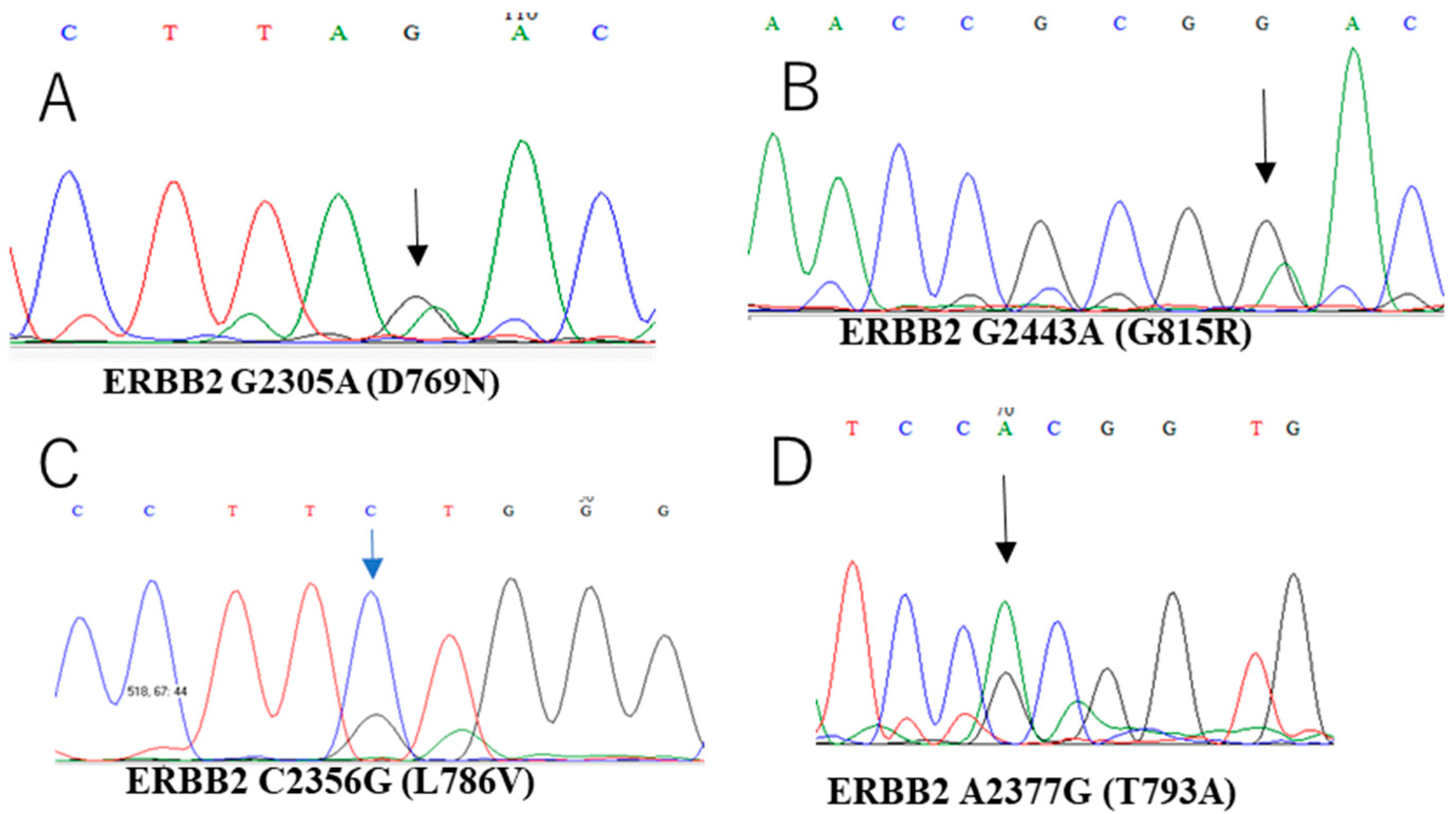
All 23 cases were assessed for mutations in KRAS, BRAF, PIK3CA, and ERBB2. The prevalence of KRAS (G12V), BRAF (V600E), ERBB2 (D769N, T793A, G815R, and L786V), and PIK3CA (T544P) mutations was 4.3% (1/23), 8.6% (2/23), 17.3% (4/23), and 8.6% (2/23), repsectively (Table 3, Figure 2).
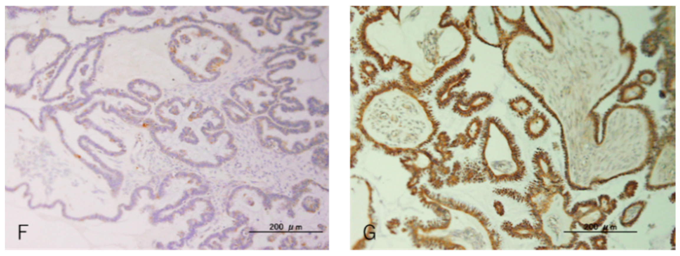
The immunohistochemical expression of ARID1A, p53, and PTEN in all samples, as surrogates for these tumor suppressor gene mutations, was analyzed. The loss of ARID1A nuclear expression was observed in one case (4.3%). It was noted that p53 expression was undetectable in two cases, and overexpression was found in four cases (26.0%) (Figure 3). PTEN was expressed in all SMBTs, and none of them lost its expression, suggesting that no PTEN mutations were found (Figure 3). The ERBB2 protein expression level was evaluated, and all of the mutant cases of ERBB2 were overexpressed (Figure 3).

4. Discussion

Ovarian cancer accounts for more deaths than all other gynecologic malignancies [17] and typically affects postmenopausal women; about 12% are diagnosed under the age of 45, with 5% under the age of 35 years [18,19,20,21,22]. Treatment for ovarian cancer usually involves a combination of surgery and chemotherapy that can impact the ovaries anatomically or functionally. Patients, particularly young patients who want to preserve their fertility, diagnosed with malignant ovarian tumors, including SMBTs, experience anxiety, anger, sadness, and depression, severely impairing their lives [23]. For this reason, fertility-sparing treatment for SMBTs with psychological care is essential.
This study first described the molecular alterations of SMBTs in the Japanese population regarding the prevalence of mutations in KRAS (4.3%), BRAF (8.6%), PIK3CA (8.6%), ERBB2 (17.3%), ARID1A (4.3%), p53 (26%) and PTEN (0%). Very recently, Wu et al. [9] performed mutational analysis on 28 SMBTs in Taiwan and reported that somatic mutations in the KRAS, PIK3CA, ARID1A, and PTEN genes were 100, 60.7, 14.0, and 3.6%, respectively. They demonstrated that KRAS was mutated in all SMBTs, whereas PIK3CA mutations occurred frequently. Compared with their report, the current frequency of these alterations in these genes was much lower. Ethnic differences seem to contribute to the incidence and prognosis of cancers. It was previously discovered that the carcinogenesis signaling pathway in low-grade serous ovarian carcinoma was different between Japanese and Western populations. The mutation of KRAS/BRAF was observed in Western countries [24,25,26], whereas PIK3CA mutation was the main driver for Japanese low-grade serous carcinoma progression [27]. Racial/ethnic differences in breast cancer incidence and outcome have also been observed. African American women are diagnosed at an advanced stage with large tumors and a higher grade than those in White women [28,29]. Differences in pharmacokinetics and toxicity of anticancer drugs between Asian and White patients have been reported [30]. Allelic variants of genes encoding drug-metabolizing enzymes are expressed with different incidences in different ethnic groups [30]. This study thus hypothesized that the difference in prevalence might be due to a difference in genetic background between the Japanese population and other ethnicities or the difference in methods between direct sequencing and next-generation sequencing. SMBTs are associated with endometriosis, and previous studies have identified this in 45–71% of patients [5,6,7,8,9], including three cases in which endometriosis was directly contiguous with SMBTs [31]. The current frequency of SMBTs with endometriosis is much lower than that reported in previous reports [5,6,7,8,9]. Therefore, it may also be because the frequency of SMBTs with concurrent endometriosis is significantly lower than that reported by Wu et al. [9].
In 2014, the World Health Organization defined SMBTs as a new histological subtype of ovarian carcinoma, but seromucinous carcinoma was not defined in endometrial carcinoma [3]. “Seromucinous Carcinoma” has been removed from the 2020 5th edition of the WHO Classification of Female Genital Tumors because this is a poorly reproducible diagnosis. Moreover, there is significant morphological overlap with endometrioid carcinoma [32]. Therefore, seromucinous carcinoma is considered a subtype of endometrioid carcinoma [33,34]. ARID1A is a tumor suppressor gene that is frequently mutated in endometriosis-related ovarian neoplasms, including clear cell and endometrioid carcinoma. Previously, a somatic mutation in ARID1A has been found in 46–57% of ovarian clear cell carcinomas [35,36], 30% of ovarian endometrioid carcinomas [34], and 40% of uterine endometrioid carcinomas [37]. Among 24 SMBTs, loss of ARID1A expression in 8/24 (33%) cases and one case with loss of ARID1A expression in synchronous endometriosis were reported [10]. A significant number of SMBTs that exhibit loss of expression of ARID1A and their common association with endometriosis reveal that these tumors are closely related to endometrioid carcinomas. However, in the current study, the loss of ARID1A was only 4.3%, which was lower than that in previous reports [5,6,7,8,9,10]. In addition, immunohistochemical staining for p53 has been found positive (mutant p53) in 26.0% (6/23) of SMBTs. Previously, mutational profiles of endometriosis-related ovarian neoplasms (ERONs) were constructed [38]. It was observed that ARID1A mutation was 95%, while p53 was 36.8%, suggesting that the occurrence of some ERONs based on p53 alterations was similar to that in the current findings in SMBTs.
Molecular alterations in SMBTs strikingly differ from those of other borderline ovarian tumors (serous and mucinous) in the ovary. Previously, Sanger sequencing was performed to determine the molecular mechanisms involved in the tumorigenesis of MBTs and SBTs. BRAF (40%) and KRAS (20%) mutations are the most frequent genetic alterations in MBTs, whereas PIK3CA (63.6%) mutations are the most responsible for the tumorigenesis in SBTs [27,39]. In contrast, the current study observed that ERBB2 was the highest prevalent mutant oncogene (17.3%) in SMBTs. This finding suggests that each borderline tumor has distinct molecular mechanisms of carcinogenesis.
The strength of this study is that it is the first molecular analysis of Japanese SMBTs. However, this study had several limitations. First, this study included a small number of patients as SMBTs are rare tumors; therefore, concrete conclusions cannot be drawn. Hence, further investigation with a larger study population is essential. Second, genetic mutations were identified using Sanger sequencing; therefore, the types of gene mutations assessed were limited. Additionally, IHC for p53, ARID1A, and PTEN was used as a surrogate marker for these tumor suppressor genes underlying the molecular derangements in SMBTs. Sanger sequencing will be necessary to confirm the IHC results in the future. Furthermore, next-generation sequencing is also needed to determine the comprehensive molecular mechanisms underlying the progression of SMBTs in Japanese patients.
The current findings suggest that alterations in KRAS/BRAF/PIK3CA and PTEN are rare carcinogenic events in Japanese SMBTs. The high frequency of positive p53 staining and a low frequency of loss of ARID1A staining suggests that the carcinogenesis of SMBTs could be related to the alteration of p53 rather than that of ARID1A. The fact that the ERBB2 oncogenic mutation is the highest event suggests its important role in the tumorigenesis of Japanese SMBTs.

Author Contributions

H.S., S.R. and K.N. drafted the manuscript. H.Y., T.I., M.I., S.S., S.N. and R.F. collected data. N.I. and Y.O. performed the pathological review. K.N. participated in this study. S.K. participated in coordinating this study. All authors have read and agreed to the published version of the manuscript.

Funding

This research was funded by JSPS KAKENHI (grant numbers 18K09229 and 18K09291).

Institutional Review Board Statement

This study was conducted in accordance with the guidelines of the Declaration of Helsinki and was approved by the Institutional Review Board of Shimane University Hospital (IRB No. 20070305-1 and No. 20070305-2, version 10; last update, 8 December 2019).

Informed Consent Statement

Written informed consent was obtained from all the patients.

Data Availability Statement

The data presented in this study are available upon request from the corresponding author (K.N.).

Conflicts of Interest

The authors declare no conflict of interest.

References

  1. Dubé, V.; Roy, M.; Plante, M.; Renaud, M.C.; Têtu, B. Mucinous Ovarian Tumors of Mullerian-Type: An Analysis of 17 Cases Including Borderline Tumors and Intraepithelial, Microinvasive, and Invasive Carcinomas. Int. J. Gynecol. Pathol. 2005, 24, 138–146. [Google Scholar] [CrossRef] [PubMed]
  2. Mikami, Y. Endometriosis-related Ovarian Neoplasms: Pathogenesis and Histopathologic Features. Diagn. Histopathol. 2014, 20, 357–363. [Google Scholar] [CrossRef]
  3. Kurman, R.J.; International Agency for Research on Cancer; World Health Organization. WHO Classification of Tumors of Female Reproductive Organs; International Agency for Research on Cancer: Lyon, France, 2014; p. 307. [Google Scholar]
  4. Rutgers, J.K.L. Mullerian Mucinous/mixed Epithelial (Seromucinous) Ovarian Tumors. AJPS 2016, 21, 206–213. [Google Scholar]
  5. Rutgers, J.L.; Scully, R.E. Ovarian Mixed-Epithelial Papillary Cystadenomas of Borderline Malignancy of Mullerian Type. A Clinicopathologic Analysis. Cancer 1988, 61, 546–554. [Google Scholar] [CrossRef]
  6. Lee, K.R.; Nucci, M.R. Ovarian Mucinous and Mixed Epithelial Carcinomas of Mullerian (endocervical-like) Type: A Clinicopathologic Analysis of Four Cases of an Uncommon Variant Associated with Endometriosis. Int. J. Gynecol. Pathol. 2003, 22, 42–51. [Google Scholar] [CrossRef]
  7. Nagamine, M.; Mikami, Y. Ovarian Seromucinous Tumors: Pathogenesis, Morphologic Spectrum, and Clinical Issues. Diagnostics 2020, 10, 77. [Google Scholar] [CrossRef] [Green Version]
  8. Nagai, Y.; Kishimoto, T.; Nikaido, T.; Nishihara, K.; Matsumoto, T.; Suzuki, C.; Ogishima, T.; Kuwahara, Y.; Hurukata, Y.; Mizunuma, M.; et al. Squamous Predominance in Mixed-Epithelial Papillary Cystadenomas of Borderline Malignancy of Mullerian Type Arising in Endometriotic Cysts: A Study of Four Cases. Am. J. Surg. Pathol. 2003, 27, 242–247. [Google Scholar] [CrossRef]
  9. Wu, R.C.; Chen, S.J.; Chen, H.C.; Tan, K.T.; Jung, S.M.; Lin, C.Y.; Chao, A.S.; Huang, K.G.; Chou, H.H.; Chang, T.C.; et al. Comprehensive Genomic Profiling Reveals Ubiquitous KRAS Mutations and Frequent PIK3CA Mutations in Ovarian Seromucinous Borderline Tumor. Mod. Pathol. 2020, 33, 2534–2543. [Google Scholar] [CrossRef]
  10. Wu, C.H.; Mao, T.L.; Vang, R.; Ayhan, A.; Wang, T.L.; Kurman, R.J.; Shih, I.E.M. Endocervical-Type Mucinous Borderline Tumors are Related to Endometrioid Tumors Based on Mutation and Loss of Expression of ARID1A. Int. J. Gynecol. Pathol. 2012, 31, 297–303. [Google Scholar] [CrossRef]
  11. Kim, K.R.; Choi, J.; Hwang, J.E.; Baik, Y.A.; Shim, J.Y.; Kim, Y.M.; Robboy, S.J. Endocervical-Like (Müllerian) Mucinous Borderline Tumours of the Ovary are Frequently Associated with the KRAS Mutation. Histopathology 2010, 57, 587–596. [Google Scholar] [CrossRef]
  12. Nakayama, K.; Takebayashi, Y.; Namiki, T.; Tamahashi, N.; Nakayama, S.; Uchida, T.; Miyazaki, K.; Fukumoto, M. Comprehensive Allelotype Study of Ovarian Tumors of Low Malignant Potential: Potential Differences in Pathways Between Tumors with and without Genetic Predisposition to Invasive Carcinoma. Int. J. Cancer 2001, 94, 605–609. [Google Scholar] [CrossRef] [PubMed] [Green Version]
  13. Nonomura, Y.; Nakayama, K.; Nakamura, K.; Razia, S.; Yamashita, H.; Ishibashi, T.; Ishikawa, M.; Sato, S.; Nakayama, S.; Otsuki, Y.; et al. Ovarian Endometrioid and Clear Cell Carcinomas with Low Prevalence of Microsatellite Instability: A Unique Subset of Ovarian Carcinomas Could Benefit from Combination Therapy with Immune Checkpoint Inhibitors and Other Anticancer Agents. Healthcare 2022, 10, 694. [Google Scholar] [CrossRef] [PubMed]
  14. Nakayama, K.; Takebayashi, Y.; Nakayama, S.; Hata, K.; Fujiwaki, R.; Fukumoto, M.; Miyazaki, K. Prognostic Value of Overexpression of p53 in Human Ovarian Carcinoma Patients Receiving Cisplatin. Cancer Lett. 2003, 192, 227–235. [Google Scholar] [CrossRef]
  15. Katagiri, A.; Nakayama, K.; Rahman, M.T.; Rahman, M.; Katagiri, H.; Nakayama, N.; Ishikawa, M.; Ishibashi, T.; Iida, K.; Kobayashi, H.; et al. Loss of ARID1A Expression is Related to Shorter Progression-free Survival and Chemoresistance in Ovarian Clear Cell Carcinoma. Mod. Pathol. 2012, 25, 282–288. [Google Scholar] [CrossRef] [Green Version]
  16. Nakamura, H.; Saji, H.; Ogata, A.; Hosaka, M.; Hagiwara, M.; Kawasaki, N.; Kato, H. Correlation between Encoded Protein Overexpression and Copy Number of the HER2 Gene with Survival in non-small Cell Lung Cancer. Int. J. Cancer 2003, 103, 61–66. [Google Scholar] [CrossRef]
  17. Fishman, D.A.; Cohen, L.; Blank, S.V.; Shulman, L.; Singh, D.; Bozorgi, K.; Tamura, R.; Timor-Tritsch, I.; Schwartz, P.E. The Role of Ultrasound Evaluation in the Detection of Early-stage Epithelial Ovarian Cancer. Am. J. Obstet. Gynecol. 2005, 192, 1214–1221. [Google Scholar] [CrossRef] [PubMed]
  18. Rasool, N.; Rose, P.G. Fertility-preserving Surgical Procedures for Patients with Gynecologic Malignancies. Clin. Obstet. Gynecol. 2010, 53, 804–814. [Google Scholar] [CrossRef]
  19. DiSaia, P.J. Conservative Management of the Patient with Early Gynecologic Cancer. CA Cancer J. Clin. 1989, 39, 135–154. [Google Scholar] [CrossRef]
  20. Huber, D.; Cimorelli, V.; Usel, M.; Bouchardy, C.; Rapiti, E.; Petignat, P. How many Ovarian Cancer Patients are Eligible for Fertility-sparing Surgery. Eur. J. Obstet. Gynecol. Reprod. Biol. 2013, 170, 270–274. [Google Scholar] [CrossRef]
  21. Franasiak, J.M.; Scott, R.T. Demographics of Cancer in the Reproductive Age Female. In Cancer and Fertility; Sabanegh, J.E., Ed.; Humana Press: Cham, Switzerland, 2016; pp. 11–19. [Google Scholar] [CrossRef]
  22. Gerstl, B.; Sullivan, E.; Vallejo, M.; Koch, J.; Johnson, M.; Wand, H.; Webber, K.; Ives, A.; Anazodo, A. Reproductive Outcomes Following Treatment for a Gynecological Cancer Diagnosis: A Systematic Review. J. Cancer Surviv. 2019, 13, 269–281. [Google Scholar] [CrossRef]
  23. La Rosa, V.L.; Garzon, S.; Gullo, G.; Fichera, M.; Sisti, G.; Gallo, P.; Riemma, G.; Schiattarella, A. Fertility Preservation in Women Affected by Gynaecological Cancer: The Importance of an Integrated Gynaecological and Psychological Approach. Ecancermedicalscience 2020, 14, 1035. [Google Scholar] [CrossRef] [PubMed]
  24. Wong, K.K.; Tsang, Y.T.; Deavers, M.T.; Mok, S.C.; Zu, Z.; Sun, C.; Malpica, A.; Wolf, J.K.; Lu, K.H.; Gershenson, D.M. BRAF Mutation is Rare in Advanced-stage Low-grade Ovarian Serous Carcinomas. Am. J. Pathol. 2010, 177, 1611–1617. [Google Scholar] [CrossRef] [PubMed]
  25. Jones, S.; Wang, T.L.; Kurman, R.J.; Nakayama, K.; Velculescu, V.E.; Vogelstein, B.; Kinzler, K.W.; Papadopoulos, N.; Shih, I.E.M. Low-grade Serous Carcinomas of the Ovary Contain Very Few Point Mutations. J. Pathol. 2012, 226, 413–420. [Google Scholar] [CrossRef] [PubMed]
  26. Singer, G.; Kurman, R.J.; Chang, H.W.; Cho, S.K.; Shih, I.E.M. Diverse Tumorigenic Pathways in Ovarian Serous Carcinoma. Am. J. Pathol. 2002, 160, 1223–1228. [Google Scholar] [CrossRef] [Green Version]
  27. Ishibashi, T.; Nakayama, K.; Razia, S.; Ishikawa, M.; Nakamura, K.; Yamashita, H.; Dey, P.; Iida, K.; Kurioka, H.; Nakayama, S.; et al. High Frequency of PIK3CA Mutations in Low-Grade Serous Ovarian Carcinomas of Japanese Patients. Diagnostics 2019, 10, 13. [Google Scholar] [CrossRef] [PubMed] [Green Version]
  28. Ghafoor, A.; Jemal, A.; Ward, E.; Cokkinides, V.; Smith, R.; Thun, M. Trends in Breast Cancer by Race and Ethnicity. CA Cancer J. Clin. 2003, 53, 342–355. [Google Scholar] [CrossRef] [Green Version]
  29. Li, C.I.; Malone, K.E.; Daling, J.R. Differences in Breast Cancer Hormone Receptor Status and Histology by Race and Ethnicity Among Women 50 years of Age and Older. Cancer Epidemiol. Biomarkers Prev. 2002, 11, 601–607. [Google Scholar]
  30. Phan, V.H.; Moore, M.M.; McLachlan, A.J.; Piquette-Miller, M.; Xu, H.; Clarke, S.J. Ethnic Differences in Drug Metabolism and Toxicity from Chemotherapy. Expert Opin. Drug Metab. Toxicol. 2009, 5, 243–257. [Google Scholar] [CrossRef]
  31. Hamada, T.; Kiyokawa, T.; Nomura, K.; Hano, H. Immunohistochemical Analysis of Reserve Cell-like Cells of Ovarian Müllerian Mucinous/Mixed Epithelial Borderline Tumor. Int. J. Gynecol. Pathol. 2008, 27, 199–206. [Google Scholar] [CrossRef]
  32. Rambau, P.F.; McIntyre, J.B.; Taylor, J.; Lee, S.; Ogilvie, T.; Sienko, A.; Morris, D.; Duggan, M.A.; McCluggage, W.G.; Koöbel, M. Morphologic reproducibility, genotyping, and immunohistochemical profiling do not support a category of seromucinous carcinoma of the ovary. Am. J. Surg. Pathol. 2017, 41, 685–695. [Google Scholar] [CrossRef]
  33. Kobel, M.; Kim, K.-R.; McCluggage, W.L.; Shih, I.; Single, N. Seromucinous Carcinoma. Tumors of Ovary. In WHO Classification of Female Genital Tumors, 5th ed.; Cheung, A.N., Ellenson, L.H., Gilks, C.B., Kim, K.-R., Kong, C.S., Lax, S.F., Longacre, T.A., Malpica, A., McCluggage, W.G., Olive, E., et al., Eds.; WHO Classification of Tumors Editorial Board, International Agency for Research on Cancer (IARC): Lyon, France, 2020; Volume 70. [Google Scholar]
  34. Kobel, M.; Huntsman, D.G.; Lim, D.; McCluggage, W.G.; Rabban, T.T.; Shih, I. Endometrioid carcinoma of the ovary. Tumors of Ovary. In WHO Classification of Female Genital Tumors, 5th ed.; Cheung, A.N., Ellenson, L.K., Gillks, C.B., Kim, K.-R., Kong, C.S., Lax, S.F., Longacre, T.A., Malpica, A., McCluggage, W.G., Olive, E., et al., Eds.; WHO Classification of Tumors Editorial Board, International Agency for Research on Cancer (IARC): Lyon, France, 2020; Volume 70. [Google Scholar]
  35. Jones, J.M.; Cui, X.S.; Medina, D.; Donehower, L.A. Heterozygosity of p21WAF1/CIP1 Enhances Tumor Cell Proliferation and Cyclin D1-associated Kinase Activity in a Murine Mammary Cancer Model. Cell Growth Differ. 1999, 10, 213–222. [Google Scholar]
  36. Wiegand, K.C.; Shah, S.P.; Al-Agha, O.M.; Zhao, Y.; Tse, K.; Zeng, T.; Senz, J.; McConechy, M.K.; Anglesio, M.S.; Kalloger, S.E.; et al. ARID1A Mutations in Endometriosis-associated Ovarian Carcinomas. N. Engl. J. Med. 2010, 363, 1532–1543. [Google Scholar] [CrossRef] [Green Version]
  37. Guan, B.; Mao, T.L.; Panuganti, P.K.; Kuhn, E.; Kurman, R.J.; Maeda, D.; Chen, E.; Jeng, Y.M.; Wang, T.L.; Shih, I.E.M. Mutation and Loss of Expression of ARID1A in Uterine Low-grade Endometrioid Carcinoma. Am. J. Surg. Pathol. 2011, 35, 625–632. [Google Scholar] [CrossRef] [PubMed] [Green Version]
  38. Ishikawa, M.; Nakayama, K.; Nakamura, K.; Ono, R.; Sanuki, K.; Yamashita, H.; Ishibashi, T.; Minamoto, T.; Iida, K.; Razia, S.; et al. Affinity-purified DNA-based mutation profiles of endometriosis-related ovarian neoplasms in Japanese patients. Oncotarget 2018, 9, 14754–14763. [Google Scholar] [CrossRef] [PubMed]
  39. Ohnishi, K.; Nakayama, K.; Ishikawa, M.; Ishibashi, T.; Yamashita, H.; Nakamura, K.; Minamoto, T.; Iida, K.; Razia, S.; Ishikawa, N.; et al. Mucinous Borderline Ovarian Tumors with BRAFV600E Mutation may have Low Risk for Progression to Invasive Carcinomas. Arch. Gynecol. Obstet. 2020, 302, 487–495. [Google Scholar] [CrossRef] [PubMed]
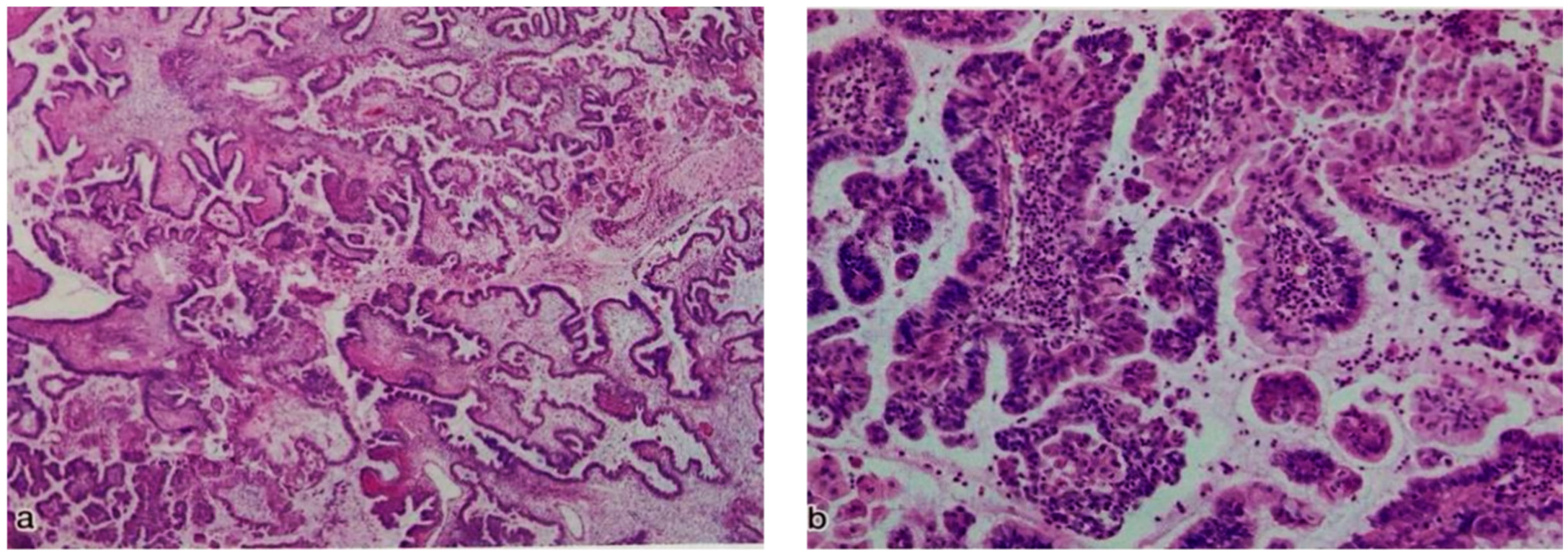
Figure 1. Representative histological characteristics of seromucinous borderline tumor (SMBT). (a) Low magnification (×10); (b) high magnification (×20).
Curroncol 29 00294 g001
Figure 2. Chromatograms of ERBB2 mutation statuses in SMBTs. Each SMBT showed mutations (A) D769N (2305G>A), (B) G815R (2443G>A), (C) L786V (2356C>G), and (D) T793A (2377A>G) in the ERBB2 gene, respectively.
Curroncol 29 00294 g002
Figure 3. Representative immunohistochemical staining of ARID1A, PTEN, p53 and ERBB2 in SMBTs. Normal (A) and loss of ARID1A expression (B). Normal PTEN (C); overexpression (D) and loss of p53 expression (E); low (F) and overexpression (G) of ERBB2 in SMBTs.
Curroncol 29 00294 g003aCurroncol 29 00294 g003b
Table 1. Clinical information of the 23 SMBTs.
Case NO.AgeStageSite of TumorTumor Size (CM)Surgical TypeEndometriosis
157IARight17BSO+TAH+omentechtomy+pelviclymphadenectomyNo
266IARight15BSO+omentechtomyNo
318IALeft10LSONo
449IALeft10BSO+TAH+omentechtomyNo
528IARight17RSO+omentechtomyNo
677IARight6BSO+TLHNo
737IALeft6.5BSO+TAH+omentechtomyNo
841IARight9BSO+TAH+omentechtomyYes
937IARight19BSO+TAH+omentechtomy+pelviclymphadenectomyNo
1047IALeft22BSO+TAH+omentechtomyNo
1176IARightNDRSONo
1254IALeftNDLSONo
1345IARightNDBSOYes
1458IALeftNDBSO+TAHYes
1531IARight21RSONo
1667IIALeft9.5BSO+TAH+pelviclymphadenectomyNo
1747IARight12RSONo
1826IARight31RSONo
1983IALeft3.7BSO+TAHNo
2072IALeft9.2BSONo
2176IARight5BSO+TAH+omentechtomyNo
2240IARight30RSONo
2360IALeft8.8LSONo
BSO: Bilateral salpingo-oophorectomy; LSO: left salpingo-oophorectomy; RSO: right salpingo-oophorectomy; TAH: total abdominal hysterectomy, ND: no data available.
Table 2. Mutation profile of 23 SMBTs.
Case NO.p53 (IHC)ARID1A (IHC)PTEN (IHC)KRASBRAFPIK3CA-E9PIK3CA-E20ERBB2ERBB2 (IHC)
1NormalNormalNormal----D769N. c.2305G>AHigh
2LossNormalNormal-----High
3LossNormalNormal-----High
4OverexpressionNormalNormalG12V, c.35G>T----Low
5OverexpressionNormalNormal-V600E, c.1799T>A---High
6NormalNormalNormal-----High
7NormalNormalNormal-----ND
8NormalNormalNormal-----Low
9NormalNormalNormal-----Low
10NormalNormalNormal-----Low
11NormalNormalNormal-----High
12NormalLossNormal-----Low
13NormalNormalNormal-----ND
14NormalNormalNormal-V600E, c.1799T>A--T793A, c.2377A>GHigh
15NormalNormalNormal-----High
16OverexpressionNormalNormal-----Low
17NormalNormalNormal-----Low
18NormalNormalNormal----G815R, c.2443G>AHigh
19NormalNormalNormal----L786V, c.2356 C>GHigh
20NormalNormalNormal-----High
21NormalNormalNormal--T544P, c.1630A>C--High
22OverexpressionNormalNormal--T544P, c.1630A>C--High
23NormalNormalNormal-----High
ND: no data available.
Table 3. Mutation frequency in the 23 SMBTs.
Gene.Frequency of Genetic Alteration
KRAS4.3% (1/23)
BRAF8.6% (2/23)
PIK3CA8.6% (2/23)
ERBB217.3% (4/23)
ARID1A4.3% (1/23)
p5326% (6/23)
PTEN0% (0/23)
Publisher’s Note: MDPI stays neutral with regard to jurisdictional claims in published maps and institutional affiliations.