Primary Myelofibrosis Occurring during Targeted Therapy for Chronic Lymphocytic Leukemia: A Report of Two Cases


 ,
, 
Abstract
:1. Introduction
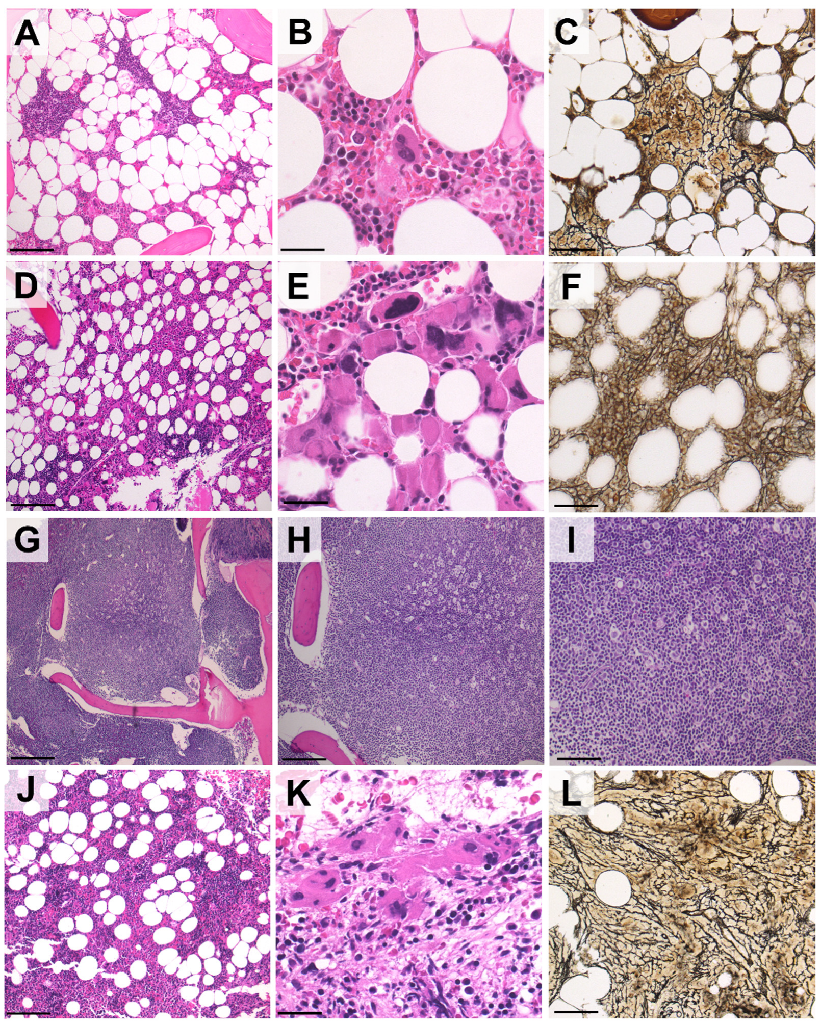
2. Case Reports
3. Conclusions
Author Contributions
Funding
Institutional Review Board Statement
Informed Consent Statement
Data Availability Statement
Acknowledgments
Conflicts of Interest
References
- Hallek, M.; Shanafelt, T.D.; Eichhorst, B. Chronic lymphocytic leukaemia. Lancet 2018, 391, 1524–1537. [Google Scholar] [CrossRef]
- Hilal, T.; Gea-Banacloche, J.C.; Leis, J.F. Chronic lymphocytic leukemia and infection risk in the era of targeted therapies: Linking mechanisms with infections. Blood Rev. 2018, 2, 387–399. [Google Scholar] [CrossRef] [PubMed]
- Zent, C.S.; Kay, N.E. Autoimmune complications in chronic lymphocytic leukaemia (CLL). Best Pract. Res. Clin. Haematol. 2010, 23, 47–59. [Google Scholar] [CrossRef] [PubMed] [Green Version]
- Royle, J.A.; Baade, P.D.; Joske, D.; Girschik, J.; Fritschi, L. Second cancer incidence and cancer mortality among chronic lymphocytic leukaemia patients: A population-based study. Br. J. Cancer 2011, 105, 1076–1081. [Google Scholar] [CrossRef] [PubMed] [Green Version]
- Holst, J.M.; Plesner, T.L.; Pedersen, M.B.; Frederiksen, H.; Møller, M.B.; Clausen, M.R.; Hansen, M.C.; Hamilton-Dutoit, S.J.; Nørgaard, P.; Johansen, P.; et al. Myeloproliferative and lymphoproliferative malignancies occurring in the same patient: A nationwide discovery cohort. Haematologica 2020, 105, 2432. [Google Scholar] [CrossRef] [PubMed]
- Marchetti, M.; Carobbio, A.; Capitoni, E.; Barbui, T. Lymphoproliferative disorders in patients with chronic myeloproliferative neoplasms: A systematic review. Am. J. Hematol. 2018, 93, 698–703. [Google Scholar] [CrossRef] [PubMed] [Green Version]
- Darawshy, F.; Ben-Yehuda, A.; Atlan, K.; Rund, D. Chronic Lymphocytic Leukemia and Myelofibrosis. Case Rep. Hematol. 2018, 2018, 7426739. [Google Scholar] [CrossRef] [PubMed] [Green Version]
- Tadmor, T.; Shvidel, L.; Aviv, A.; Ruchlemer, R.; Bairey, O.; Yuklea, M.; Herishanu, Y.; Braester, A.; Levene, N.; Vernea, F.; et al. Significance of bone marrow reticulin fibrosis in chronic lymphocytic leukemia at diagnosis: A study of 176 patients with prognostic implications. Cancer 2013, 119, 1853–1859. [Google Scholar] [CrossRef] [PubMed]
- Todisco, G.; Manshouri, T.; Verstovsek, S.; Masarova, L.; Pierce, S.A.; Keating, M.J.; Estrov, Z. Chronic lymphocytic leukemia and myeloproliferative neoplasms concurrently diagnosed: Clinical and biological characteristics. Leuk. Lymph. 2015, 57, 1054–1059. [Google Scholar] [CrossRef] [PubMed] [Green Version]
- Marchetti, M.; Ghirardi, A.; Masciulli, A. Second cancers in MPN: Survival analysis from an international study. Am. J. Hematol. 2020, 95, 295–301. [Google Scholar] [CrossRef] [PubMed]
- Visentin, A.; Imbergamo, S.; Gurrieri, C.; Frezzato, F.; Trimarco, V.; Martini, V.; Severin, F.; Raggi, F.; Scomazzon, E.; Facco, M.; et al. Major infections, secondary cancers and autoimmune diseases occur in different clinical subsets of chronic lymphocytic leukaemia patients. Eur. J. Cancer 2017, 72, 103–111. [Google Scholar] [CrossRef] [PubMed]
- Sousos, N.; Buck, G.; Rodriguez-Meira, A.; Norfo, R.; Hamblin, A.; Pezzella, F.; Davies, J.; Hublitz, P.; Psaila, B.; Mead, A.J. Rapid Emergence of Chronic Lymphocytic Leukemia During JAK2 Inhibitor Therapy in a Patient With Myelofibrosis. Hemasphere 2020, 4, e356. [Google Scholar] [CrossRef] [PubMed]
- Bond, D.A.; Huang, Y.; Fisher, J.L.; Ruppert, A.S.; Owen, D.H.; Bertino, E.M.; Rogers, K.A.; Bhat, S.A.; Grever, M.R.; Jaglowski, S.M.; et al. Second cancer incidence in CLL patients receiving BTK inhibitors. Leukemia 2020, 34, 3197–3205. [Google Scholar] [CrossRef] [PubMed]
- Niemann, C.U.; Herman, S.E.; Maric, I.; Gomez-Rodriguez, J.; Biancotto, A.; Chang, B.Y.; Martyr, S.; Stetler-Stevenson, M.; Yuan, C.M.; Calvo, K.R.; et al. Disruption of in vivo Chronic Lymphocytic Leukemia Tumor-Microenvironment Interactions by Ibrutinib—Findings from an Investigator-Initiated Phase II Study. Clin. Cancer Res. 2016, 22, 1572–1582. [Google Scholar] [CrossRef] [PubMed] [Green Version]
- Svanberg, R.; Janum, S.; Patten, P.E.M.; Ramsay, G.A.; Niemann, C.U. Targeting the tumor microenvironment in chronic lymphocytic leukemia. Haematologica 2021, 106, 2312–2324. [Google Scholar] [CrossRef] [PubMed]


Publisher’s Note: MDPI stays neutral with regard to jurisdictional claims in published maps and institutional affiliations. |
© 2022 by the authors. Licensee MDPI, Basel, Switzerland. This article is an open access article distributed under the terms and conditions of the Creative Commons Attribution (CC BY) license (https://creativecommons.org/licenses/by/4.0/).
Share and Cite
Angotzi, F.; Visentin, A.; Scarmozzino, F.; Cellini, A.; Bertorelle, R.; Pizzi, M.; Binotto, G.; Dei Tos, A.P.; Trentin, L. Primary Myelofibrosis Occurring during Targeted Therapy for Chronic Lymphocytic Leukemia: A Report of Two Cases. Curr. Oncol. 2022, 29, 1455-1460. https://doi.org/10.3390/curroncol29030122
Angotzi F, Visentin A, Scarmozzino F, Cellini A, Bertorelle R, Pizzi M, Binotto G, Dei Tos AP, Trentin L. Primary Myelofibrosis Occurring during Targeted Therapy for Chronic Lymphocytic Leukemia: A Report of Two Cases. Current Oncology. 2022; 29(3):1455-1460. https://doi.org/10.3390/curroncol29030122
Chicago/Turabian StyleAngotzi, Francesco, Andrea Visentin, Federico Scarmozzino, Alessandro Cellini, Roberta Bertorelle, Marco Pizzi, Gianni Binotto, Angelo Paolo Dei Tos, and Livio Trentin. 2022. "Primary Myelofibrosis Occurring during Targeted Therapy for Chronic Lymphocytic Leukemia: A Report of Two Cases" Current Oncology 29, no. 3: 1455-1460. https://doi.org/10.3390/curroncol29030122
APA StyleAngotzi, F., Visentin, A., Scarmozzino, F., Cellini, A., Bertorelle, R., Pizzi, M., Binotto, G., Dei Tos, A. P., & Trentin, L. (2022). Primary Myelofibrosis Occurring during Targeted Therapy for Chronic Lymphocytic Leukemia: A Report of Two Cases. Current Oncology, 29(3), 1455-1460. https://doi.org/10.3390/curroncol29030122