Article

Contralateral Neck Irradiation Can Be Omitted for Selected Lateralized Oral Cancer in Locally Advanced Stage

1
Department of Radiation Oncology, National Cheng Kung University Hospital, College of Medicine, National Cheng Kung University, Tainan 704302, Taiwan
2
Department of Ophthalmology, Chi Mei Medical Center, Tainan 71004, Taiwan
3
Institute of Computer Science and Information Engineering, National Cheng Kung University, Tainan 704302, Taiwan
4
Department of Emergency Medicine, National Cheng Kung University Hospital, College of Medicine, National Cheng Kung University, Tainan 704302, Taiwan
*
Author to whom correspondence should be addressed.
Curr. Oncol. 2022, 29(10), 6956-6967; https://doi.org/10.3390/curroncol29100547
Submission received: 25 August 2022 / Revised: 19 September 2022 / Accepted: 22 September 2022 / Published: 26 September 2022
(This article belongs to the Topic Advances in Radiotherapy and Prognosis)

Abstract

:
(1) Background: To investigate the contralateral neck failure (cRF) rates and outcomes among patients with well-lateralized locally advanced oral cavity squamous cell carcinoma (OSCC) with/without ipsilateral or bilateral neck adjuvant irradiation. (2) Methods: Patients with lateralized OSCC diagnosed between 2007 and 2017 were retrospectively enrolled. Patients who had undergone curative surgery with pathologically proven pT3/4 or pN0-2b without distant metastasis were included, while those with cross-midline, neck-level 1a involvement and positive extra-nodal extension (ENE) were excluded. The primary endpoint was the cumulative incidence of 5-year cRF as the first site of failure. The secondary endpoints included cancer-specific survival (CSS), local-regional recurrence-free survival (LRRFS), distant-metastasis-free survival (DMFS), and contralateral-regional recurrence-free survival (cRRFS). (3) Results: In total, 149 patients were analyzed with a median follow-up time of 5.2 years (range, 2.91–7.83). Pathological stages T3 and T4 were 22.7% and 56.8%, respectively. Pathologically negative and positive lymph nodes were 61.4% and 38.6%, respectively. The cumulative 5-year cRF rate was 3.6% (95% CI, 1.3–7.7%). No significant differences in the 5-year CSS, LRRFS, DMFS, and cRRFS were observed among those undergoing unilateral or bilateral neck irradiation. Five patients (3.4%) had contralateral neck recurrence, all simultaneously with local recurrence. No isolated contralateral neck recurrence was identified. (4) Conclusions: The cRF rate was acceptably low in patients with well-lateralized advanced OSCC with the initially uninvolved contralateral neck. Omitting contralateral neck irradiation with active surveillance could be considered without compromising the cure rate in locally advanced OSCC patients.

1. Introduction

In patients with oral cavity squamous cell carcinoma (OSCC), neck metastasis is an important prognostic parameter to determine the treatment outcome [1]. It is generally accepted that elective neck treatment, either dissection or radiotherapy (RT), is the usual choice for the clinically N0 neck with ≥20% probability of occult neck metastasis [2]. However, the decision to administer elective treatment to the contralateral neck, which has a low recurrence risk, depends on the discretion of the physician.
Historically, contralateral neck failure rates of 0.9–34.7% have been reported from oral carcinoma [3,4]. This wide range of rates may be attributed to various factors, including tumor extension, tumor status (e.g., T- or N-stage, histologic grade), number of lymph nodes involved, or the presence of extranodal extension (ENE) [5,6,7]. Some analyses in the literature have reported a higher contralateral neck metastasis rate with a positive ENE status and cross-midline primary OSCC [8,9], while some studies have reported a low rate of cRF (<6%) in patients with well-lateralized resected oropharyngeal/oral cavity cancer [5,10,11,12,13,14]. Another recently published prospective phase II study (n = 72) demonstrated a good control rate (97%; 95% CI: 93.4–100%) in unirradiated neck in pathologically node-negative head and neck cancer [15]. However, most of these studies contained groups with relatively early-stage disease (T1-2) and/or included cancer sites with mixed entities of head and neck cancer. In addition, no current randomized control trial could present any robust evidence for recommending contralateral neck-sparing irradiation. As a result, no standard practice has yet been developed for managing contralateral nodal-negative OSCC, especially in lateralized advanced stages.
In the past, once radiotherapy was administered, bilateral neck lymph nodes would be electively irradiated in most of the patients with contralateral nodal-negative OSCC. This treatment paradigm was empirically based on series from an old era [16,17], and nodal staging evaluation in those series was also exclusively based on clinical examination instead of advanced diagnostic techniques such as neck ultrasound, computed tomography (CT), magnetic resonance imaging (MRI), positron emission tomography scan (PET-CT), or sentinel node biopsy. More recently, these modern techniques have significantly improved the accuracy and reliability of nodal staging, reducing the possibility of occult nodal metastasis and avoiding unnecessary treatment.
This study investigates the cRF rate and outcomes in a group of patients with well-lateralized locally advanced OSCC with/without ipsilateral or bilateral neck irradiation. The study aims to corroborate the result obtained after omitting the contralateral neck irradiation field without compromising the cure rate in such OSCC patients.

2. Materials and Methods

2.1. Selection of Patients

Patients diagnosed between 2007 and 2017 with oral cavity cancer were identified. The inclusion criteria were as follows: patients diagnosed with lateralized oral cancer (buccal and cheek, gum, and retromolar subsites), curative surgery with pathologically proven pT3/4 or pN0-2b, and no metastasis before treatment. The exclusion criteria were as follows: nonlateralized-tendency oral cancer (oral tongue and floor of the mouth subsites) or unknown subsite, previous cancer or RT history before the diagnosis of oral cancer, simultaneously diagnosed other cancers, nonsquamous cell histology type, pathological proven neck-level 1a involvement, or bilateral neck lymph node involvement (stage N2c). Pathologically proven ENE of the positive lymph node was also excluded because of the high tendency of treating prophylactic contralateral neck fields by radiation oncologists. Patients with an obvious central disease or in whom the initial tumor invaded beyond the midline or was within 1 cm from the midline were also excluded. All patients had undergone the pretreatment workup that included chest X-ray, abdominal sonography, bone scan, and baseline laboratory blood test. Head and neck images (either CT or MRI at the minimum) had to be obtained before beginning the treatment. The use of PET-CT, chest CT, and neck sonogram was optional and depended on the clinician’s discretion. Tumor staging was conducted based on the American Joint Committee on Cancer (AJCC) staging system 6th and 7th Edition [18,19]; the two editions did not report any significant changes for the oral cavity cancer. This study was reviewed and approved by the Institutional Review Board (IRB) of our institute (National Cheng Kung University Hospital [IRB number: A-ER-111-181]) and conducted according to the ethical guidelines of the Declaration of Helsinki. The requirement for informed consent from the study subjects was waived due to the retrospective study design.

2.2. Treatments

Maximal curative resection to the primary tumor with uni- or bilateral neck dissection was conducted depending on the surgeon’s discretion. Surgical margins of >3 mm, 1–3 mm, and <1 mm were defined as free margin, close margin, and inadequate margin, respectively. The decision to undergo adjuvant radiotherapy was made based on the National Comprehensive Cancer Network (NCCN) guidelines. Patients with minor risks for recurrence (such as advanced T- or N-stage, perineural invasion, and lymphovascular invasion) were suggested to receive adjuvant radiotherapy, while adjuvant concurrent chemoradiotherapy was usually administered to those with a major risk for recurrence (such as an inadequate margin or positive ENE).
Once adjuvant RT was planned, CT simulation with a thermoplastic immobilization mask was performed for contouring the patients by physicians. An intensity-modulated radiation therapy (IMRT) technique was used for tailoring the treatment volume to maximize the coverage conformally while sparing the organs at risk. The IMRT field encompassed the primary surgical tumor bed, either unilateral or bilateral neck lymphatics, and some fields of the lower anterior neck using three-dimensional conformal techniques depending on the preference of the radiation oncologist. RT was given in doses of 1.8–2 Gy five times per week. The treatment guidelines included a prophylactic dose of 45–54 Gy to the elective clinical target volume (CTV), with a boost dose of 59.4–66.6 Gy to the high-risk CTV. Cisplatin-based chemotherapy was usually administered in concurrent settings (either 3-weekly 100 mg/m2 cisplatin, weekly 35–40 mg/m2 cisplatin or combined with fluorouracil was acceptable at the discretion of medical oncologists).
After treatment, the patients were regularly followed up every 3–6 months for physical and image examination. If the tumor recurred or the patients developed a second primary malignancy, salvage therapy was usually performed.

2.3. Outcomes and Statistical Analysis

The primary endpoint was to assess the cumulative 5-year cRF rate as the first site of failure. All-time cRF of all patients was also assessed from after the first definitive treatment until the last follow-up. Other endpoints, including cancer-specific survival (CSS), local-regional recurrence free survival (LRRFS), distant-metastasis free survival (DMFS), and contralateral-regional recurrence free survival (cRRFS), were also analyzed for the cohort using the Kaplan–Meier method. To test statistically significant differences between the curves, the log-rank test was used. All survival times were calculated immediately from the date of curative surgery. CSS was determined until death from the oral cavity cancer (patients dying from other cancers or causes were censored at the time of death). LRRFS and DMFS were defined to develop either local or regional recurrence and distant metastasis, respectively (any events or second primary diseases or lost follow-ups were censored at the time). The cRRFS was defined as the development of contralateral neck recurrence including, but not limited to, simultaneous local or distant failure, occurring even after second primary disease (any events or lost follow-ups were censored at the time). Univariate analyses were performed with the log-rank test to detect significant clinicopathological predictors for the cRRFS outcome. The pattern of first-site failure was documented as the local, ipsilateral, or contralateral neck, distant metastasis, or any combination thereof. All statistical analyses were performed using R software (version 3.6.1; R Foundation for Statistical Computing, Vienna, Austria). Two-tailed p values of <0.05 were considered statistically significant.

3. Results

3.1. Patient Characteristics

The patient characteristics are summarized in Table 1. In total, 149 patients (men = 145 (97.3%), women = 4 (2.7%)) were analyzed from 2007 to 2017. The median follow-up time of all patients was 5.2 years (range, 2.91–7.83 years). The median diagnostic age was 52 years (range, 47–59 years). The most common subsite was buccal and cheek (n = 95, 63.8%), followed by gum (n = 46, 30.9%) and retromolar region (n = 8, 5.4%). The proportions of pathological stages T3 and T4 were 22.7% and 56.8%, respectively, while those of pathologically negative and positive lymph nodes were 61.4% and 38.6%, respectively. All patients received neck dissection on at least one side, 18 patients received it on bilaterally (12.1%), and 131 received it on one side (87.9%). There were 5 (3.4%) patients who received PET-CT scan during the pretreatment workup. Only 15 (10.0%) patients reported having never smoked, consumed alcohol, or chewed betel quid.

3.2. Characteristic Differences between Different Treatment Approaches

A group of 44 patients received adjuvant RT with unilateral neck irradiation (UNI) and 34 with bilateral neck irradiation (BNI), while 71 patients did not receive any adjuvant RT. The no-RT group had more pN0 and free margin status than those in the adjuvant RT group (p < 0.05). In the adjuvant RT setting, no significant differences between the UNI and BNI groups were observed except for in the median diagnostic age (56 years in UNI and 50 years in BNI, p = 0.018) (Table 1). In addition, 36 patients (46.2%) of the adjuvant setting population received concurrent chemotherapy, with most concurrent chemotherapy regimens being cisplatin and fluorouracil (52.8%), then cisplatin alone (30.6%).

3.3. Cumulative 5-Year cRF Rate and Survival Analysis

No cRF was identified as the first site of failure in both UNI and BNI groups, while it was reported in 7.2% of the no-RT group (Figure 1a). In the entire study cohort, the cumulative 5-year cRF rate was 3.6% (95% CI, 1.3–7.7%). Considering cRF at any time (not limited to first recurrence), the overall cRF rate after 5 years became 6.0% (95% CI, 2.8–10.9%) (Figure 1b).
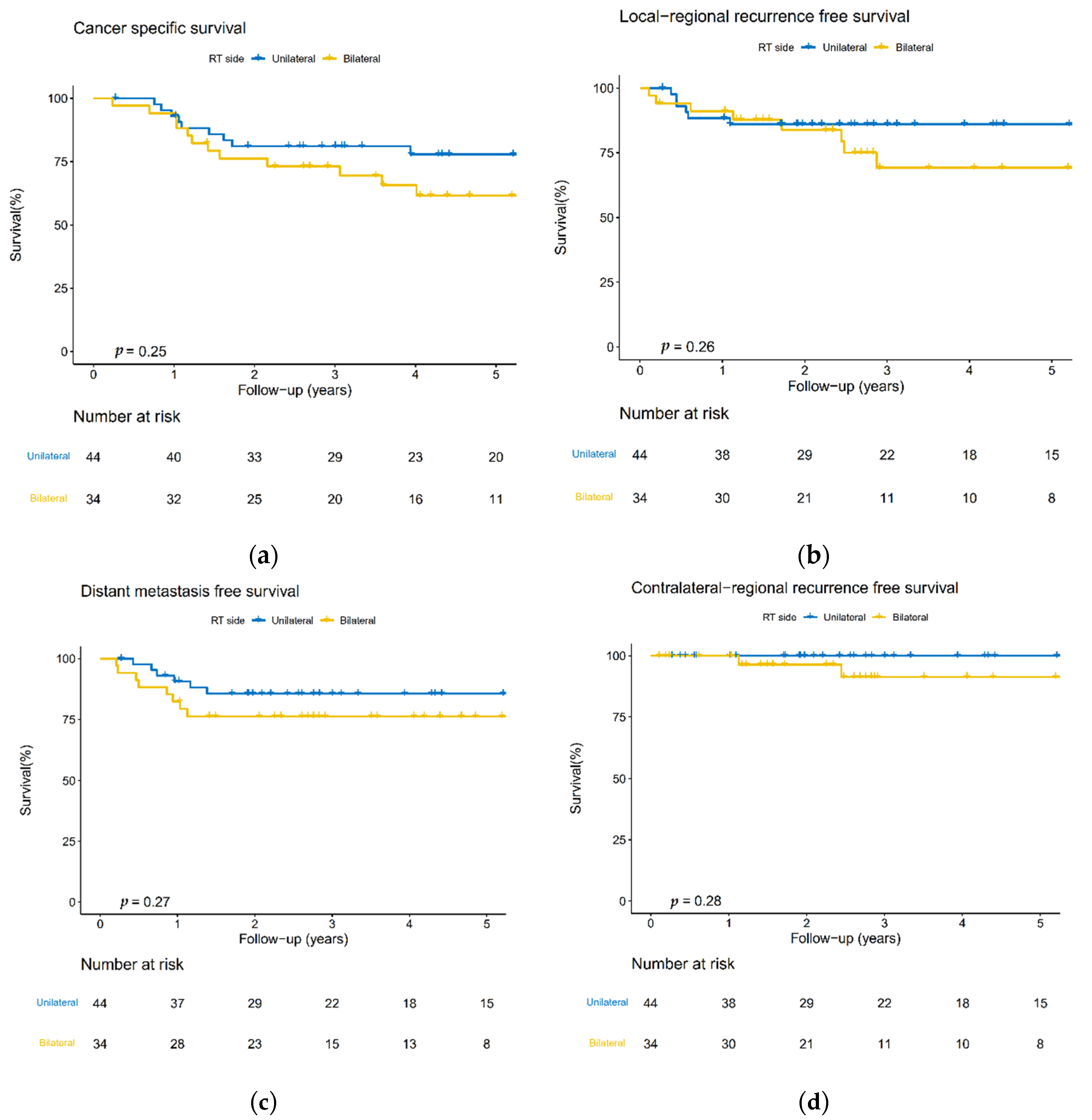
No statistically significant differences were observed for 5-year CSS, LRRFS, DMFS, and cRRFS for the UNI and BNI groups (Figure 2). The 5-year CSS was 77.9% vs. 61.6% (p = 0.25); the 5-year LRRFS was 86.0% vs. 69.2% (p = 0.26); the 5-year DMFS was 85.6% vs. 76.3% (p = 0.27); and the 5-year cRRFS was 100% vs. 91.4% (p = 0.28). Univariate analysis did not detect any significant clinicopathological predictors for cRF (Table 2).

3.4. Disease-Failure Pattern

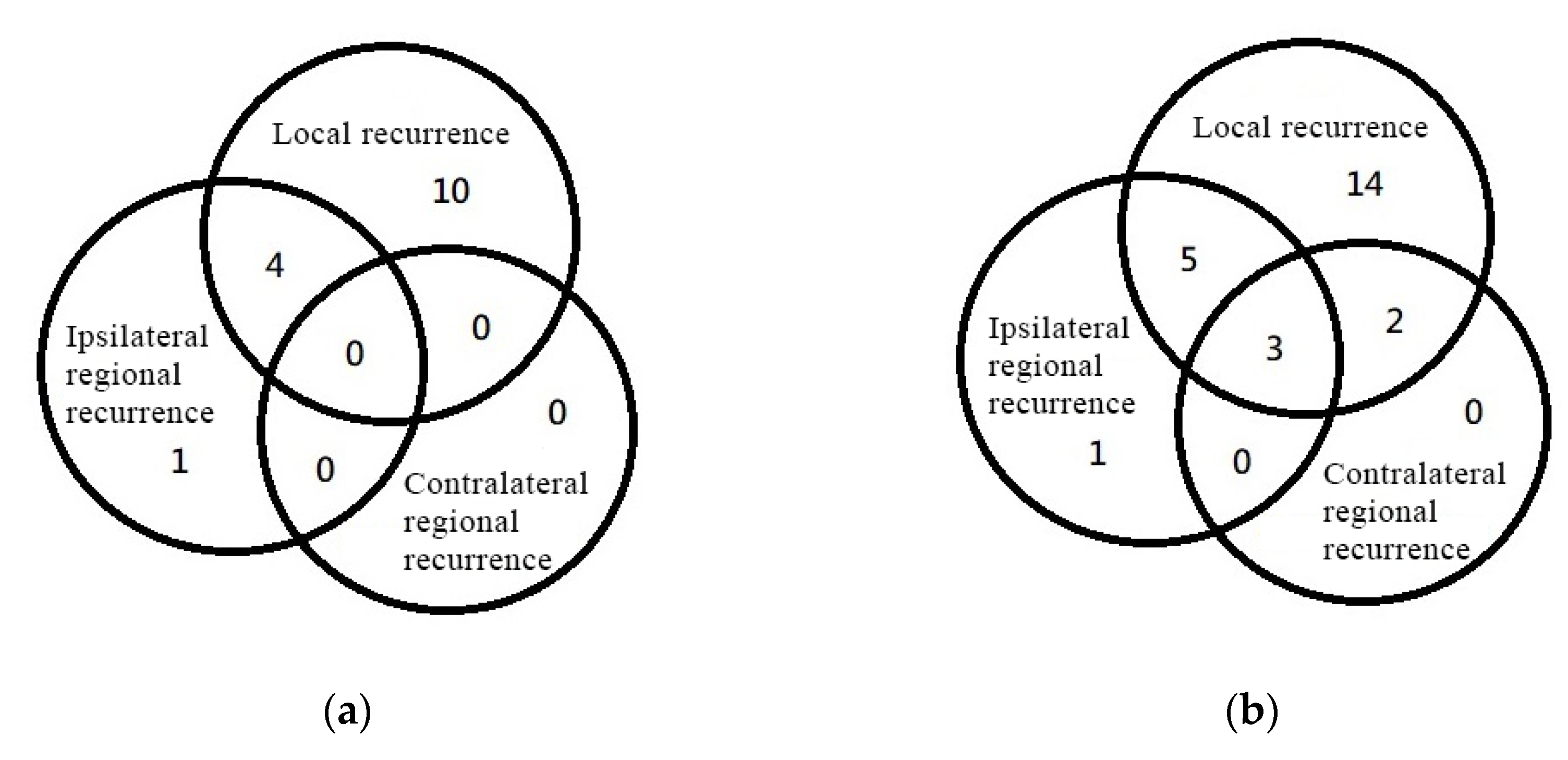
No contralateral neck recurrence was identified in the UNI and BNI groups (Figure 3a). Among the 15 patients (19.2%) who reported locoregional relapse, 2 were without any disease-free status, 9 had in-field recurrence (including the only one with isolated ipsilateral neck recurrence), 2 showed marginal recurrence (1 of them received delayed RT due to poor wound condition), and the remaining 2 patients showed distant metastasis before local recurrence.
No isolated contralateral neck recurrence was identified in the entire cohort. Five patients (3.4%) reported contralateral neck recurrence, all simultaneously with local recurrence (Figure 3b). Locoregional relapse was noted in 25 patients (16.8%), most with local failure only (56% of all relapsed patients). There was only one case of isolated ipsilateral neck recurrence (0.7%). In total, 12 patients (8.1%) had distant metastasis, 4 had combined local or regional relapse (2.7%), and 8 had distant metastasis only (5.4%).

4. Discussion

No consensus has yet been reached and no robust evidence is available on the benefits of contralateral neck irradiation for patients with contralateral nodal-negative OSCC. Furthermore, RT-associated acute and long-term toxicities are significantly impacted by the treatment volume in patients with head and neck cancer, especially those receiving trimodality therapy (radical surgery plus concurrent chemoradiotherapy). With improved radiation techniques, IMRT enables tailored treatment, maximizing target volume coverage while limiting the doses to normal tissues. As radiation oncologists aim to achieve a balance between the radiation toxicity and treatment outcome, the chance to omit the unnecessary treatment field without compromising the cure rate needs further investigation.
Recent retrospective data reported a low contralateral neck recurrence rate in patients with head and neck cancers. Table 3 lists a literature review of contralateral neck failure rate in primary head and neck cancers, particularly cancer at the oral cavity sites [5,11,12,13,14,15,20]. Unlike most of the historical analysis of mixed head and neck cancers and relatively early-stage OSCC, our study focused on a homogeneous patient series with well-lateralized and more advanced-stage OSCC to avoid confounding interactions between different origins of head and neck cancers. According to the results, this study demonstrates a comparably low cRF with that of patients with high-consistency advanced-stage OSCC.
In our cohort, the 5-yr cRF rate was as low as 3.6%. No cRF was identified in patients who received neck irradiation, either UNI or BNI. Hence, omitting the contralateral N0 neck might be a reasonable approach in such patients. This strategy was corroborated by previous studies. Vergeer et al. investigated 123 patients with oral cavity cancer (85%) and oropharyngeal cancer without contralateral neck irradiation and reported a cRF rate of 5.7% [5]; however, their group included 7% patients with close/cross midline disease, which could possibly increase the cRF. Wirtz et al. reported a cRF rate of 6.1%, mainly in the oropharynx (52.8%), with extended data for oral cavity (38%) and hypopharynx (10%) [12]; however, 73.1% of their patients received contralateral neck dissection, which could have been an overtreatment. Another phase II study demonstrated a low cRF rate of 2.8% for resected head and neck cancers. Similarly, 71% of patients had a cross-midline disease and up to 92% of patients received bilateral neck dissection before adjuvant RT [15]. Notably, our cohort, comprising 79.2% of patients with the T3/4 tumor, still revealed a reliably low contralateral neck recurrence rate, even though nearly 90% of them did not undergo contralateral neck dissection. Although contralateral neck dissection could be actively performed to detect occult contralateral nodal metastasis, there were certain short-term and long-term impacts on the quality of life of the patients [21].
Tumor-dependent factors that predicted contralateral neck recurrence are not well known and are still under investigation. The Sentinel European Node Trial included patients with lateralized, early T, and N0 tumors and demonstrated that the positive contralateral sentinel node was detected in only 1.9% of cases but in up to 6% of cases of midline tumor [22]. Al Mamgani et al. found midline involvement as the most significant correlation with cRF in a pooled analysis (12.12% with midline involved vs. 1.71% with free midline, p = 0.001) [23]. It is generally accepted that there is a high frequency of lymphatic vessels crossing the midline in certain tumor localizations (e.g., part of the oral cavity, tongue, and floor of the mouth) [24]. ENE is also regarded as a predictor of cRF. Two large retrospective studies identified ENE as a strongly independent risk factor for 5-yr cRF (HR: 12.978, 95% CI: 1.328–126.829, p = 0.028) and for cRF in patients showing local recurrence (HR: 4.957, 95% CI: 1.763–13.934, p = 0.002) [8,9]. As a result, one should be very cautious before deciding to omit contralateral neck irradiation in cases of midline crossing primary and ENE-positive status cancers. However, these patients were initially excluded from our study cohort because midline crossing primary and ENE-positive status cancers usually drive more extensive treatments (e.g., elective contralateral neck dissection and/or prophylactic contralateral neck field irradiation) by clinical physicians. A prompt discussion should be conducted at a multidisciplinary tumor board when treating such patients with N0 neck.
Though the univariate analysis conducted herein did not show any significant clinicopathological predictors for cRF, some cRF predictors have been previously reported (Table 3). Vergeer et al. showed that the number of nodes involved in the ipsilateral neck is a prognostic factor for cRF [5]. Hence, a higher N stage might impact the cRF rate. Liu et al. reported that a tumor depth of invasion (DOI) of >10 mm is a significant predictor in small (T1–2) lateralized OSCCs (HR: 6.7, 95% CI 1.4–32.3, p = 0.02) [11], which indicates that a higher T stage from patients diagnosed with early T1/2 in the AJCC 7th staging system can be revised to T3/4 in the DOI-incorporated edition of the AJCC 8th staging system. It might also impact the cRF rate. As our study mainly used the AJCC 6th and 7th staging systems, some T1-2N0 cases that were earlier excluded from our analysis might get upstaged if the AJCC 8th staging system is used. However, except for the influence of DOI, there were still no factors significantly associated with cRF in our study, possibly owing to the limited number of cRF cases.
Notably, contralateral neck recurrence was detected as the first site of failure in five patients in our cohort, all of whom also simultaneously exhibited local recurrence. No isolated contralateral neck recurrence was observed. The failure pattern between the local failure and the cRF of other studies was also reviewed in Table 3. Wirtz reported simultaneous local and contralateral regional failure in 42% of patients with cRF [12]. Contreras reported that half of cRF occurred simultaneously with local failure and the other half occurred following the local failure [15]. Though these studies did not conduct any statistical tests of the correlation between local failure and cRF, the results implied that contralateral neck should be more closely examined or followed up when local failure was found. It is generally considered that patients with a history of dissection or RT in the neck may have aberrant lymphatic drainage caused by the disruption of lymphatic channels. This concept was verified and tested by SLNB in a new study, given the result of unexpected drainage pattern variability in 30% of patients with cT1-2N0 OSCC [25]. Hence, SLNB has become an emerging technique that can benefit the staging of the contralateral negative neck and avoid the overtreatment of the contralateral neck in the future [26]. Achieving better local control might still be a priority in clinical situations for patients with a low risk of contralateral nodal recurrence.
The study has several limitations. First, the study could have a selection bias because of its retrospective nature; patients with more advanced stage or risks are likely to receive more intense treatments such as bilateral neck dissection or irradiation. Second, distance of the primary tumor to the midline was likely not measured with standardization. Third, the multivariable analysis should be considered exploratory because of the low number of cRF events, which limits its statistical power. Fourth, toxicity profiles were not assessed in this retrospective study. Although we could not compare the side effects between UNI to BNI in this study, we believed that eliminating coverage of one side of neck volume would significantly decrease the exposure dose to the neighboring normal organs and tissues, leading to fewer possible associated side effects. Furthermore, for a group of highly consistent subsites of OSCC, our results are only applicable to well-lateralized buccal, cheek, gum, and retromolar OSCC populations; therefore, extrapolating the results to ENE-positive, tumor cross-midline, or other head and neck cancers is not applicable. Nonetheless, this study shows a reliably low cRF rate in well-lateralized OSCC with locally advanced stage and provides support for the possibility of omitting treatment to the contralateral neck in this group. At present, we are designing a more rigorous study to prospectively validate these results.

5. Conclusions

The rate of cRF was acceptably low in patients with well-lateralized OSCC, even with advanced stage of initially uninvolved contralateral neck. Local and ipsilateral recurrence remained the main pattern of relapse, underlining the importance of RT to the primary and ipsilateral lymph nodes. Omitting contralateral neck irradiation with active surveillance could be safely considered without compromising the cure rate in patients with such locally advanced OSCC.

Author Contributions

Conceptualization, Y.-J.C. and Y.-H.W.; Data curation, Y.-J.C., T.-H.P. and C.-H.H.; Formal analysis, Y.-J.C., H.-Y.L. and M.-H.T.; Funding acquisition, Y.-J.C.; Investigation, Y.-J.C. and H.-Y.L.; Methodology, H.-Y.L. and M.-H.T.; Project administration, Y.-H.W.; Resources, Y.-H.W.; Software, H.-Y.L.; Supervision, H.-Y.L. and Y.-H.W.; Validation, Y.-H.W.; Visualization, H.-Y.L.; Writing—original draft, Y.-J.C.; Writing—review & editing, Y.-J.C., H.-Y.L., M.-H.T. and Y.-H.W.. All authors have read and agreed to the published version of the manuscript.

Funding

The research was supported by grants NCKUH-11103011 from the National Cheng Kung University Hospital and in part by the Higher Education Sprout Project of the Ministry of Education to the Headquarters of University Advancement at National Cheng Kung University.

Institutional Review Board Statement

This study was reviewed and approved by the Institutional Review Board (IRB) of our institute (National Cheng Kung University Hospital Institutional Review Board [IRB number: A-ER-111-181]) and conducted according to the ethical guidelines of the Declaration of Helsinki.

Informed Consent Statement

The requirement for informed consent from the study subjects was waived due to the retrospective study design.

Data Availability Statement

The datasets used and/or analysed during the current study are available on reasonable request from the corresponding author.

Acknowledgments

We acknowledge the kind assistance of Lin for providing the statistical consulting to this work.

Conflicts of Interest

The authors declare no conflict of interest.

References

  1. Shingaki, S.; Takada, M.; Sasai, K.; Bibi, R.; Kobayashi, T.; Nomura, T.; Saito, C. Impact of lymph node metastasis on the pattern of failure and survival in oral carcinomas. Am. J. Surg. 2003, 185, 278–284. [Google Scholar] [CrossRef]
  2. Weiss, M.H.; Harrison, L.B.; Isaacs, R.S. Use of decision analysis in planning a management strategy for the stage N0 neck. Arch. Otolaryngol. Head Neck Surg. 1994, 120, 699–702. [Google Scholar] [CrossRef]
  3. Kurita, H.; Koike, T.; Narikawa, J.N.; Sakai, H.; Nakatsuka, A.; Uehara, S.; Kobayashi, H.; Kurashina, K. Clinical predictors for contralateral neck lymph node metastasis from unilateral squamous cell carcinoma in the oral cavity. Oral Oncol. 2004, 40, 898–903. [Google Scholar] [CrossRef]
  4. Koo, B.S.; Lim, Y.C.; Lee, J.S.; Choi, E.C. Management of contralateral N0 neck in oral cavity squamous cell carcinoma. Head Neck 2006, 28, 896–901. [Google Scholar] [CrossRef]
  5. Vergeer, M.R.; Doornaert, P.A.H.; Jonkman, A.; Kaanders, J.H.A.M.; van den Ende, P.L.A.; de Jong, M.A.; Leemans, C.R.; Slotman, B.J.; Langendijk, J.A. Ipsilateral Irradiation for Oral and Oropharyngeal Carcinoma Treated with Primary Surgery and Postoperative Radiotherapy. Int. J. Radiat. Oncol. Biol. Phys. 2010, 78, 682–688. [Google Scholar] [CrossRef] [PubMed]
  6. Chung, E.-J.; Oh, J.-I.; Choi, K.-Y.; Lee, D.-J.; Park, I.-S.; Kim, J.-H.; Rho, Y.-S. Pattern of cervical lymph node metastasis in tonsil cancer: Predictive factor analysis of contralateral and retropharyngeal lymph node metastasis. Oral Oncol. 2011, 47, 758–762. [Google Scholar] [CrossRef]
  7. Capote-Moreno, A.; Naval, L.; Muñoz-Guerra, M.F.; Sastre, J.; Rodríguez-Campo, F.J. Prognostic factors influencing contralateral neck lymph node metastases in oral and oropharyngeal carcinoma. J. Oral Maxillofac. Surg. Off. J. Am. Assoc. Oral Maxillofac. Surg. 2010, 68, 268–275. [Google Scholar] [CrossRef] [PubMed]
  8. Feng, Z.; Niu, L.X.; Yuan, Y.; Peng, X.; Guo, C.B. Risk factors and treatment of contralateral neck recurrence for unilateral oral squamous cell carcinoma: A retrospective study of 1482 cases. Oral Oncol. 2014, 50, 1081–1088. [Google Scholar] [CrossRef]
  9. Liao, C.T.; Huang, S.F.; Chen, I.H.; Chang, J.T.; Wang, H.M.; Ng, S.H.; Hsueh, C.; Lee, L.Y.; Lin, C.H.; Cheng, A.J.; et al. Risk stratification of patients with oral cavity squamous cell carcinoma and contralateral neck recurrence following radical surgery. Ann. Surg. Oncol. 2009, 16, 159–170. [Google Scholar] [CrossRef] [PubMed]
  10. Rackley, T.P.; Namelo, W.C.; Palaniappan, N.; Cole, N.; Owens, D.M.J.; Evans, M. Unilateral radiotherapy for surgically resected lateralized squamous cell carcinoma of the tonsil. Head Neck 2017, 39, 17–23. [Google Scholar] [CrossRef]
  11. Liu, H.Y.; Tam, L.; Woody, N.M.; Caudell, J.; Reddy, C.A.; Ghanem, A.; Schymick, M.; Joshi, N.; Geiger, J.; Lamarre, E.; et al. Failure rate in the untreated contralateral node negative neck of small lateralized oral cavity cancers: A multi-institutional collaborative study. Oral Oncol. 2021, 115, 105190. [Google Scholar] [CrossRef] [PubMed]
  12. Wirtz, M.M.; Temming, S.; Kocher, M.; Kunze, S.; Semrau, R. Low risk of contralateral lymph node recurrence in lateralized head and neck carcinoma after postoperative ipsilateral radiotherapy. Strahlenther. Und Onkol. Organ Der Dtsch. Rontgenges. 2020, 196, 474–484. [Google Scholar] [CrossRef]
  13. O’Steen, L.; Amdur, R.J.; Morris, C.G.; Hitchcock, K.E.; Mendenhall, W.M. Challenging the Requirement to Treat the Contralateral Neck in Cases With >4 mm Tumor Thickness in Patients Receiving Postoperative Radiation Therapy for Squamous Cell Carcinoma of the Oral Tongue or Floor of Mouth. Am. J. Clin. Oncol. 2019, 42, 89–91. [Google Scholar] [CrossRef] [PubMed]
  14. Nobis, C.-P.; Otto, S.; Grigorieva, T.; Alnaqbi, M.; Troeltzsch, M.; Schöpe, J.; Wagenpfeil, S.; Ehrenfeld, M.; Wolff, K.-D.; Kesting, M.R. Elective neck dissection in unilateral carcinomas of the tongue: Unilateral versus bilateral approach. J. Cranio Maxillofac. Surg. 2017, 45, 579–584. [Google Scholar] [CrossRef] [PubMed]
  15. Contreras, J.A.; Spencer, C.; DeWees, T.; Haughey, B.; Henke, L.E.; Chin, R.I.; Paniello, R.; Rich, J.; Jackson, R.; Oppelt, P.; et al. Eliminating Postoperative Radiation to the Pathologically Node-Negative Neck: Long-Term Results of a Prospective Phase II Study. J. Clin. Oncol. Off. J. Am. Soc. Clin. Oncol. 2019, 37, 2548–2555. [Google Scholar] [CrossRef] [PubMed]
  16. Lindberg, R. Distribution of cervical lymph node metastases from squamous cell carcinoma of the upper respiratory and digestive tracts. Cancer 1972, 29, 1446–1449. [Google Scholar] [CrossRef]
  17. Fletcher, G.H. Elective irradiation of subclinical disease in cancers of the head and neck. Cancer 1972, 29, 1450–1454. [Google Scholar] [CrossRef]
  18. Greene, F.L.; Page, D.L.; Fleming, I.D.; Fritz, A.; Balch, C.M.; Haller, D.G.; Fleming, I.D.; Morrow, M.; Fritz, A.G. (Eds.) AJCC Cancer Staging Manual, 6th ed.; Springer: New York, NY, USA, 2002. [Google Scholar]
  19. Edge, S.B.; Compton, C.C. The American Joint Committee on Cancer: The 7th edition of the AJCC cancer staging manual and the future of TNM. Ann. Surg. Oncol. 2010, 17, 1471–1474. [Google Scholar] [CrossRef]
  20. Waldram, R.; Taylor, A.E.; Whittam, S.; Iyizoba-Ebozue, Z.; Murray, L.; Frood, R.; Cardale, K.; Dyker, K.E.; Murray, P.; Ramasamy, S.; et al. Evaluation of Locoregional Recurrence Patterns Following Adjuvant (Chemo)Radiotherapy for Oral Cavity Carcinoma. Clin. Oncol. R. Coll. Radiol. 2020, 32, 228–237. [Google Scholar] [CrossRef]
  21. Shah, S.; Har-El, G.; Rosenfeld, R.M. Short-term and long-term quality of life after neck dissection. Head Neck 2001, 23, 954–961. [Google Scholar] [CrossRef]
  22. Schilling, C.; Stoeckli, S.J.; Haerle, S.K.; Broglie, M.A.; Huber, G.F.; Sorensen, J.A.; Bakholdt, V.; Krogdahl, A.; von Buchwald, C.; Bilde, A.; et al. Sentinel European Node Trial (SENT): 3-year results of sentinel node biopsy in oral cancer. Eur. J. Cancer 2015, 51, 2777–2784. [Google Scholar] [CrossRef] [PubMed]
  23. Al-Mamgani, A.; Verheij, M.; van den Brekel, M.W.M. Elective unilateral nodal irradiation in head and neck squamous cell carcinoma: A paradigm shift. Eur. J. Cancer 2017, 82, 1–5. [Google Scholar] [CrossRef] [PubMed]
  24. Werner, J.A.; Dünne, A.A.; Myers, J.N. Functional anatomy of the lymphatic drainage system of the upper aerodigestive tract and its role in metastasis of squamous cell carcinoma. Head Neck 2003, 25, 322–332. [Google Scholar] [CrossRef] [PubMed]
  25. Den Toom, I.J.; Boeve, K.; van Weert, S.; Bloemena, E.; Brouwers, A.H.; Hoekstra, O.S.; de Keizer, B.; van der Vegt, B.; Willems, S.M.; Leemans, C.R.; et al. High rate of unexpected lymphatic drainage patterns and a high accuracy of the sentinel lymph node biopsy in oral cancer after previous neck treatment. Oral Oncol. 2019, 94, 68–72. [Google Scholar] [CrossRef]
  26. Mahieu, R.; den Toom, I.J.; Boeve, K.; Lobeek, D.; Bloemena, E.; Donswijk, M.L.; de Keizer, B.; Klop, W.M.C.; Leemans, C.R.; Willems, S.M.; et al. Contralateral Regional Recurrence in Lateralized or Paramedian Early-Stage Oral Cancer Undergoing Sentinel Lymph Node Biopsy—Comparison to a Historic Elective Neck Dissection Cohort. Front. Oncol. 2021, 11, 644306. [Google Scholar] [CrossRef]
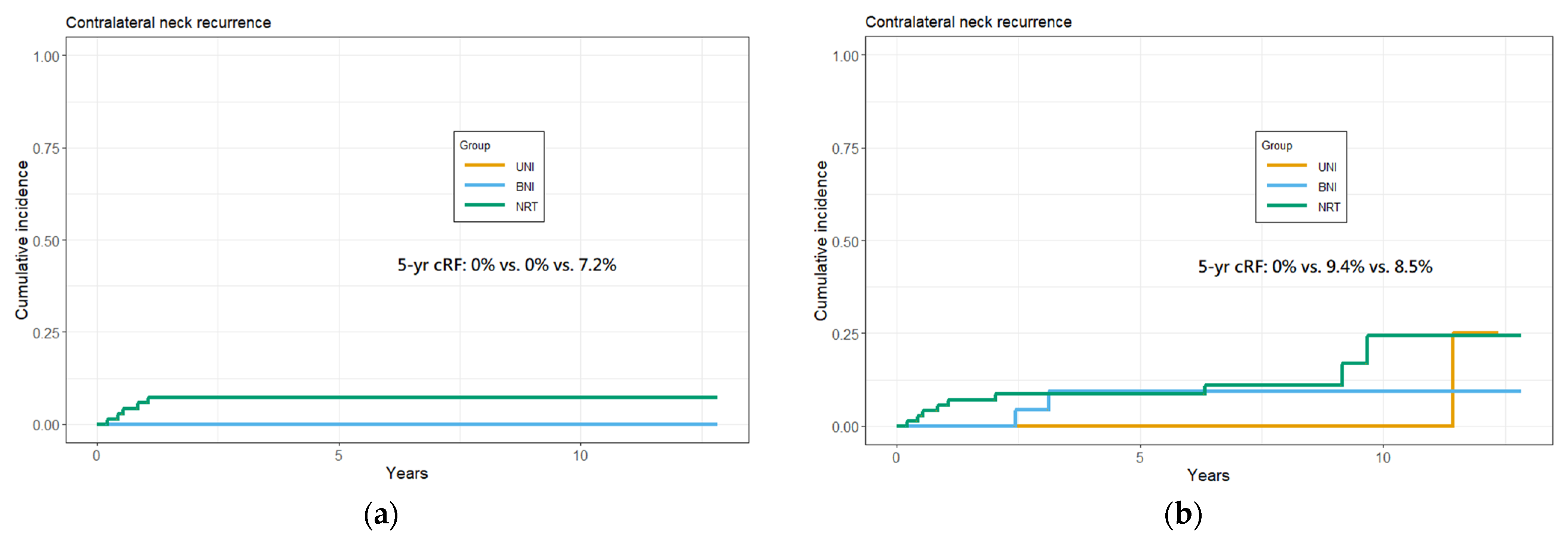
Figure 1. Cumulative 5-year cRF rate among patients in different groups. (a) Calculated with cRF as the first failure site. (b) Calculated with all-time cRF, which does not have to be the first failure site.
Curroncol 29 00547 g001
Figure 2. Kaplan–Meier plots of the 5-year (a) cancer-specific survival (CSS); (b) local-regional recurrence-free survival (LRRFS); (c) distant metastasis-free survival (DMFS), and (d) contralateral-regional recurrence-free survival (cRRFS) rates between the unilateral and bilateral neck irradiation groups.
Curroncol 29 00547 g002
Figure 3. Locoregional failure patterns among (a) Fifteen patients with recurrent tumors in patients with neck irradiation; (b) Twenty-five patients with recurrent tumors in the entire cohort.
Curroncol 29 00547 g003
Table 1. Characteristics of patients.
CovariateTotalUNI GroupBNI Groupp-ValueNo Adjuvant RT Groupp-Value
No. of cases1494434 71
Sex (%)
Female4 (2.7)1 (2.3)1 (2.9)1.0002 (2.8)0.979
Male145 (97.3)43 (97.7)33 (97.1) 69 (97.2)
Diagnosed age (median [IQR])52.0 [47.0, 59.0]56.0 [47.8, 62.0]50.0 [45.3, 55.8]0.01852.0 [47.0, 61.0]0.075
Cancer site (%)
Buccal + cheek95 (63.8)31 (70.5)25 (73.5)0.74439 (54.9)0.243
Retromolar8 (5.4)3 (6.8)1 (2.9) 4 (5.6)
Gum46 (30.9)10 (22.7)8 (23.5) 28 (39.4)
AJCC pT (%)
110 (6.7)3 (6.8)3 (8.8)0.6454 (5.6)0.336
221 (14.1)6 (13.6)7 (20.6) 8 (11.3)
332 (21.5)10 (22.7)4 (11.8) 18 (25.4)
4A78 (52.3)22 (50.0)16 (47.1) 40 (56.3)
4B8 (5.4)3 (6.8)4 (11.8) 1 (1.4)
AJCC pN (%)
097 (65.1)27 (61.4)15 (44.1)0.17155 (77.5)0.001
127 (18.1)5 (11.4)9 (26.5) 13 (18.3)
2B25 (16.8)12 (27.3)10 (29.4) 3 (4.2)
AJCC pStage (%)
346 (30.9)9 (20.5)10 (29.4)0.42527 (38.0)0.079
4A95 (63.8)32 (72.7)20 (58.8) 43 (60.6)
4B8 (5.4)3 (6.8)4 (11.8) 1 (1.4)
ND side (%)
Bilateral18 (12.1)5 (11.4)7 (20.6)0.5346 (8.5)0.247
Left69 (46.3)23 (52.3)16 (47.1) 30 (42.3)
Right62 (41.6)16 (36.4)11 (32.4) 35 (49.3)
Number of node examed
(median [IQR])
27.0 [16.0, 35.0]29.0 [23.8, 36.3]32.5 [22.0, 44.8]0.45220.0 [13.5, 33.0]0.001
Histology grade (%)
174 (49.7)18 (40.9)21 (61.8)0.09035 (49.3)0.323
268 (45.6)23 (52.3)13 (38.2) 32 (45.1)
37 (4.7)3 (6.8)0 (0.0) 4 (5.6)
Tumor size (median [IQR])40.0 [27.0, 50.0]40.0 [25.8, 50.0]37.0 [30.0, 45.0]0.84040.0 [28.5, 50.0]0.800
PNI (%)
Negative104 (69.8)27 (61.4)22 (64.7)0.94755 (77.5)0.144
Positive45 (30.2)17 (38.6)12 (35.3) 16 (22.5)
LVI (%)
Negative121 (81.2)37 (84.1)23 (67.6)0.15061 (85.9)0.068
Positive28 (18.8)7 (15.9)11 (32.4) 10 (14.1)
Surgical margin (%)
Free122 (81.9)30 (68.2)27 (79.4)0.27165 (91.5)0.016
Close13 (8.7)8 (18.2)2 (5.9) 3 (4.2)
Positive14 (9.4)6 (13.6)5 (14.7) 3 (4.2)
Adjuvant treatment (%)
Negative71 (47.7)NANA 71 (100.0)
RT alone42 (28.2)24 (54.5)18 (52.9) NA
CCRT36 (24.2)20 (45.5)16 (47.1) NA
Concurrent CT regimen (%)
Cisplatin11 (30.6)6 (30.0)5 (31.3)0.640NA
PF19 (52.8)11 (55.0)8 (50.0) NA
PF+ Erbitux1 (2.8)0 (0.0)1 (6.3) NA
Unknown5 (13.9)3 (15.0)2 (12.5) NA
Smoking (%)
Negative24 (17.1)7 (15.9)3 (9.7)0.66214 (21.5)0.342
Positive116 (82.9)37 (84.1)28 (90.3) 51 (78.5)
Betel nut chewing (%)
Negative31 (22.1)8 (18.2)4 (12.9)0.77019 (29.2)0.147
Positive109 (77.9)36 (81.8)27 (87.1) 46 (70.8)
Alcohol drinking (%)
Negative52 (36.9)18 (40.9)8 (25.8)0.26826 (39.4)0.347
Positive59 (63.1)26 (59.1)23 (74.2) 40 (60.6)
Abbreviations: IQR, interquartile range; UNI, unilateral neck irradiation; BNI, bilateral neck irradiation; AJCC, American joint committee on Cancer; ND, neck dissection; PNI, perineural invasion; LVI, lymphovascular invasion; RT, radiotherapy; CCRT, concurrent chemoradiation therapy; CT, chemotherapy; PF, cisplatin and fluorouracil; NA, not applicable; p, p-value.
Table 2. Univariate analysis of factors associated with cRF.
Univariate AnalysisHazard Ratio (95% CI)p-Value
pT Stage (T3–4 vs. T1–2)0.367 (0.06–2.20)0.272
pN number (positive vs. negative)3.117 (0.52–18.67)0.213
PNI (presence vs. absence)1.753 (0.29–10.51)0.539
LVI (presence vs. absence)1.190 (0.13–10.65)0.876
Margin (close vs. negative)3.369 (0.35–32.40)0.293
Margin (positive vs. negative)3.212 (0.33–30.96)0.313
Abbreviations: cRF, contralateral neck failure; PNI, perineural invasion; LVI, lymphovascular invasion; CI, confidence interval.
Table 3. Literature review of failure rates in the node-negative contralateral neck of oral cavity cancers.
StudynPrimary SiteLateralized
Primary
NDpT StatuspN Status Adjuvant TxRT Neck SideRate of cRFPredictors of cRF
Present study149BUC (63.8%)
GUM (30.9%)
RMT (5.4%)
YUni (88%)
Bil (12%)
T1 (6.7%)
T2 (14.1%)
T3 (21.5%)
T4 (57.7%)
N+ (34.9%)
N0 (65.1%)
NRT (47.7%)
RT (28.2%)
CRT (24.2%)
56.4% UNI
43.6% BNI
5 year rate 3.6%
Crude rate 3.4%
None
(100% SLRF)
Liu (2021) [11]176OT (82%)
FOM (18%)
YUni (100%)T1 (68%)
T2 (32%)
N+ (12%)
N0 (81%)
UNK (7%)
NRT (83%)
RT (17%)
100% PTB ± UNI
0% BNI
2 year rate 3.6%
5 year rate 4.3%
Crude rate 5%
DOI > 10 mm
(22% SLRF; 22% LFTR; 56% iCLF)
Waldram (2020) [20]101OT (52%)
FOM (17%)
BUC (7%)
GUM (15%)
RMT (10%)
NRUni (69%)
Bil (27%)
T1 (11%)
T2 (40%)
T3 (9%)
T4 (40%)
N+ (63%)
N0 (37%)
RT (75%)
CRT (25%)
43% UNI
53% BNI
Crude rate 5%NR
Contreras (2019) [15]72Oral cavity (20%)
Oropharynx (51%)
Hypopharynx (6%)
Larynx (22%)
No, 71% involved/cross midline Uni (8%)
Bil (92%)
T1-T2 (49%)
T3-T4 (51%)
N2-N3 (58%)
N0-N1 (42%)
RT (53%)
CRT (47%)
24% PTB
76% UNI
0% BNI
5 year contralateral neck control 94.5%
Crude rate 2.8%
NR
(50% SLRF; 50% LFTR)
Wirtz (2019) [12]197Oral cavity (38%)
Oropharynx (53%)
Hypopharynx (10%)
YUni (27%)
Bil (73%)
T0 (1%)
T1 (36%)
T2 (41%)
T3 (10%)
T4 (11%)
N+ (80.7%)
N0 (19.3%)
RT (70%)
CRT (30%)
100% UNI
0% BNI
5 year contralateral neck control 94.6%
Crude rate 6.1%
None
(42% SLRF)
O’steen (2019) [13]32OT (72%)
FOM (28%)
YUni (78%)
Bil (22%)
T1 (47%)
T2 (41%)
T3 (9%)
T4 (3%)
N+ (19%)
N0 (81%)
RT (62%)
CRT (38%)
41% PTB
59% UNI
0% BNI
Crude rate 0%NR
Nobis (2017) [14]150OT
(100%)
YUni (70%)
Bil (30%)
T1 (71%)
T2 (29%)
N+ (23%)
N0 (77%)
NRT (75%)
RT (19%)
CRT (6%)
NRCrude rate 2.7%NR
Vergeer (2010) [5]123Oral cavity (85%)
-OT (25%)
-FOM (8%)
-BUC (19%)
-GUM (48%)
Oropharynx (15%)
No, 7% close/
cross midline
Uni (83%)
Bil (0%)
T1 (22%)
T2 (35%)
T3 (11%)
T4 (33%)
N+ (41%)
N0 (59%)
RT (100%)10% PTB
90% UNI
0% BNI
5 year contralateral
neck control 92%
Crude rate 5.7%
Number of positive nodes
(14% SLRF)
Abbreviations: BUC, buccal; RMT, retromolar trigon; OT, oral tongue; FOM, floor of mouth; NR, not reported; ND, neck dissection; Uni, unilateral; Bil, bilateral; UNK, unknown; Tx, treatment; RT, radiotherapy; CRT, concurrent chemoradiation therapy; NRT, no radiotherapy; UNI, unilateral neck irradiation; BNI, bilateral neck irradiation; PTB, primary tumor bed; cRF, contralateral neck failure; SLRF, simultaneous local recurrence failure; LFTR, local failure then neck recurrence; iCLF, isolated contralateral failure; DOI, depth of invasion.
Publisher’s Note: MDPI stays neutral with regard to jurisdictional claims in published maps and institutional affiliations.