Novel Mutations in a Lethal Case of Lymphomatous Adult T Cell Lymphoma with Cryptic Myocardial Involvement

 and
and 
Abstract
1. Introduction
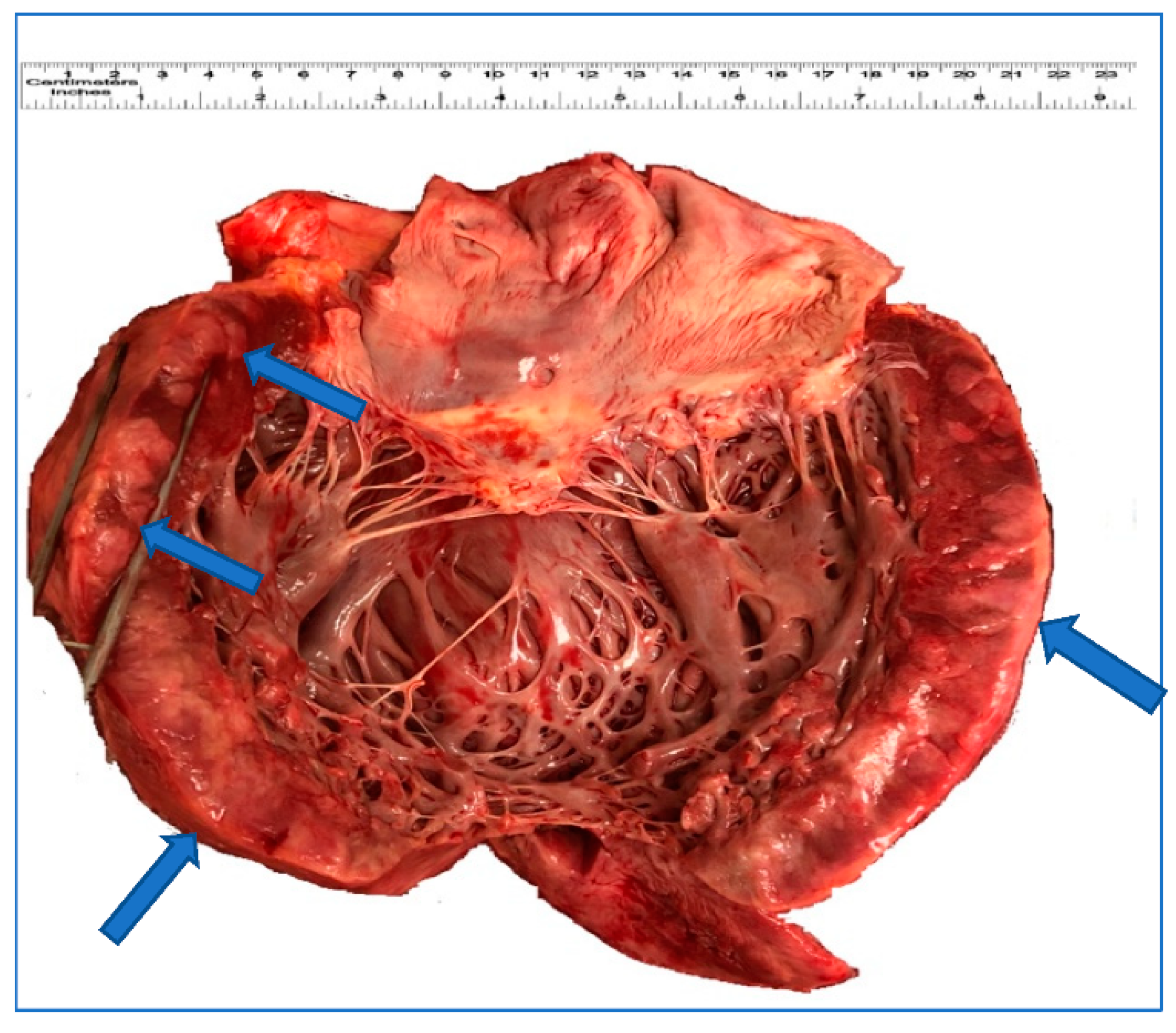
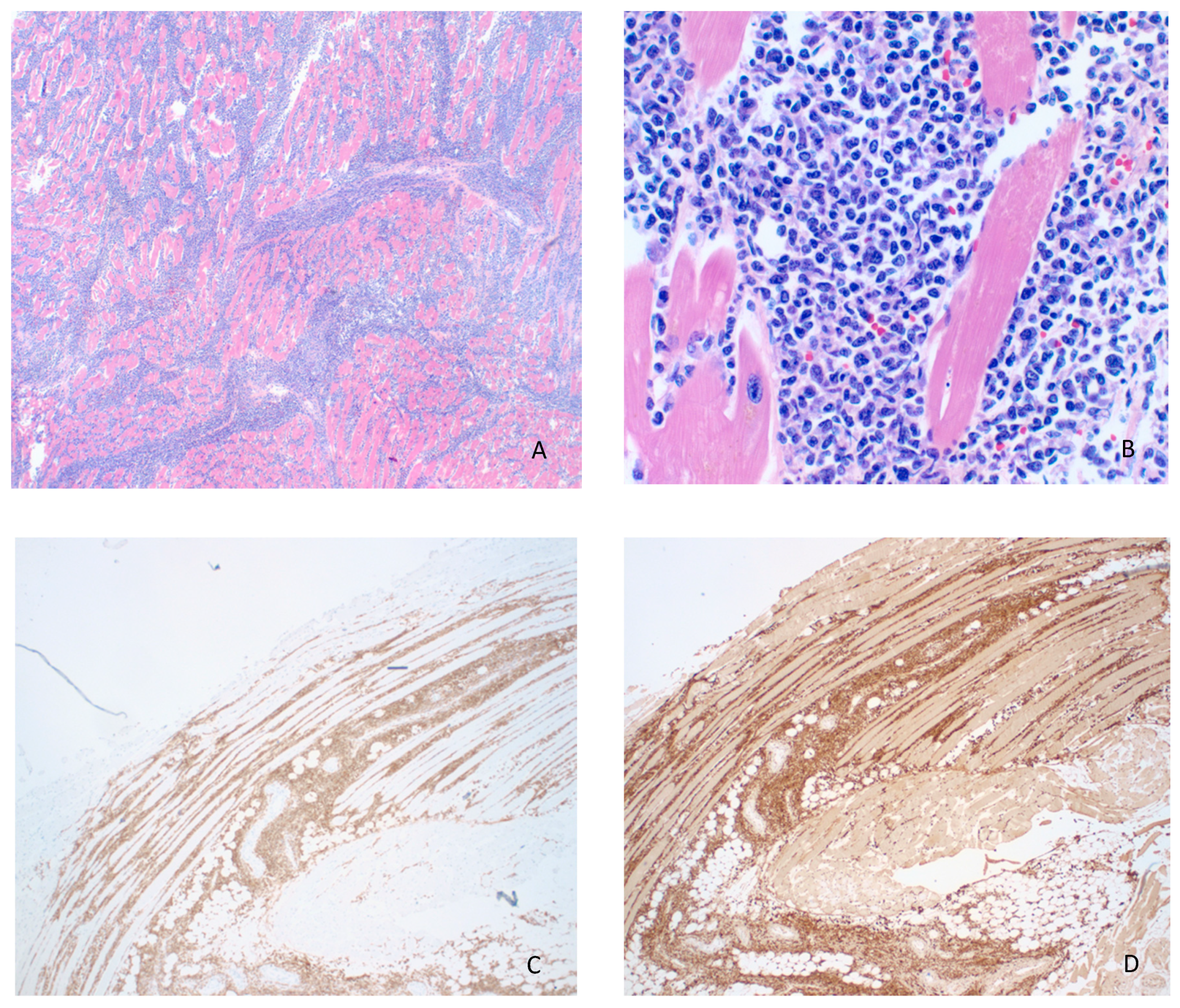
2. Result
3. Discussion
Author Contributions
Funding
Conflicts of Interest
References
- Ohshima, K.; Jaffe, E.; Yoshino, T.; Siebert, R. Adult T-cell leukemia/lymphoma. In World Health Organization Classification of Tumours, Revised 4th ed.; Swerdlow, S.H., Campo, C.E., Harris, N.L., Eds.; IARC press: Lyon, France, 2017; pp. 363–367. [Google Scholar]
- Ishitsuka, K.; Tamura, K. Human T-cell leukaemia virus type I and adult T-cell leukaemia-lymphoma. Lancet Oncol. 2014, 15, e517–e526. [Google Scholar] [CrossRef]
- Shimoyama, M. Diagnostic criteria and classification of clinical subtypes of adult T-cell leukaemia-lymphoma. Br. J. Haematol. 1991, 79, 428–437. [Google Scholar] [CrossRef] [PubMed]
- Furihata, M.; Ido, E.; Iwata, J.; Sonobe, H.; Ohtsuki, Y.; Takata, J.; Chikamori, T.; Doi, Y. Adult T cell leukemia/lymphoma with massive involvement of cardiac muscle and valves. Pathol. Int. 1998, 48, 221–224. [Google Scholar] [CrossRef] [PubMed]
- Iemura, A.; Yano, H.; Kojiro, M.; Nouno, R.; Kouno, K. Massive cardiac involvement of adult T-cell leukemia/lymphoma. An autopsy case. Arch. Pathol. Lab. Med. 1991, 115, 1052–1054. [Google Scholar] [PubMed]
- O’Mahony, D.; Debnath, I.; Janik, J.; Aisner, D.; Jaffe, E.; Waldmann, T.; Morris, J.C. Cardiac involvement with human T-cell lymphotrophic virus type-1-associated adult T-cell leukemia/lymphoma: The NIH experience. Leuk. Lymphoma 2008, 49, 439–446. [Google Scholar] [CrossRef] [PubMed]
- Tajima, K.; Hinuma, Y. Epidemiology of HTLV-I/II in Japan and in the world. In Advances in Adult T-Cell Leukemia and HTLV-1 Research (Gann Monograph on Cancer Research); Takatsuki, K., Hinuma, Y., Yoshida, M., Eds.; Japan Scientific Societies Press: Tokyo, Japan, 1992; pp. 129–149. [Google Scholar]
- Matsuoka, M.; Jeang, K.-T. Human T-cell leukaemia virus type 1 (HTLV-1) infectivity and cellular transformation. Nat. Rev. Cancer 2007, 7, 270–280. [Google Scholar] [CrossRef] [PubMed]
- Franchini, G. Molecular mechanisms of human T-cell leukemia/lymphotropic virus type I infection. Blood 1995, 86, 3619–3639. [Google Scholar] [CrossRef] [PubMed]
- Satou, Y.; Yasunaga, J.-I.; Yoshida, M.; Matsuoka, M. HTLV-I basic leucine zipper factor gene mRNA supports proliferation of adult T cell leukemia cells. Proc. Natl. Acad. Sci. USA 2006, 103, 720–725. [Google Scholar] [CrossRef] [PubMed]
- Kataoka, K.; Nagata, Y.; Kitanaka, A.; Shiraishi, Y.; Shimamura, T.; Yasunaga, J.-I.; Totoki, Y.; Chiba, K.; Sato-Otsubo, A.; Nagae, G.; et al. Integrated molecular analysis of adult T cell leukemia/lymphoma. Nat. Genet. 2015, 47, 1304–1315. [Google Scholar] [CrossRef] [PubMed]
- Shah, U.A.; Chung, E.Y.; Giricz, O.; Pradhan, K.; Kataoka, K.; Gordon-Mitchell, S.; Bhagat, T.D.; Mai, Y.; Wei, Y.; Ishida, E.; et al. North American ATLL has a distinct mutational and transcriptional profile and responds to epigenetic therapies. Blood 2018, 132, 1507–1518. [Google Scholar] [CrossRef] [PubMed]
- Paul, S.; Schaefer, B.C. A new look at T cell receptor signaling to nuclear factor-κB. Trends Immunol. 2013, 34, 269–281. [Google Scholar] [CrossRef] [PubMed]
- DeRosa, P.; Nava, V. Comment on Aasebø, E.; et al. The Progression of Acute Myeloid Leukemia from First Diagnosis to Chemoresistant Relapse: A Comparison of Proteomic and Phosphoproteomic Profiles. Cancers 2020, 12, 1466. Cancers 2020, 12, 2461. [Google Scholar] [CrossRef] [PubMed]
- Liu, C.-X.; Li, Y.; Obermoeller-McCormick, L.M.; Schwartz, A.L.; Bu, G. The Putative Tumor Suppressor LRP1B, a Novel Member of the Low Density Lipoprotein (LDL) Receptor Family, Exhibits Both Overlapping and Distinct Properties with the LDL Receptor-related Protein. J. Biol. Chem. 2001, 276, 28889–28896. [Google Scholar] [CrossRef] [PubMed]
- Yeh, C.-H.; Bai, X.T.; Moles, R.; Ratner, L.; Waldmann, T.A.; Watanabe, T.; Nicot, C. Mutation of epigenetic regulators TET2 and MLL3 in patients with HTLV-I-induced acute adult T-cell leukemia. Mol. Cancer 2016, 15, 1–7. [Google Scholar] [CrossRef]
- Renella, R.; Carnevale, J.; Schneider, K.A.; Hornick, J.L.; Rana, H.Q.; Janeway, K.A. Exploring the association of succinate dehydrogenase complex mutations with lymphoid malignancies. Fam. Cancer 2014, 13, 507–511. [Google Scholar] [CrossRef] [PubMed]
- Schatz, J.H.; Horwitz, S.M.; Teruya-Feldstein, J.; Lunning, M.A.; Viale, A.; Huberman, K.; Socci, N.D.; Lailler, N.; Heguy, A.; Dolgalev, I.; et al. Targeted mutational profiling of peripheral T-cell lymphoma not otherwise specified highlights new mechanisms in a heterogeneous pathogenesis. Leukemia 2014, 29, 237–241. [Google Scholar] [CrossRef] [PubMed]
- Feigenbaum, L.; Fujita, K.; Collins, F.S.; Jay, G. Repression of the NF1 gene by Tax may expain the development of neurofibromas in human T-lymphotropic virus type 1 transgenic mice. J. Virol. 1996, 70, 3280–3285. [Google Scholar] [CrossRef] [PubMed]
- Salahshor, S.; Woodgett, J.R. The links between axin and carcinogenesis. J. Clin. Pathol. 2005, 58, 225–236. [Google Scholar] [CrossRef] [PubMed]
- Bellon, M.; Ko, N.L.; Lee, M.-J.; Yao, Y.; Waldmann, T.A.; Trepel, J.B.; Nicot, C. Adult T-cell leukemia cells overexpress Wnt5a and promote osteoclast differentiation. Blood 2013, 121, 5045–5054. [Google Scholar] [CrossRef] [PubMed]


| Gene | Somatic Mutation | Allele Frequency (%) |
|---|---|---|
| AXIN1 | R712Q | 44.94 |
| BARD1 | R749K | 24.75 |
| CBL | H42_L43insH | 17.61 |
| CTNNB1 | I315V | 76.45 |
| CUX1 | R44W | 36.15 |
| CUX1 | P102T | 26.84 |
| DNMT3A | S199R | 39.04 |
| FAS | D228fs*2 | 45.37 |
| FGFR2 | S431L | 26.61 |
| LRP1B | D1063N | 68.28 |
| LRP1B | Y2560C | 46.64 |
| NF1 | S665F | 31.97 |
| SDHD | F34C | 29.79 |
| SMO | G24A | 48.98 |
| STAG2 | I771M | 40.98 |
| TP53 | H193L | 70.02 |
Publisher’s Note: MDPI stays neutral with regard to jurisdictional claims in published maps and institutional affiliations. |
© 2021 by the authors. Licensee MDPI, Basel, Switzerland. This article is an open access article distributed under the terms and conditions of the Creative Commons Attribution (CC BY) license (http://creativecommons.org/licenses/by/4.0/).
Share and Cite
Hashemi Zonouz, T.; Abdulbaki, R.; Bandyopadhyay, B.C.; Nava, V.E. Novel Mutations in a Lethal Case of Lymphomatous Adult T Cell Lymphoma with Cryptic Myocardial Involvement. Curr. Oncol. 2021, 28, 818-824. https://doi.org/10.3390/curroncol28010079
Hashemi Zonouz T, Abdulbaki R, Bandyopadhyay BC, Nava VE. Novel Mutations in a Lethal Case of Lymphomatous Adult T Cell Lymphoma with Cryptic Myocardial Involvement. Current Oncology. 2021; 28(1):818-824. https://doi.org/10.3390/curroncol28010079
Chicago/Turabian StyleHashemi Zonouz, Taraneh, Rami Abdulbaki, Bidhan C. Bandyopadhyay, and Victor E. Nava. 2021. "Novel Mutations in a Lethal Case of Lymphomatous Adult T Cell Lymphoma with Cryptic Myocardial Involvement" Current Oncology 28, no. 1: 818-824. https://doi.org/10.3390/curroncol28010079
APA StyleHashemi Zonouz, T., Abdulbaki, R., Bandyopadhyay, B. C., & Nava, V. E. (2021). Novel Mutations in a Lethal Case of Lymphomatous Adult T Cell Lymphoma with Cryptic Myocardial Involvement. Current Oncology, 28(1), 818-824. https://doi.org/10.3390/curroncol28010079