Article

The Impact of Radiotherapy on the Incidence of Secondary Malignancies: A Pan-Cancer Study in the US SEER Cancer Registries

Department of Plastic and Burns Surgery, Sichuan University, Chengdu 610041, China
*
Author to whom correspondence should be addressed.
These authors contributed equally to this manuscript.
Curr. Oncol. 2021, 28(1), 301-316; https://doi.org/10.3390/curroncol28010035
Submission received: 9 November 2020 / Revised: 28 November 2020 / Accepted: 9 December 2020 / Published: 8 January 2021

Abstract

:
The population of cancer patients with second primary malignancies (SPMs) is rapidly growing. The relationship between radiotherapy and SPMs for some types of tumors is unknown or debated. In this study, we identify 24 types of first primary malignancies (FPMs) between 2004 and 2015 in the Surveillance, Epidemiology, and End Results (SEER) database. Patients in the radiotherapy group were matched to those in the no radiotherapy group with a matching ratio of 1:1. After propensity-score matching (PSM), additional competing risk regression analyses were performed to calculate the efficacy of radiotherapy to SPMs in the PSM-adjusted population. In addition, the Fine and Gray model was utilized in the primary cohorts, and stratified analyses were performed based on surgery. This study includes a total of 2,831,789 eligible patients with tumors diagnosed from 2004 to 2015 in the SEER 18 database, amongst whom 100,194 (3.5%) patients developed SPMs. We observe higher risks of SPMs associated with radiotherapy in several types of tumors in the PSM-adjusted populations (small bowel adenocarcinoma, small cell lung carcinoma, prostate adenocarcinoma, urinary bladder transitional cell carcinoma, invasive ductal breast carcinoma, invasive lobular breast carcinoma, and Hodgkin lymphoma). The results in the PSM-adjusted populations were consistent with outcomes in the multivariable competing risk models. Meanwhile, in subgroup analyses stratified by surgery, some other types of tumor (except for those with positive results in the PSM-adjusted cohorts) with radiotherapy were also associated with a higher prevalence of SPMs in the subgroups of surgical treatment (pancreatic adenocarcinoma, rectal adenocarcinoma, lung adenocarcinoma and follicular thyroid carcinoma in the surgery subgroups). The impact of radiotherapy on the incidence of secondary malignancies is distinct in different types of cancer. These findings merit further investigation and may ultimately impact treatment decision-making for tumor management.

1. Introduction

Radiotherapy remains one of the major treatment modalities for cancers. More than 50% of all cancer patients need radiotherapy at some point in the treatment process [1]. Though radiotherapy can significantly improve the rate of local tumor control, its effect on overall long-term survival is controversial in some types of tumors. One of the major concerns of radiotherapy is that patients with tumors receiving radiotherapy may have higher risks of developing secondary primary malignancies (SPMs) [2,3,4,5,6,7].
Compared with the general population, approximately 14% of patients diagnosed with a common cancer developed a secondary cancer [8]. Except for some genetic and environmental risk factors, treatment methods were also associated with the occurrence of secondary malignancies [1]. Though controversies still existed, some previous studies have observed independent associations of radiotherapy and SPMs in some tumor types, including tumors originated from the prostate, [6,9,10] breast, [7,11,12,13] rectum [14], and uterus [15,16]. However, previous publications have not involved some common cancer types, such as tumors from the esophagus, stomach, and urinary bladder. Gonzalez et al. have performed a comprehensive and representative study related to the impact of radiotherapy on secondary cancers [8]. However, they failed to include all the tumor sites of the prior tumors, and they classified patients by tumor sites instead of pathological types. This study also did not utilize competing risk regression models (such as the Fine and Gray model) considering death and secondary malignancies as competing risks. In addition, they did not include patients after 2002, and some radiotherapy techniques, such as intensity-modulated radiotherapy (IMRT), have not been commonly used at that time [17].
Owing to the shortages and controversies of previous studies, in this study, utilizing the Surveillance, Epidemiology, and End Results (SEER) database, a newest pan-cancer study was carried out to explore the impact of radiotherapy on the occurrence of secondary malignancies.

2. Material and Methods

Data from the SEER 18 registry database for 2004 through 2015 was extracted for the present study. The SEER program collects data regarding cancer incidence, patient demographics, tumor parameters, patient treatment, and prognosis from 18 population-based cancer registries, covering approximately 28% of the US population (seer.cancer.gov/about/overview.html). Primary cancer site and histology were coded based on the third edition of the International Classification of Diseases for Oncology (ICD-O-3) in SEER 18. We identified 2,831,789 patients with 24 types of first primary malignancies (FPMs) according to ICD-O-3 site code and histologic code from the SEER database (Supplementary Table S1). We excluded patients without histologic diagnosis and patients without complete survival data and follow-up information. In addition, SPMs diagnosed within one year of the FPMs were excluded to eliminate what were probably multiple primary cancers. SPMs with tumor sites identical to those of their corresponding FPMs were excluded. Additionally, SPMs with histologic features similar to their corresponding FPMs were examined carefully to confirm that the recurrent tumors of FPMs would not be considered as SPMs. The 24 types of FPMs included esophageal adenocarcinoma, esophageal squamous cell carcinoma, gastric adenocarcinoma, hepatocellular carcinoma, intrahepatic cholangiocarcinoma, pancreatic adenocarcinoma, small bowel adenocarcinoma, colon adenocarcinoma (divided into six subgroups [appendix, cecum, ascending colon, transverse colon, descending colon, and sigmoid colon] based on tumor site), rectal adenocarcinoma, lung adenocarcinoma, lung squamous cell carcinoma, small cell lung carcinoma, large cell lung carcinoma, renal cell carcinoma, prostate adenocarcinoma, urinary bladder transitional cell carcinoma, papillary thyroid carcinoma, follicular thyroid carcinoma, invasive ductal breast carcinoma, invasive lobular breast carcinoma, ovarian epithelial carcinoma, Hodgkin lymphoma (nodal), non-Hodgkin lymphoma (nodal) and melanoma (skin).
We extracted data of demographic and clinicopathological characteristics from SEER, including gender, age, race, tumor grade (differentiation), tumor location, American Joint Committee on Cancer (AJCC) stage, chemotherapy, radiotherapy, and surgery. Race was divided into three groups (white, black, and others). Tumor grade was divided into four groups: Well-differentiated (grade I), moderately-differentiated (grade II), poorly-differentiated (grade III), and un-differentiated (grade IV). The Tumor Node Metastasis (TNM) stage was based on the AJCC (6th edition) staging system. SPMs were treated as time-to-event data, and the time to an SPM was calculated as the period between the date of diagnosis of the FPMs and the date of the diagnosis of the SPMs.

Statistical Analysis

Patients were divided into two groups based on whether they had received radiotherapy as part of their cancer treatment or not. The radiotherapy techniques included external beam, brachytherapy, or a combination of external beam and brachytherapy. Most of the patients with thyroid cancer received radioisotopes, while not the external beam radiation (Table 1). To explore the association of radiotherapy with the incidence of SPMs, competing-risks regression models (the Fine and Gray method) were utilized in the original cohorts. In this method, death and SPMs were considered as competing events. In the competing risk regression models, variables, including gender, age, race, tumor differentiation (grade), were adjusted in model 1 and variables, including gender, age, race, tumor differentiation (grade), AJCC-TNM stage, surgery, and chemotherapy (if available), were adjusted in model 2. Owing to the limitations of Fine and Gray models, the proportional sub-distribution hazard assumption in the competing risk model is often impossible to hold over a long time of follow-up [18], thus in some multivariable models, Cox models were utilized to calculate the effect sizes of radiotherapy vs. no radiotherapy. In Cox models, SPMs were deemed as the only event.
Propensity scores were used to minimize the effect of bias caused by differences in clinicopathologic parameters between the two groups. Propensity scores were estimated according to the following variables: Gender, age, race, tumor differentiation (grade), tumor location, AJCC-TNM stage, surgery, and chemotherapy. With a matching ratio of 1:1, patients in the radiotherapy group were matched to those in the no radiotherapy group with the closest estimated propensity score within 0.02 of the standard deviation of the logit-transformed propensity score. After propensity-score matching (PSM), the variables of the PSM cohorts were then compared to confirm that the baseline features were well-balanced between the two groups. Based on the cohorts produced by PSM methods, an additional competing risk regression analysis was performed to calculate the efficacy of radiotherapy on incidences of SPMs.
A p value less than 0.05 was deemed statistically significant. All statistical analyses were performed by R (http://www.R-project.org) and EmpowerStats software 2.0 (www.empowerstats.com, X&Y solutions, Inc. Boston, MA, USA).

3. Results

3.1. Patient Characteristics

This study enrolled a total of 2,831,789 patients meeting the inclusion criteria with tumors diagnosed from 2004 to 2015 in the SEER 18 registry, amongst whom 100,194 (3.5%) patients developed a second primary malignancy. The proportions of FPMs and SPMs with radiotherapy in terms of the 24 cancer types (based on tumor sites and histologic types) were shown in Table 1. Patients with follicular thyroid carcinoma (54.9%), papillary thyroid carcinoma (48.8%), invasive ductal breast carcinoma (48.4%), invasive lobular carcinoma (44.6%), rectal adenocarcinoma (42.0%), and esophageal adenocarcinoma (21.0%) had the highest radiotherapy rates among patients with FPMs. In patients with SPMs, these five tumor types still had the highest radiotherapy rates (Table 1). In the total cohort, the date of FPMs to the date of the newly diagnosed SPMs was distinct across different types of tumor (median range from 29 to 60 months, mean from 38.8 to 63.7 months).
Figure 1 displays the patterns of SPMs (proportions of the tumor sites) based on the primary pathological tumor types. The patterns of adenocarcinoma and squamous carcinoma were different. The five most common sites of SPMs were lung and bronchus (28%), prostate (11%), stomach (5%), tongue (5%) and breast (5%) for esophageal squamous carcinoma, and prostate (20%), lung and bronchus (16%), stomach (11%), urinary bladder (7%) and kidney (6%) for esophageal adenocarcinoma, respectively. The most common types of SPMs were also different among patients with lung squamous cell carcinoma, lung adenocarcinoma, large cell lung carcinoma, and small cell lung carcinoma (shown in Figure 1 in detail). Notably, the most common types and prevalence of SPMs for invasive ductal breast carcinoma and invasive lobular carcinoma, and for follicular thyroid carcinoma and papillary thyroid carcinoma were similar. The patterns of SPMs were similar for patients with small bowel adenocarcinoma and colorectal adenocarcinoma (the three most common sites of SPMs were lung and bronchus, prostate and breast for the two types of tumor). The proportions of SPMs for the other FPMs were shown in Figure 1 in detail.

3.2. Association between Radiotherapy and Incidence of SPMs in Non-Adjusted Competing Risk Models

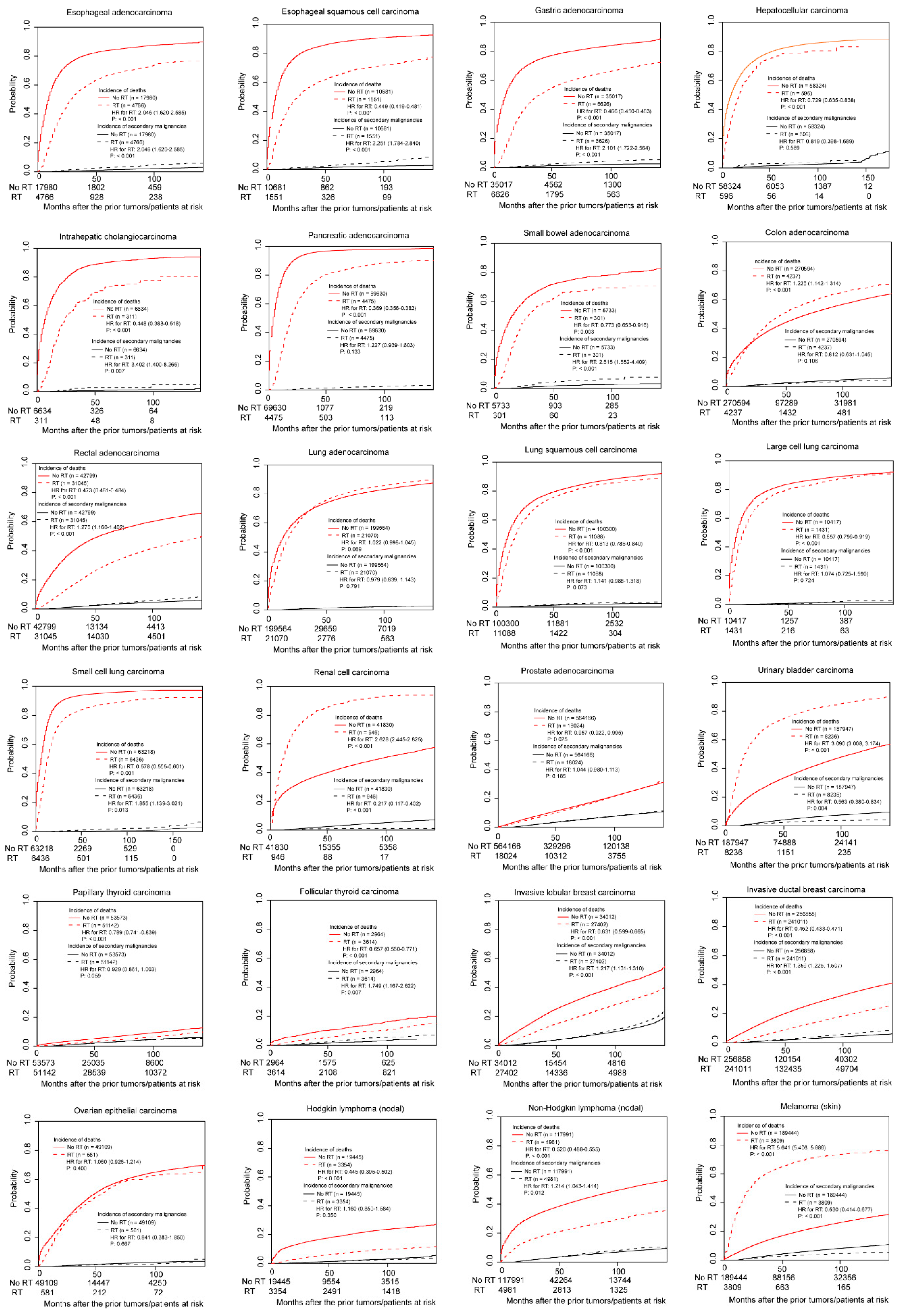
Figure 2 shows the results of the non-adjusted Fine and Gray models. Radiotherapy was associated with more SPMs in patients with esophageal adenocarcinoma, esophageal squamous cell carcinoma, gastric adenocarcinoma, intrahepatic cholangiocarcinoma, small bowel adenocarcinoma, rectal adenocarcinoma, small cell lung carcinoma, follicular thyroid carcinoma, invasive lobular breast carcinoma, invasive ductal breast carcinoma, and non-Hodgkin lymphoma (all p < 0.05). In contrast, radiotherapy was related to fewer SPMs in patients with renal cell carcinoma, urinary bladder transitional cell carcinoma, and skin melanoma (all p < 0.05). No associations between radiotherapy and SPMs were observed in the other tumor types, including hepatocellular carcinoma, pancreatic adenocarcinoma, colon adenocarcinoma (Supplementary Figure S1), lung adenocarcinoma, lung squamous cell carcinoma, large cell lung carcinoma, prostate adenocarcinoma, papillary thyroid carcinoma, ovarian epithelial carcinoma, and Hodgkin lymphoma (all p > 0.05).

3.3. Association between Radiotherapy and Incidence of SPMs in PSM Cohorts

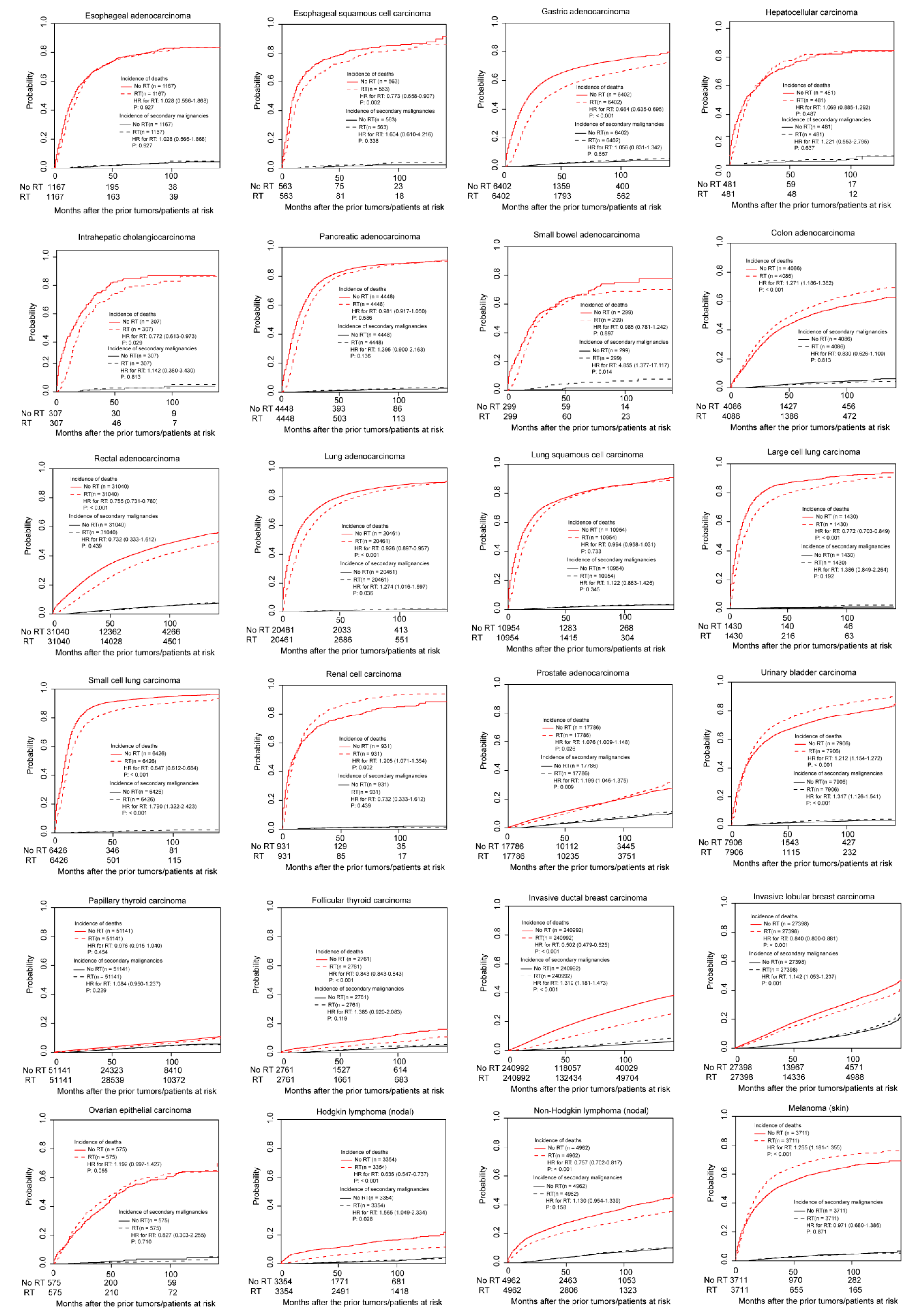
The impact of radiotherapy on the incidence of SPMs was different across cancers. In the PSM-adjusted population, according to the outcomes of the survival analysis (Kaplan-Meier) considering the competing-risks in event occurrence, the results could be divided into two groups: (I) Patients receiving radiotherapy were associated with more SPMs and (II) Patients who received radiotherapy showed similar SPM incidences. Group I included the following tumor types: Small bowel adenocarcinoma (p = 0.014), small cell lung carcinoma (<0.001), prostate adenocarcinoma (p = 0.009), urinary bladder transitional cell carcinoma (p < 0.001), invasive ductal breast carcinoma (p < 0.001), invasive lobular breast carcinoma (p = 0.001) and Hodgkin lymphoma (nodal; p = 0.028) (Table 2). The other tumor types were assigned to Group II, due to the similar SPM incidences for patients receiving or not receiving radiotherapy. Notably, in supplementary Figure S1, there were still no associations between radiotherapy and SPMs for patients with colon adenocarcinoma stratified by tumor site (cecum, appendix, ascending colon, transverse colon, descending colon, and sigmoid colon). Figure 3 displays the cumulative incidences of death and secondary malignancy of the 24 types of tumor in the PSM cohorts.

3.4. Association between Radiotherapy and Incidence of SPMs in Multivariable Competing Risk Models

As shown in Table 3, in the primary cohorts, we adjusted different variables in two models. Model II was also adjusted for tumor stage and treatment (surgery and chemotherapy) except for variables in model I (age, sex, race, and tumor grade). The effect and significance in most of the results in model 2 were consistent with the results in the PSM-adjusted population. Patients with small bowel adenocarcinoma (p = 0.004), small cell lung carcinoma (p = 0.043), prostate adenocarcinoma (p < 0.001), urinary bladder transitional cell carcinoma (p < 0.001), and invasive lobular carcinoma (p < 0.001) still had higher incidence of SPMs after radiotherapy. However, due to the data limitation (failed to meet the proportional distribution hazard assumption in both Cox and Fine and Gray models), there were no results for patients with invasive ductal breast carcinoma after adjusting the confounding factors. In addition, in the primary cohorts, a higher prevalence of SPMs was observed in patients with pancreatic adenocarcinoma after radiotherapy (p = 0.019). In both models, there was no significant difference in SPM rate for Hodgkin lymphoma (nodal) after radiotherapy or not (p = 0.351). Notably, for patients with several types of tumor (esophageal tumors, gastric adenocarcinoma, intrahepatic cholangiocarcinoma, ascending colon adenocarcinoma, lung adenocarcinoma, lung squamous cell carcinoma, renal cell carcinoma, non-Hodgkin lymphoma (nodal) and melanoma), the significant differences in model I were not observed in model II after adjusting additional covariates, including treatment and tumor stage.

3.5. Stratified Analyses

In supplementary Table S2, we performed subgroup analyses based on surgical treatment for patients whose major treatment method was surgical resection (exclude lymphoma and small cell lung carcinoma). In the surgery subgroups, the small bowel adenocarcinoma (p = 0.013), prostate adenocarcinoma (p < 0.001), urinary bladder transitional cell carcinoma (p < 0.001), and invasive lobular breast carcinoma (p < 0.001) still showed consistent effect and significance with the previous multivariable and PSM-adjusted cohort analyses. However, in the no-surgery subgroups, owing to the sparse data (limited number of patients with SPMs), no reasonable results related to these tumor types were yielded by the multivariable models.
In the surgery subgroups, several types of tumor with no significance in the PSM-adjusted population became significant (Supplementary Table S2). These types of tumor included pancreatic adenocarcinoma (p = 0.023), rectal adenocarcinoma (p < 0.001), lung adenocarcinoma (p = 0.003) and follicular thyroid carcinoma (p = 0.010) in the surgery subgroups. More SPMs were observed after radiotherapy for patients with these types of tumor.

4. Discussion

Radiotherapy has been deemed as a double-edged sword, since it is a well-established treatment modality for solid cancers, but is simultaneously likely to induce new primary cancers years after the radiation in a small number of patients. In this study, we analyzed the effect of radiotherapy on the incidence of SPMs in 24 cancer types. In the PSM-adjusted population, several tumor types who underwent radiotherapy showed a higher prevalence of SPMs. These types of tumor include small bowel adenocarcinoma, small cell lung carcinoma, prostate adenocarcinoma, urinary bladder transitional cell carcinoma, invasive ductal breast carcinoma, invasive lobular breast carcinoma, and Hodgkin lymphoma. In the surgical treatment subgroup, the effect of several other types of tumor (pancreatic adenocarcinoma, rectal adenocarcinoma, lung adenocarcinoma, and follicular thyroid carcinoma) became significant in the multivariable models (Supplementary Table S2). All these types of tumors should be with caution when carrying out radiotherapy, and the radiological doses should be carefully managed.
We performed stratified analyses based on surgery because patients receiving surgical treatment represented a specific population, usually with more favorable performance status and earlier tumor stage [8,14,19]. In addition, patients undergoing surgery usually had a longer survival compared to those without surgery, thus more patients in the surgery subgroup had SPMs during the follow-up. Consequently, the impact of radiotherapy on the incidence of SPMs for patients with or without surgery may be different. Actually, patients with combined surgery and radiotherapy can achieve a higher local tumor control rate and better long-term survival. Surgery and radiotherapy were often performed simultaneously in many types of tumors [20,21,22,23]. In other words, most of the patients without surgery also had not received radiotherapy, thus, we cannot yield a reasonable effect of radiotherapy based on the sparse data (few patients received radiotherapy) in the no surgery subgroup in some types of tumor (Supplementary Table S2).
There were several advantages of this study compared to previously published literature on this topic. Firstly, we explored the effect of radiotherapy on SPMs based on the pathological types of FPMs in corresponding tumor sites. It is more reasonable than previous studies, which only utilized sites to classify tumor types. Patients with different pathological types may have different responses to radiotherapy (e.g., small and non-small lung carcinoma; adenocarcinoma and squamous cell carcinoma) [24,25]. Secondly, this study is a systematic and comprehensive study of many types of tumors that were received radiotherapy with data from the SEER database, thus we can compare risks across first tumor types and evaluate common patterns of risk. In contrast, previous studies did not explore the overall effect of radiotherapy on SPMs. Most of the publications, including several studies of the SEER database, focused on a single FPM or a single SPM [3,6,7,9,13,15,19,23,26,27,28]. Third, the strength of this study lies in that the outcomes were confirmed by adequate statistical analyses in large population-based cohorts. PSM analysis considering competing risk allowed balancing of many covariates (e.g., age, sex, race, tumor grade, and stage) without the common statistical concerns for regression analysis. To avoid selection bias of PSM methods, we also utilized Fine and Gray models in the primary cohorts, adjusting the confounding factors. Additionally, we conducted a stratified analysis by surgery to validate the conclusions.
Previous studies have demonstrated higher risks of SPMs for patients with breast tumors, prostate tumors, and Hodgkin lymphoma receiving radiotherapy [1,8]. The conclusions in this study were consistent with the literature. However, some inconsistent phenomena were also observed in the present study. An increase in the prevalence of SPMs would be the most important late effect that could occur in patients with thyroid cancers treated with radioisotopes [29,30]. However, in this study, we did not observe a higher risk for SPMs associated with radioisotopes (in both PSM and multivariable models) in patients with papillary thyroid carcinoma. The possible reasons for this discrepancy were the different sample size and different follow-up of the study populations. Additionally, in future studies, maybe it is necessary to provide physicians with quantitative information on the risk of developing SPMs after radioisotope treatment, which will make the decision to treat a patient with radioisotopes incorporating a careful risk-benefit analysis. Except for thyroid tumors, a previous study by Warschkow et al. demonstrated that the risk of SPMs was slightly decreased after radiation in patients with rectal adenocarcinoma after resection [14]. The opposite conclusion in this study (increased risk of SPMs after radiotherapy in patients with rectal adenocarcinoma after surgery) may be caused by the increased use of IMRT in recent years, [31,32,33] which may increase the chance of SPMs. The study of Warschkow et al. have included patients in the era before IMRT was widely used. For patients with other types of tumors, there were only a few existed publications related to this topic. In the present study, for the first time, we presented the newest landscape for the impact of radiotherapy on SPMs across 24 cancer types, which is significant to guide the clinical decision-making for managing tumors.
Notably, for different tumor types, the role of radiation was distinct. For example, surgical resection was the predominant treatment method for early-stage prostate, bladder, and breast cancers, and radiotherapy was only the adjuvant therapy for some late-stage patients with these types of tumor. However, for small cell lung cancer, [34] radiotherapy played an important role in managing cases with both limited and extensive stages of tumor. Consequently, though radiotherapy had a significant impact on the incidence of SPMs in all of the above tumor types, the instructional clinical values were different. For surgery-based tumors, early-stage prostate, bladder, and breast cancers, radiotherapy should be carried out with caution, and informed consent should be obtained. In contrast, for small cell lung cancer, radiotherapy should be used as one of the major treatment regimens, and the optimal doses and courses of treatment should be explored in future studies.
Admittedly, there are several limitations to this study. Like other observational researches, the main drawback of the data from SEER is the lack of treatment randomization, which may bias our results. We utilized both PSM and multivariable competing risk models to adjust confounders. And the stratified analysis based on surgery was performed to minimize potential bias. In addition, we did not have information on the doses and modalities of radiotherapy, which may impact the second cancer risk. However, in this study, we included patients after 2004, and some techniques, including IMRT, was introduced and commonly used. The second limitation for this study is that we used two types of models for multivariable analysis, due to the sparse data. Actually, the results from both Cox and Fine and Gray models were similar in cancer types that both methods were available (data are not shown). In the PSM-adjusted population, Fine and Gray models can be used in all the tumor types, which made the results more reliable and explicable. The third limitation lies in that we failed to perform more subgroup analyses based on other potential interactive factors, including age of diagnosis, time to the prior cancer, and AJCC stage. More studies were needed to illustrate the effect and significance of radiotherapy among some specific populations. Regarding fourth limitation, we demonstrated a higher risk of SPMs of patients with small cell lung carcinoma and small bowel adenocarcinoma after radiotherapy. Actually, for patients with these types of tumor, only a small proportion of patients have received radiotherapy in the SEER database, thus, the conclusions related to the benefit and long-term side effect of radiotherapy for these types of tumors should also be validated in further studies. Finally, given the difficulty to identify the regional organs that were influenced by the radiations, the total number of SPMs after radiotherapy was used as the endpoint in this study, which may have an impact on the conclusions. However, emerging studies indicated that the biological effects of radiotherapy were not limited to targeted regions. As a result of signal transmission from an irradiated cell, a plethora of biological phenomena occurred in non-irradiated cells (the radiation-induced bystander effect). The mechanisms underlying it should be totally illustrated in further studies. Based on this theory, in this study, we still used the total number of SPMs as the observed endpoint.

5. Conclusions

We present a comprehensive and newest pan-cancer study illustrating the effect of radiotherapy on the incidence of SPMs. To our knowledge, this is the first pan-cancer study exploring the role of radiotherapy in different pathological types of tumors. We have observed higher risks of SPMs in several types of tumors in the PSM-adjusted populations. Meanwhile, in subgroup analyses stratified by surgery, some types of tumor were also associated with the prevalence of SPMs in the surgery subgroups except for those with positive results in the PSM-adjusted cohorts. All these findings were clinically important for the selection of radiotherapy in managing cancers. However, further studies are still needed to validate the conclusions in the present study.

Supplementary Materials

The following are available online at https://www.mdpi.com/1718-7729/28/1/35/s1. Figure S1: The cumulative incidences of death and secondary primary malignancy in colon adenocarcinoma divided by tumor sites before and after propensity score matching. Table S1: Codes for patient selection based on the International Classification of Diseases for Oncology, third edition (ICD-O-3), Table S2: Stratified analyses according to surgery.

Author Contributions

W.L. proposed the study; Y.Z. and W.L. performed the research and wrote the first draft; H.X. revised this manuscript. X.X. is the guarantor; all authors contributed to the design and interpretation of the study and to further drafts. All authors have read and agreed to the published version of the manuscript.

Funding

This research received no external funding.

Conflicts of Interest

The authors declare no conflict of interest.

References

  1. Dracham, C.B.; Shankar, A.; Madan, R. Radiation induced secondary malignancies: A review article. Radiat. Oncol. J. 2018, 36, 85–94. [Google Scholar] [CrossRef] [PubMed]
  2. Abou-Antoun, T.; Mikhael, R.; Massoud, M.; Chahine, G.; Saad, A. Effects of Radiotherapy on the Risk of Developing Secondary Malignant Neoplasms in Hodgkin’s Lymphoma Survivors. Asian Pac. J. Cancer Prev. 2016, 17, 749–754. [Google Scholar] [CrossRef] [PubMed] [Green Version]
  3. Andrade-Campos, M.M.; Lievano, P.; Espinosa-Lara, N.; Soro-Alcubierre, G.; Grasa-Ulrich, J.M.; Lopez-Gomez, L.; Baringo, T.; Giraldo, P. Long-term complication in follicular lymphoma: Assessing the risk of secondary neoplasm in 242 patients treated or not with 90-yttrium-ibritumomab-tiuxetan. Eur. J. Haematol. 2016, 97, 576–582. [Google Scholar] [CrossRef] [PubMed]
  4. Balcer-Kubiczek, E.K.; Eley, J.G. Secondary Malignancies in the Era of High-Precision Radiation Therapy. Crit. Rev. Oncog. 2018, 23, 93–112. [Google Scholar] [CrossRef]
  5. Kebudi, R.; Ozdemir, G.N. Secondary Neoplasms in Children Treated for Cancer. Curr. Pediatric Rev. 2017, 13, 34–41. [Google Scholar] [CrossRef]
  6. Lee, Y.C.; Hsieh, C.C.; Li, C.Y.; Chuang, J.P.; Lee, J.C. Secondary Cancers after Radiation Therapy for Primary Prostate or Rectal Cancer. World J. Surg. 2016, 40, 895–905. [Google Scholar] [CrossRef]
  7. Mendes, B.M.; Trindade, B.M.; Fonseca, T.C.F.; de Campos, T.P.R. Assessment of radiation-induced secondary cancer risk in the Brazilian population from left-sided breast-3D-CRT using MCNPX. Br. J. Radiol. 2017, 90, 20170187. [Google Scholar] [CrossRef]
  8. Berrington de Gonzalez, A.; Curtis, R.E.; Kry, S.F.; Gilbert, E.; Lamart, S.; Berg, C.D.; Stovall, M.; Ron, E. Proportion of second cancers attributable to radiotherapy treatment in adults: A cohort study in the US SEER cancer registries. Lancet Oncol. 2011, 12, 353–360. [Google Scholar] [CrossRef] [Green Version]
  9. Kawada, K.; Hida, K.; Sakai, Y. Advantages of transperineal approach for secondary rectal cancer following prior radiotherapy to prostate cancer–video vignette. Colorectal Dis. 2017. [Google Scholar] [CrossRef]
  10. Shiota, M.; Yokomizo, A.; Takeuchi, A.; Inokuchi, J.; Tatsugami, K.; Ohga, S.; Sasaki, T.; Nakamura, K.; Honda, H.; Eto, M. Smoking effect on secondary bladder cancer after external beam radiotherapy for prostate cancer. Jpn. J. Clin. Oncol. 2016, 46, 952–957. [Google Scholar] [CrossRef] [Green Version]
  11. Fogliata, A.; De Rose, F.; Franceschini, D.; Stravato, A.; Seppala, J.; Scorsetti, M.; Cozzi, L. Critical Appraisal of the Risk of Secondary Cancer Induction From Breast Radiation Therapy with Volumetric Modulated Arc Therapy Relative to 3D Conformal Therapy. Int. J. Radiat. Oncol. Biol. Phys. 2018, 100, 785–793. [Google Scholar] [CrossRef] [PubMed]
  12. Chao, P.J.; Lee, H.F.; Lan, J.H.; Guo, S.S.; Ting, H.M.; Huang, Y.J.; Chen, H.C.; Lee, T.F. Propensity-score-matched evaluation of the incidence of radiation pneumonitis and secondary cancer risk for breast cancer patients treated with IMRT/VMAT. Sci. Rep. 2017, 7, 13771. [Google Scholar] [CrossRef] [PubMed]
  13. Winaikosol, K.; Surakunprapha, P. Rapidly developed Secondary Cutaneous Squamous cell Carcinoma after Post-Surgical Radiation Therapy for Breast Cancer. J. Med. Assoc. Thail. 2016, 99, S173–S176. [Google Scholar]
  14. Warschkow, R.; Guller, U.; Cerny, T.; Schmied, B.M.; Plasswilm, L.; Putora, P.M. Secondary malignancies after rectal cancer resection with and without radiation therapy: A propensity-adjusted, population-based SEER analysis. Radiother. Oncol. J. Eur. Soc. Ther. Radiol. Oncol. 2017, 123, 139–146. [Google Scholar] [CrossRef] [PubMed]
  15. Chaturvedi, A.K.; Engels, E.A.; Gilbert, E.S.; Chen, B.E.; Storm, H.; Lynch, C.F.; Hall, P.; Langmark, F.; Pukkala, E.; Kaijser, M.; et al. Second cancers among 104,760 survivors of cervical cancer: Evaluation of long-term risk. J. Natl. Cancer Inst. 2007, 99, 1634–1643. [Google Scholar] [CrossRef] [PubMed] [Green Version]
  16. Creutzberg, C.L.; Nout, R.A.; Lybeert, M.L.M.; Wárlám-Rodenhuis, C.C.; Jobsen, J.; Mens, J.M.; Lutgens, L.C.H.W.; Pras, E.; van de Poll-Franse, L.V.; van Putten, W.L.J. Fifteen-year radiotherapy outcomes of the randomized PORTEC-1 trial for endometrial carcinoma. Int. J. Radiat. Oncol. Biol. Phys. 2011, 81, e631–e638. [Google Scholar] [CrossRef] [Green Version]
  17. Journy, N.; Mansouri, I.; Allodji, R.S.; Demoor-Goldschmidt, C.; Ghazi, D.; Haddy, N.; Rubino, C.; Veres, C.; Zrafi, W.S.; Rivera, S.; et al. Volume effects of radiotherapy on the risk of second primary cancers: A systematic review of clinical and epidemiological studies. Radiother. Oncol. J. Eur. Soc. Ther. Radiol. Oncol. 2018, 131, 150–159. [Google Scholar] [CrossRef]
  18. De Glas, N.A.; Kiderlen, M.; Vandenbroucke, J.P.; de Craen, A.J.; Portielje, J.E.; van de Velde, C.J.; Le Cessie, S. Performing Survival Analyses in the Presence of Competing Risks: A Clinical Example in Older Breast Cancer Patients. J. Natl. Cancer Inst. 2016, 108, djv366. [Google Scholar] [CrossRef] [Green Version]
  19. Wissing, M.D.; Azoulay, L. Postoperative Pelvic Radiotherapy in Patients With Endometrial Cancer May Increase the Risk for Secondary Pelvic Cancers: A Post Hoc Analysis of Results From the TME, PORTEC-1, and PORTEC-2 Trials. J. Clin. Oncol. Off. J. Am. Soc. Clin. Oncol. 2017, 35, 1861–1862. [Google Scholar] [CrossRef]
  20. Abraha, I.; Aristei, C.; Palumbo, I.; Lupattelli, M.; Trastulli, S.; Cirocchi, R.; de Florio, R.; Valentini, V. Preoperative radiotherapy and curative surgery for the management of localised rectal carcinoma. Cochrane Database Syst. Rev. 2018, 10, CD002102. [Google Scholar] [CrossRef]
  21. Zhang, N.; Fei, Q.; Gu, J.; Yin, L.; He, X. Progress of preoperative and postoperative radiotherapy in gastric cancer. World J. Surg. Oncol. 2018, 16, 187. [Google Scholar] [CrossRef] [PubMed]
  22. Crehange, G.; Mabrut, J.Y.; Rouffiac, M. Surgery after upfront radiochemotherapy for locally advanced esophageal cancer: To do or not to do? Cancer Radiother. J. Soc. Fr. Radiother. Oncol. 2018, 22, 540–545. [Google Scholar]
  23. Vyfhuis, M.A.L.; Rice, S.; Remick, J.; Mossahebi, S.; Badiyan, S.; Mohindra, P.; Simone, C.B., 2nd. Reirradiation for locoregionally recurrent non-small cell lung cancer. J. Thorac. Dis. 2018, 10, S2522–S2536. [Google Scholar] [CrossRef] [PubMed]
  24. Deng, H.Y.; Wang, W.P.; Wang, Y.C.; Hu, W.P.; Ni, P.Z.; Lin, Y.D.; Chen, L.Q. Neoadjuvant chemoradiotherapy or chemotherapy? A comprehensive systematic review and meta-analysis of the options for neoadjuvant therapy for treating oesophageal cancer. Eur. J. Cardio Thorac. Surg. Off. J. Eur. Assoc. Cardio Thorac. Surg. 2017, 51, 421–431. [Google Scholar] [CrossRef] [PubMed]
  25. Horner-Rieber, J.; Bernhardt, D.; Dern, J.; Konig, L.; Adeberg, S.; Paul, A.; Heussel, C.P.; Kappes, J.; Hoffmann, H.; Herth, F.J.P.; et al. Histology of non-small cell lung cancer predicts the response to stereotactic body radiotherapy. Radiother. Oncol. J. Eur. Soc. Ther. Radiol. Oncol. 2017, 125, 317–324. [Google Scholar] [CrossRef]
  26. Lee, H.F.; Lan, J.H.; Chao, P.J.; Ting, H.M.; Chen, H.C.; Hsu, H.C.; Lee, T.F. Radiation-induced secondary malignancies for nasopharyngeal carcinoma: A pilot study of patients treated via IMRT or VMAT. Cancer Manag. Res. 2018, 10, 131–141. [Google Scholar] [CrossRef] [Green Version]
  27. Yamanaka, R.; Abe, E.; Sato, T.; Hayano, A.; Takashima, Y. Secondary Intracranial Tumors Following Radiotherapy for Pituitary Adenomas: A Systematic Review. Cancers 2017, 9, 103. [Google Scholar] [CrossRef] [Green Version]
  28. Conway, J.L.; Connors, J.M.; Tyldesley, S.; Savage, K.J.; Campbell, B.A.; Zheng, Y.Y.; Hamm, J.; Pickles, T. Secondary Breast Cancer Risk by Radiation Volume in Women with Hodgkin Lymphoma. Int. J. Radiat. Oncol. Biol. Phys. 2017, 97, 35–41. [Google Scholar] [CrossRef]
  29. Tulchinsky, M.; Binse, I.; Campenni, A.; Dizdarevic, S.; Giovanella, L.; Jong, I.; Kairemo, K.; Kim, C.K. Radioactive Iodine Therapy for Differentiated Thyroid Cancer: Lessons from Confronting Controversial Literature on Risks for Secondary Malignancy. J. Nucl. Med. 2018, 59, 723–725. [Google Scholar] [CrossRef] [Green Version]
  30. Gillis, C.C.; Chang, E.H.; Al-Kharazi, K.; Pickles, T. Secondary malignancy following radiotherapy for thyroid eye disease. Rep. Pract. Oncol. Radiother. 2016, 21, 156–161. [Google Scholar] [CrossRef] [Green Version]
  31. Elson, J.K.; Kachnic, L.A.; Kharofa, J.R. Intensity-modulated radiotherapy improves survival and reduces treatment time in squamous cell carcinoma of the anus: A National Cancer Data Base study. Cancer 2018, 124, 4383–4392. [Google Scholar] [CrossRef] [PubMed] [Green Version]
  32. Kilic, S.; Kilic, S.S.; Hsueh, W.D.; Eloy, J.A.; Baredes, S.; Woo Park, R.C.; Mahmoud, O. Radiotherapy modality as a predictor of survival in hypopharyngeal cancer. Head Neck 2018, 40, 2441–2448. [Google Scholar] [CrossRef] [PubMed]
  33. Zhu, Q.; Hu, H.; Tang, L.Q.; You, R.; Zhao, J.J.; Weng, D.S.; Pan, Q.Z.; Chen, C.L.; Zhou, Z.Q.; Tang, Y.; et al. Weekly versus triweekly cisplatin plus intensity-modulated radiotherapy in locally advanced nasopharyngeal carcinoma: A propensity score analysis with a large cohort. J. Cancer 2018, 9, 3447–3455. [Google Scholar] [CrossRef] [PubMed] [Green Version]
  34. Gensheimer, M.F.; Loo, B.W., Jr. Optimal Radiation Therapy for Small Cell Lung Cancer. Curr. Treat. Opt. Oncol. 2017, 18, 21. [Google Scholar] [CrossRef]
Figure 1. The proportion of the sites of the second primary malignancies of the 24 types of primary cancers.
Curroncol 28 00035 g001
Figure 2. The cumulative incidences of death and secondary primary malignancy in the non-adjusted primary cohorts.
Curroncol 28 00035 g002
Figure 3. The cumulative incidences of death and secondary primary malignancy in the PSM-adjusted populations.
Curroncol 28 00035 g003
Table 1. Patient characteristics.
Prior CancerNumber of Patients with FPMsNumber of Patients with SPMsTime to a Secondary Malignancy
Total NumberRadiation *No Radiation *Total NumberRadiation *No Radiati on *Total Cohort **Radiation **No Radiation **
Esophagus
Adenocarcinoma22,7464766 (21.0)17,980 (79.0)390137 (35.1)253 (64.9)47.6 (40.5)44.8 (37.0)49.1 (42.0)
Squamous cell carcinoma12,2321551 (12.7)10,681 (87.3)23058 (25.2)172 (74.8)45.9 (37.0)53.9 (47.5)43.2 (36.0)
Gastric adenocarcinoma41,6436626 (15.9)35,017 (84.1)719204 (28.4)515 (71.6)43.7 (36.0)45.6 (40.5)42.9 (34.0)
Liver
Hepatocellular carcinoma58,920596 (1.0)58,324 (99.0)98716 (1.6)971 (98.4)46.0 (34.0)28.7 (20.0)46.3 (34.0)
Intrahepatic cholangiocarcinoma6945311 (4.5)6634 (95.5)517 (13.7)44 (86.3)42.9 (34.0)38.1 (29.0)43.6 (35.5)
Pancreatic adenocarcinoma74,1054475 (6.0)69,630 (94.0)24481 (33.2)163 (66.8)38.8 (29.0)43.1 (31.0)36.6 (28.0)
Small bowel adenocarcinoma6034301 (5.0)5733 (95.0)11714 (12.0)103 (88.0)43.0 (36.0)42.7 (34.5)43.0 (36.0)
Colon adenocarcinoma
Cecum62,641827 (1.3)61,594 (98.7)200626 (1.3)1980 (98.7)47.7 (41.0)39.2 (22.0)47.9 (42.0)
Appendix6382118 (1.8)6264 (98.2)1066 (5.7)102 (94.3)43.3 (35.5)30.2 (27.5)43.8 (35.5)
Ascending colon54,343456 (0.8)53,887 (99.2)19678 (0.4)1959 (99.6)48.5 (41.0)60.2 (36.2)48.5 (41.0)
Transverse colon26,637189 (0.7)26,448 (99.3)9016 (0.7)895 (99.3)48.2 (40.0)64.7 (32.1)48.1 (40.0)
Descending colon16,721225 (1.3)16,496 (98.7)5264 (0.8)522 (99.2)50.4 (43.0)51.0 (45.0)50.4 (53.0)
Sigmoid colon74,6422099 (2.8)72,543 (97.2)256759 (2.3)2508 (97.7)50.4 (44.0)48.5 (38.0)50.4 (44.0)
Rectal adenocarcinoma73,83531,045 (42.0)42,790 (58.0)24951183 (47.4)1312 (52.6)50.6 (45.0)52.9 (47.0)48.5 (42.0)
Lung
Adenocarcinoma220,63421,070 (9.5)199,564 (90.5)3046182 (6.0)2764 (94.0)44.6 (37.0)41.3 (32.5)44.9 (38.0)
Squamous cell carcinoma111,38811,088 (10.0)100,300 (90.0)1669211 (12.6)1458 (87.4)41.6 (35.0)41.3 (36.0)41.6 (35.0)
Small cell carcinoma69,6546436 (9.2)63,218 (90.8)33270 (21.1)262 (78.9)52.3 (42.0)51.7 (41.0)52.4 (42.0)
Large cell carcinoma11,8481431 (12.1)10,417 (87.9)15523 (14.8)132 (85.2)48.6 (45.0)65.4 (75.0)45.6 (42.0)
Renal cell carcinoma42,776946 (2.2)41,830 (97.8)14748 (0.5)1466 (99.5)51.1 (45.0)31.6 (28.0)51.2 (45.0)
Prostate adenocarcinoma582,19018,024 (3.1)564,166 (96.9)30,836976 (3.2)29860 (96.8)62.2 (59.0)61.7 (58.0)62.2 (59.0)
Urinary bladder transitional cell carcinoma196,1838236 (4.2)187,947 (95.8)9969214 (2.1)9755 (97.9)46.5 (39.0)38.8 (34.5)46.7 (40.0)
Thyroid
Papillary thyroid carcinoma104,71551,142 (48.8)53,573 (51.2)26261342 (51.1)1284 (48.9)48.6 (43.0)48.8 (44.0)48.4 (43.0)
Follicular thyroid carcinoma65783614 (54.9)2964 (45.1)191129 (67.5)62 (32.5)51.6 (47.0)50.9 (47.0)53.0 (47.5)
Breast
Invasive ductal carcinoma497,869241,011 (48.4)286,858 (51.6)15,2368749 (57.4)6487 (42.6)55.0 (50.0)56.4 (52.0)53.0 (47.0)
Invasive lobular carcinoma61,41427,402 (44.6)34,012 (55.4)44502139 (48.1)2311 (51.9)63.7 (60.0)66.9 (64.0)60.8 (55.0)
Ovarian epithelial carcinoma49,690581 (1.2)49,109 (98.8)93210 (1.1)922 (98.9)46.9 (40.0)58.9 (43.0)46.8 (40.0)
Lymphoma
Hodgkin lymphoma (nodal)22,7993354 (14.7)19,445 (85.3)45575 (16.5)380 (83.5)55.7 (51.0)56.8 (52.0)55.5 (50.5)
Non-Hodgkin lymphoma (nodal)122,9724981 (4.1)117,991 (95.9)5652320 (5.7)5332 (94.3)49.9 (43.0)55.7 (53.0)49.6 (43.0)
Melanoma (skin)193,2533809 (2.0)189,444 (98.0)9865109 (1.1)9756 (98.9)47.6 (40.0)38.4 (31.0)47.7 (41.0)
* The number and proportion of patients with or without radiation. ** The mean (median) time of prior cancer to newly diagnosed cancer (months). Radioisotopes: 95.5% (n = 48,820); Beam radiation: 2.1% (n = 1059). Radioisotopes: 93.7% (n = 3388); Beam radiation: 3.5% (n = 125). FPMs, first primary malignancies; SPMs, second primary malignancies.
Table 2. The risk for secondary malignancies after radiation in pan-cancers after PSM.
Prior CancerFine and Gray Models in PSM Cohorts
NumberEventsHR (95%CI)p Values
Esophagus
Adenocarcinoma2334571.028 (0.566–1.868)0.927
Squamous cell carcinoma1126211.604 (0.610–4.216)0.338
Gastric adenocarcinoma12,8043711.056 (0.831–1.342)0.657
Hepatocellular carcinoma962241.221 (0.553–2.795)0.637
Intrahepatic cholangiocarcinoma614131.142 (0.380–3.430)0.813
Pancreatic adenocarcinoma88961341.395 (0.900–2.163)0.136
Small bowel adenocarcinoma598174.855 (1.377–17.117)0.014
Colon adenocarcinoma
Cecum1562471.507 (0.944–2.406)0.086
Appendix21461.878 (0.333–10.582)0.475
Ascending colon852170.660 (0.248–1.758)0.406
Transverse colon362190.478 (0.177–1.293)0.146
Descending colon422100.652 (0.175–2.428)0.523
Sigmoid colon3890991.209 (0.782–1.870)0.393
Rectal adenocarcinoma62,08024040.984 (0.904–1.071)0.706
Lung
Adenocarcinoma40,9224521.179 (0.933–1.490)0.169
Squamous carcinoma21,9083911.122 (0.883–1.426)0.345
Small cell carcinoma12,8521091.790 (1.322–2.423)<0.001
Large cell carcinoma2860371.386 (0.849–2.264)0.192
Renal cell carcinoma1862200.732 (0.333–1.612)0.439
Prostate adenocarcinoma3557217831.199 (1.046–1.375)0.009
Urinary bladder transitional
cell carcinoma
15,8124051.317 (1.126–1.541)<0.001
Thyroid
Papillary thyroid carcinoma102,28225911.084 (0.950–1.237)0.229
Follicular thyroid carcinoma55221401.385 (0.920–2.083)0.119
Breast
Invasive ductal carcinoma481,984150701.319 (1.181–1.473)<0.001
Invasive lobular carcinoma54,79640741.142 (1.053–1.237)0.001
Ovarian epithelial cancer 1150240.827 (0.303–2.255)0.710
Lymphoma
Hodgkin lymphoma (nodal)67081241.565 (1.049–2.334)0.028
Non-Hodgkin lymphoma (nodal)99246081.130 (0.954–1.339)0.158
Melanoma (skin)74222280.971 (0.680–1.386)0.871
Propensity score matching was based on age, sex, race, tumor grade, American Joint Committee on Cancer (AJCC) stage, chemotherapy, surgery, and year of diagnosis if available.
Table 3. The risk for secondary malignancies after radiation in multivariable models.
Prior CancerFine and Gray Models
Model IModel II
NumberEventsHR (95%CI)p ValueNumberEventsHR (95%CI)p Value
Esophagus
Adenocarcinoma22,7463901.895 (1.516–2.369)<0.00122,6503880.856 (0.524–1.399)0.535
Squamous cell carcinoma12,2322302.085 (1.668–2.607)<0.00112,1942300.694 (0.458–1.053)0.086
Gastric adenocarcinoma41,6437191.898 (1.547–2.329)<0.00141,5237180.954 (0.764–1.190)0.674
Hepatocellular carcinoma58,920 9870.494 (0.186–1.316)0.15948,2926741.056 (0.555–2.008)0.869
Intrahepatic cholangiocarcinoma6945514.165 (1.255–13.828)0.0206894500.700 (0.203–2.415)0.572
Pancreatic adenocarcinoma74,1052441.274 (0.963–1.684) *0.09073,8372431.479 (1.067–2.048) *0.019
Small bowel adenocarcinoma60331172.511 (1.310–4.813) 0.00660211172.669 (1.369–5.206)0.004
Colon adenocarcinoma
Cecum62,64120060.955 (0.659–1.385)0.80960,36719881.168 (0.790–1.727)0.436
Appendix63821062.199 (0.854–5.666)0.10347891052.008 (0.732–5.509)0.176
Ascending colon54,34319670.443 (0.201–0.975)0.04352,51719460.625 (0.284–1.374)0.242
Transverse colon26,6379010.987 (0.451–2.163)0.97425,7538861.049 (0.484–2.274)0.904
Descending colon16,7215260.548 (0.148–2.021)0.36616,0845180.591 (0.171–2.040)0.406
Sigmoid colon74,64225670.834 (0.605–1.450)0.26871,72324990.956 (0.682–1.339)0.791
Rectal adenocarcinoma73,83524950.957 (0.883–1.037) *0.28573,65424911.070 (0.942–1.216) *0.298
Lung
Adenocarcinoma220,63430461.163 (1.026–1.318) *0.018220,10730451.076 (0.943–1.227) *0.277
Squamous carcinoma111,38816691.232 (1.047–1.450)0.012110,97116651.077 (0.877–1.322)0.480
Small cell carcinoma69,6543321.776 (1.016–3.106)0.04469,3523311.459 (1.013–2.103)0.043
Large cell carcinoma11,8481551.000 (0.638–1.568)0.99811,8041540.836 (0.520–1.344)0.460
Renal cell carcinoma42,77614740.242 (0.120–0.489)<0.00142,63014740.502 (0.200–1.261)0.143
Prostate adenocarcinoma582,190308361.110 (1.041–1.183) *0.001579,521307121.216 (1.138–1.300) *<0.001
Urinary bladder transitional cell carcinoma196,183 99691.477 (1.288–1.694) *<0.001195,77799481.757 (1.516–2.037) *<0.001
Thyroid
Papillary thyroid carcinoma104,715 26261.067 (0.987–1.152) *0.102104,68026261.028 (0.948–1.114) *0.504
Follicular thyroid carcinoma6578 1911.566 (1.155–2.123) *0.00465741911.553 (1.134–2.127) *0.006
Breast (women)
Invasive ductal carcinoma497,869 15236/ 497,31215229/
Invasive lobular carcinoma61,414 44501.373 (1.218–1.547)<0.00161,33644441.108 (1.108–1.108)<0.001
Ovarian epithelial cancer 49,690 9320.841 (0.389–1.819)0.66049,6369310.744 (0.330–1.677)0.475
Lymphoma
Hodgkin lymphoma (nodal)22,799 4550.824 (0.642–1.057) *0.12822,7994550.872 (0.653–1.164) *0.351
Non-Hodgkin lymphoma (nodal)122,97256521.230 (1.058–1.430)0.007122,97256521.090 (0.948–1.254)0.228
Melanoma (skin)193,253 98651.425 (1.180–1.722) *< 0.001192,95498551.109 (0.912–1.348) *0.302
Model I: Fine and Gray models adjusting for age, sex, race, and tumor grade if available; Model II: Fine and Gray models adjusting for age, sex, race, tumor grade, AJCC-TNM stage, chemotherapy, and surgery if available. * Cox proportional hazards regression analyses were used. Sex was not adjusted in breast tumors (women) and prostate tumors (men). In lymphoma, tumor site was also adjusted in model II, while surgery was not adjusted. In small cell lung carcinoma, surgery was not adjusted in model II.
Publisher’s Note: MDPI stays neutral with regard to jurisdictional claims in published maps and institutional affiliations.