Next Article in Journal
Special Communication
Previous Article in Journal
LAA Occluders for All Patients with Atrial Fibrillation - An Overreaching Statement
 
 
Cardiovascular Medicine is published by MDPI from Volume 28 Issue 1 (2025). Previous articles were published by another publisher in Open Access under a CC-BY (or CC-BY-NC-ND) licence, and they are hosted by MDPI on mdpi.com as a courtesy and upon agreement with Editores Medicorum Helveticorum (EMH).
Article

Right Ventricle Metastasis of Pulmonary Sarcomatoid Carcinoma

by
Verena Charlotte Wilzeck
1,
Tilo Niemann
2 and
Christpher Hansi
1,*
1
Department of Cardiology, Cantonal Hospital Baden, CH-5404 Baden, Switzerland
2
Department of Radiology, Cantonal Hospital Baden, Baden, Switzerland
*
Author to whom correspondence should be addressed.
Cardiovasc. Med. 2017, 20(3), 466; https://doi.org/10.4414/cvm.2017.00466 (registering DOI)
Submission received: 15 December 2016 / Revised: 15 January 2017 / Accepted: 15 February 2017 / Published: 15 March 2017
A 79-year-old female was diagnosed with a pulmonary sarcomatoid carcinoma (PSC) of the left lower lobe (stage IB) and underwent an open lobectomy and systematic lymph node dissection. Because of the early tumour stage, the molecular genetic characteristics of the tumour and the patient’s advanced age, adjuvant chemotherapy was not given. The patient was followed up every 3 months and, except for slight tiredness and dyspnoea, she did well.
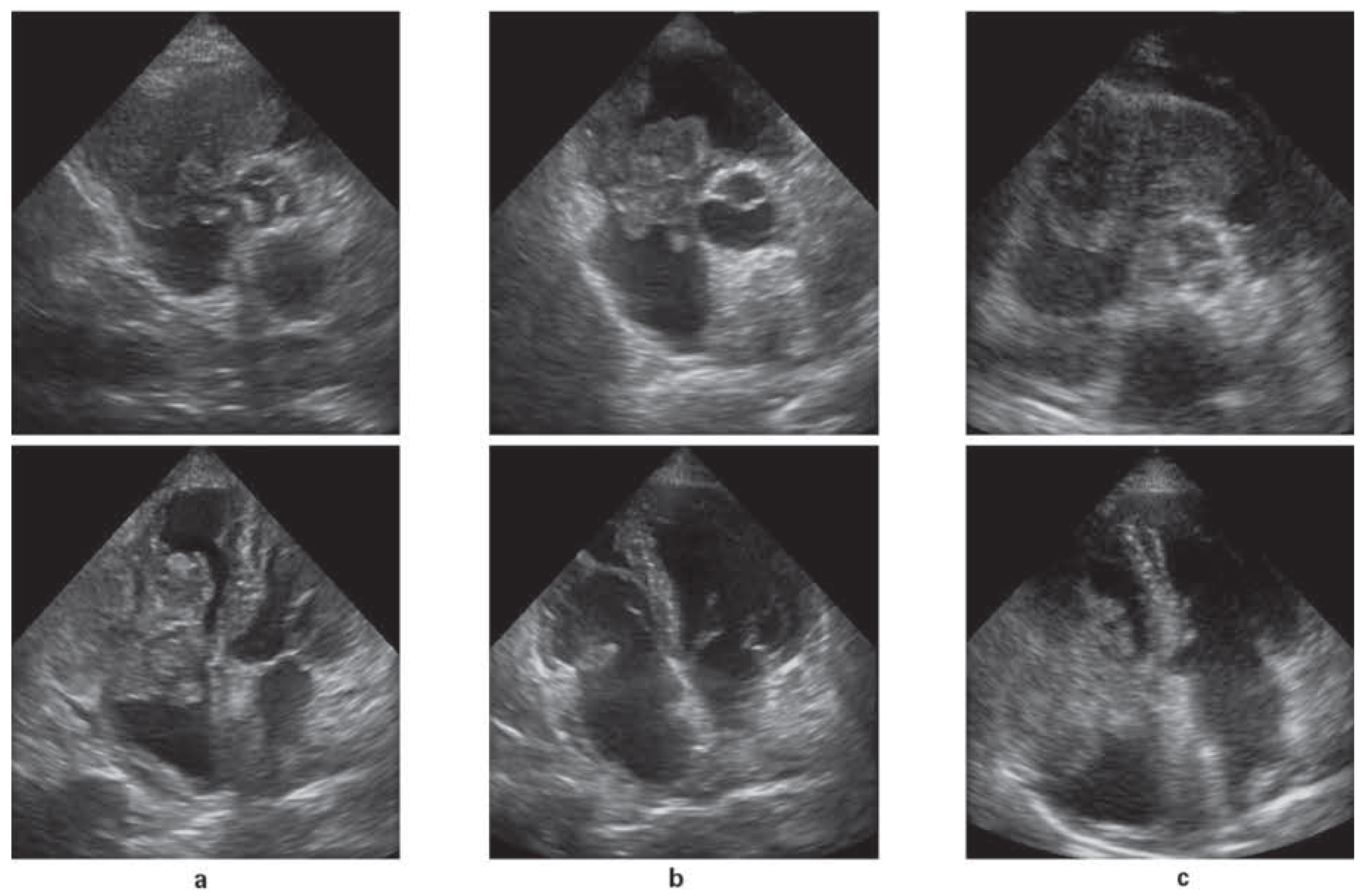
One year later, the patient went to her general practitioner (GP) because of weakness, weight loss, diarrhoea and exertional dyspnoea. The GP sent her for the annually scheduled computed tomography scan (CT), which showed a soft tissue density mass measuring 6×6 cm in the right ventricle of the heart, pulmonary embolism in a right segmental artery and suspect lesions in the right upper lobe of the lung and both adrenal glands. Echocardiography revealed that the right ventricular mass was almost completely filling the ventricle including the tricuspid valve (Figure 1a).
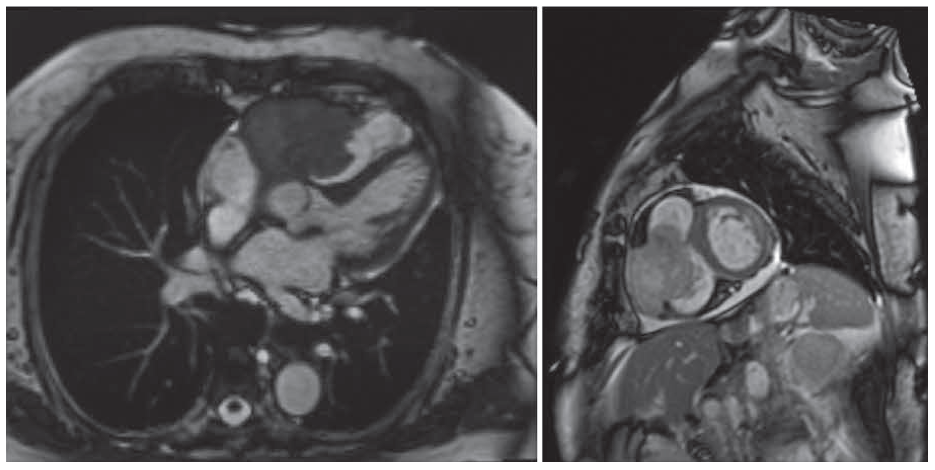
There was no sign of right heart failure. Cardiac magnetic resonance imaging showed that the mass involved the myocardium, originating from the anterior wall of the heart (Figure 2). The most likely dierential diagnosis included a metastasis or a primary tumour. Because of the moderate contrast uptake, an entire thrombus was unlikely.
The case was discussed on an interdisciplinary basis. On the basis of the patient’s history, an intracardiac recurrence of the sarcomatoid carcinoma was very likely. Because initial molecular genetic analysis had shown an activating EGFR mutation, tyrosine kinase inhibitor therapy with erlotinib was initiated. The risk of an imminent pericardial tamponade or myocardial rupture was discussed and surgical options were evaluated, but due to the high risk, no surgical procedure seemed reasonable. Seven days after initiating erlotinib the echocardiography already showed a shrunken right ventricular mass and another 5 days later the tricuspid valve became visible again (Figure 1b), leading to a moderate to severe tricuspid insufficiency. The patient was discharged in a good general condition and erlotinib treatment was continued.
In the following weeks the patient suered from Clostridium difficile colitis and erlotinib had to be stopped intermittently. The tumour mass in the right ventricle regrew immediately (Figure 1c). Shortly after, CT showed a new big tumour mass in the central abdomen and progression of the metastases of the adrenal glands.
Heart metastases have an incidence at least 100 times higher than that of primary tumours of the heart [1]. Usually, cardiac metastases are small and multiple; intracavitary growth of single large tumour lesions is rare. Pulmonary sarcomatoid carcinoma (PSC) is a rare, poorly dierentiated subtype of non-small-cell lung cancer and generally runs an aggressive clinical course with a poor prognosis. PSC metastasises via lymph and blood vessel routes to the same anatomical sites as other non-small-call lung cancers [2]; heart metastases of PSC are very rare. According to the literature there is only one case report of PSC involving the heart: Campagnoli et al. reported a case of PSC with left atrial extension through the pulmonary vein [3].

Conflicts of Interest

No financial support and no other potential conflict of interest relevant to this article was reported.

References

  1. Burke, A.; Virmani, R. Tumors of the Cardiovascular System, Atlas of Tumor Pathology; 3rd Series , Fascicle 16; Armed Forces Institute of Pathology: Washington, DC, USA, 1996. [Google Scholar]
  2. Pelosi, G.; Sonzogni, A.; De Pas, T.; Galetta, D.; Veronesi, G.; Spaggiari, L.; et al. Review article: pulmonary sarcomatoid carcinomas: a practical overview. Int J SurgPathol. 2010, 18, 103–120. [Google Scholar] [CrossRef] [Scilit] [PubMed]
  3. Campagnoli; Tania, M.; et al. Pulmonary Sarcomatoid Carcinoma with Left Atrial Extension and Atrial Fibrillation. Archives of Medicine. 2015, 7. Available from: http://www.archivesofmedicine.com/medicine/pulmonary-sarcomatoid-carcinoma-withleft-atrialextension-and-atrial-fibrillation.
Figure 1. TTE parasternal short-axis and apical four-chamber view at a) day 0, b) day 12 and c) day 53 showing an astonishing decrease of tumour mass on Erlotinib treatment with tumour relapse after treatment had been stopped intermittently.
Cardiovascmed 20 00466 g001
Figure 2. Cardiac-MRI (T2w TRUFI, 4-chamber view left and short axis right) showing the predominant hypointens mass with central necrotic component in the right ventricle involving the tricuspid valve.
Cardiovascmed 20 00466 g002