Next Article in Journal
Richtlinien 2005 Zur Nachkontrolle Von Patienten Mit Implantierten Defibrillatoren
Previous Article in Journal
Successful Follow-Up Twenty Years After Heart Transplantation
 
 
Cardiovascular Medicine is published by MDPI from Volume 28 Issue 1 (2025). Previous articles were published by another publisher in Open Access under a CC-BY (or CC-BY-NC-ND) licence, and they are hosted by MDPI on mdpi.com as a courtesy and upon agreement with Editores Medicorum Helveticorum (EMH).
Interesting Images

Another Case of Typical Hypertrophic Cardiomyopathy?

by
Dagmar I. Keller
1,
Uwe Füllhaas
2,
Michael Mihatsch
3 and
Stefan Osswald
1,*
1
Cardiology Department, University Hospital Basel, Basel, Switzerland
2
Kardiologie-Praxis, Liestal, Switzerland
3
Pathology Department, University Hospital Basel, Basel, Switzerland
*
Author to whom correspondence should be addressed.
Cardiovasc. Med. 2006, 9(7), 276; https://doi.org/10.4414/cvm.2006.01186
Submission received: 30 May 2006 / Revised: 30 June 2006 / Accepted: 30 July 2006 / Published: 30 August 2006

Case report

A 51-year-old male patient developed rapidly progressive shortness of breath on exertion and prelung oedema. His past history was notable for right kidney agenesis, but negative for arterial hypertension. Also, the family history was negative for sudden cardiac deaths. The ECG on admission is shown in Figure 1.
(1)
What is your diagnosis: hypertrophic cardiomyopathy, hypertensive cardiomyopathy or something else?
(2)
Which examination will help to establish the diagnosis?

Explanatory answers

(1.) The 12-lead-ECG is highly suggestive for left ventricular hypertrophy (LVH) with a typical strain pattern as well as signs of left atrial hypertrophy. However, there is no left axis deviation as one would expect in a case of severe LVH. PQ and QTc intervals are within normal limits (Figure 1). Transthoracic echocardiography revealed severe left ventricular thickening of the interventricular septum (19 mm) with involvement of the apex and the posterior wall (12 mm) without LVOT obstruction. Systolic left ventricular function was preserved, but diastolic dysfunction was present. There was a tendency towards hypertrophy of the right ventricular free wall as well, however, pulmonary hypertension could be ruled out.
(2.) Since clinical and echo findings could not rule out any infiltrative disease, catheterguided cardiac biopsy was performed, which revealed severe amyloidosis of the heart (Figure 2). Histology (Figure 2A) showed interstitial deposition of amyloid with additional replacement of muscle fibers by amyloid (CAB staining). In panel 2B, the amyloid depositions were visualised in the polarised light (Kongo red staining), and in Figure 2C, by immuno-histochemic staining with antibodies against light chain lambda proteins.

ECG findings

In familial hypertrophic cardiomyopathy, apart from voltage criteria for left ventricular hypertrophy similar to those in hypertensive cardiomyopathy, additional more specific signs may be found depending on the localisation of hypertrophy. In asymmetric septal hypertrophy, typically Q-waves in leads I, II, aVF, aVL, V4–V6 may be seen, whereas in the apical form (type Yamaguchi), giant negative T-waves predominantly in the inferolateral leads may be hallmarks of the disease. The typical ECG findings of cardiac amyloidosis are usually lowvoltage amplitudes in the peripheral leads, and depending on the degree of His-Purkinje involvement, also atrio-ventricular block, which both were not present in our patient. Rarely, anteroseptal Q-waves similar to those observed in familial hypertrophic obstructive cardiomyopathy or in patients with status post anterior wall myocardial infarction may be observed [1,2]. The ECG pattern of our case, however, is not described as typical for amyoloidosis.

References

  1. Braunwald’s Heart Disease: A Textbook of Cardiovascular Medicine, 7th ed.; Zipes, D.P., Libby, P., Bonow, R.O., Braunwald, E., Eds.; Elsevier Saunders: Philadelphia, PA, USA, 2005; pp. 1683–1685. ISBN 0-7216-0479-X. [Google Scholar]
  2. The ECG: A Two Step Approach to Diagnosis; Gertsch, M., Ed.; Springer: Berlin/Heidelberg, Germany, 2004; pp. 268–278. [Google Scholar]
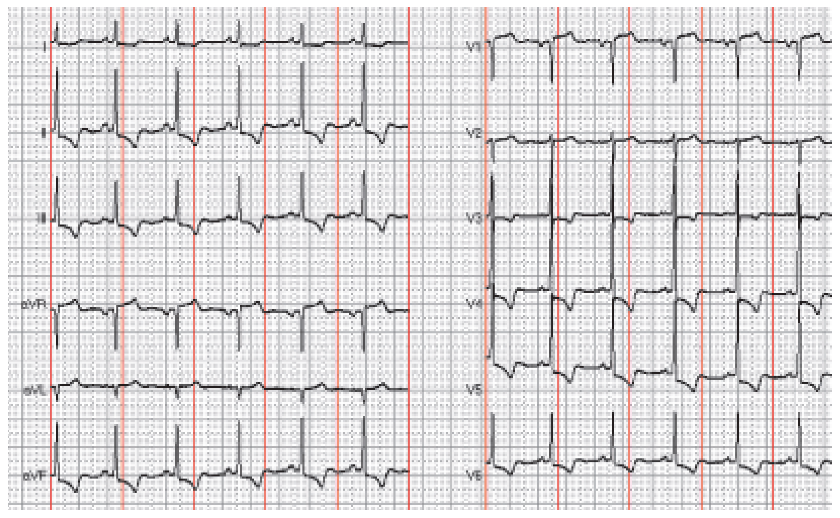
Figure 1. 12-lead-ECG at rest. See text for details.
Cardiovascmed 09 00276 g001
Figure 2. Specimen of right ventricular endomyocardial biopsy. See text for details.
Cardiovascmed 09 00276 g002