Next Article in Journal
Arterielle Hypertonie und Hypotrophe Beine bei Einem 10jährigen Bub
Previous Article in Journal
Palpitations Anciennes, Malaises Récents
 
 
Cardiovascular Medicine is published by MDPI from Volume 28 Issue 1 (2025). Previous articles were published by another publisher in Open Access under a CC-BY (or CC-BY-NC-ND) licence, and they are hosted by MDPI on mdpi.com as a courtesy and upon agreement with Editores Medicorum Helveticorum (EMH).
Interesting Images

Valve Prosthesis in the Tricuspid Position: An Useasy Relationship

1
Service de cardiologie, Hôpitaux Universitaires de Genève, Genève, Switzerland
2
Service de chirurgie cardio-vasculaire, Hôpitaux Universitaires de Genève, Genève, Switzerland
*
Author to whom correspondence should be addressed.
Cardiovasc. Med. 2006, 9(4), 167; https://doi.org/10.4414/cvm.2006.01163
Submission received: 28 January 2006 / Revised: 28 February 2006 / Accepted: 28 March 2006 / Published: 28 April 2006

Case report

A 27-year-old woman from Madagascar was referred to our institution because of right heart failure. At the age of 12 she had presented with severe tricuspid regurgitation consecutive to endocarditis of the tricuspid valve with a perforation of a sinus of Valsalva into the right ventricle. After a 6-week course of antibiotics the patient was sent to surgery. The tricuspid valve was not amenable to repair; thus, the surgeons replaced the tricuspid valve with a Porcine Bioprosthesis Medtronic® no. 29 and sutured the perforated sinus of Valsalva. During the post-operative period transient complete heart block was noted. Subsequently this conduction anomaly subsided; the patient was discharged and was lost to follow-up.
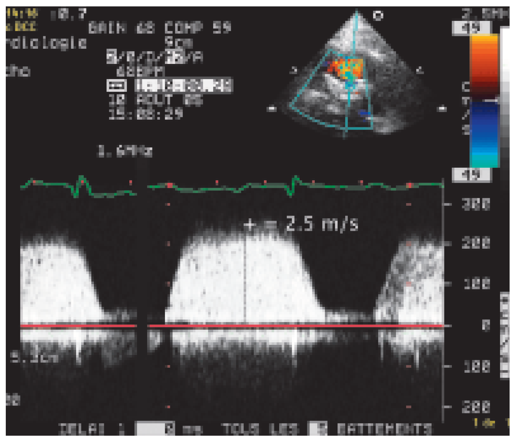
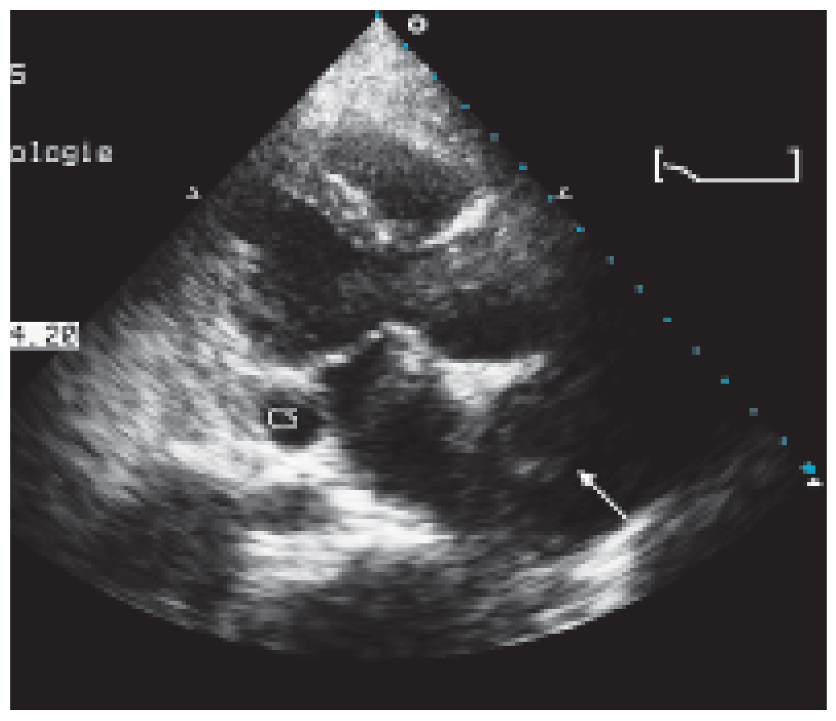
Fifteen years after her valve surgery the patient came again to medical attention because of dyspnea, ankle oedema and palpitations. On examination she appeared unwell, BP was 110/75 mm Hg, her pulse was irregularly irregular at 100 bpm. The jugular venous pressure was raised to the angle of the jaw and she had pitting oedema above the knees. On cardiac auscultation a 3/6 diastolic murmur increasing with inspiration was heard. The abdominal right upper quadrant was very tender to palpation and the liver was enlarged. The chest X-ray showed cardiomegaly and the ECG documented atrial fibrillation. Transthoracic echocardiography showed a calcified bioprosthesis in the tricuspid position (Figure 1) with very high gradients on continuous Doppler examination (Figure 2) and a markedly dilated right atrium. On the parasternal and the modified parasternal views a dilated coronary sinus was seen (Figure 3) consistent with severely elevated right atrial pressures. Two large heterogeneous masses filling the right atrium, one of which extending into the superior vena cava were interpreted as thrombi (Figure 3 and Figure 4).
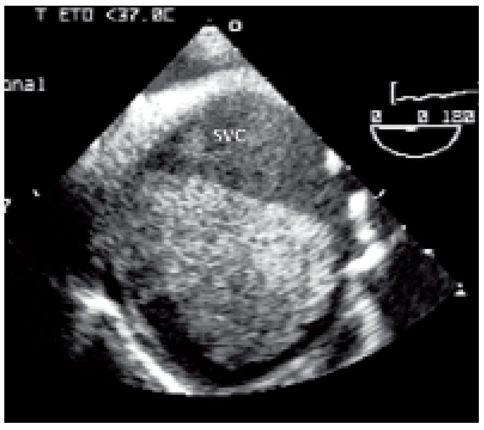
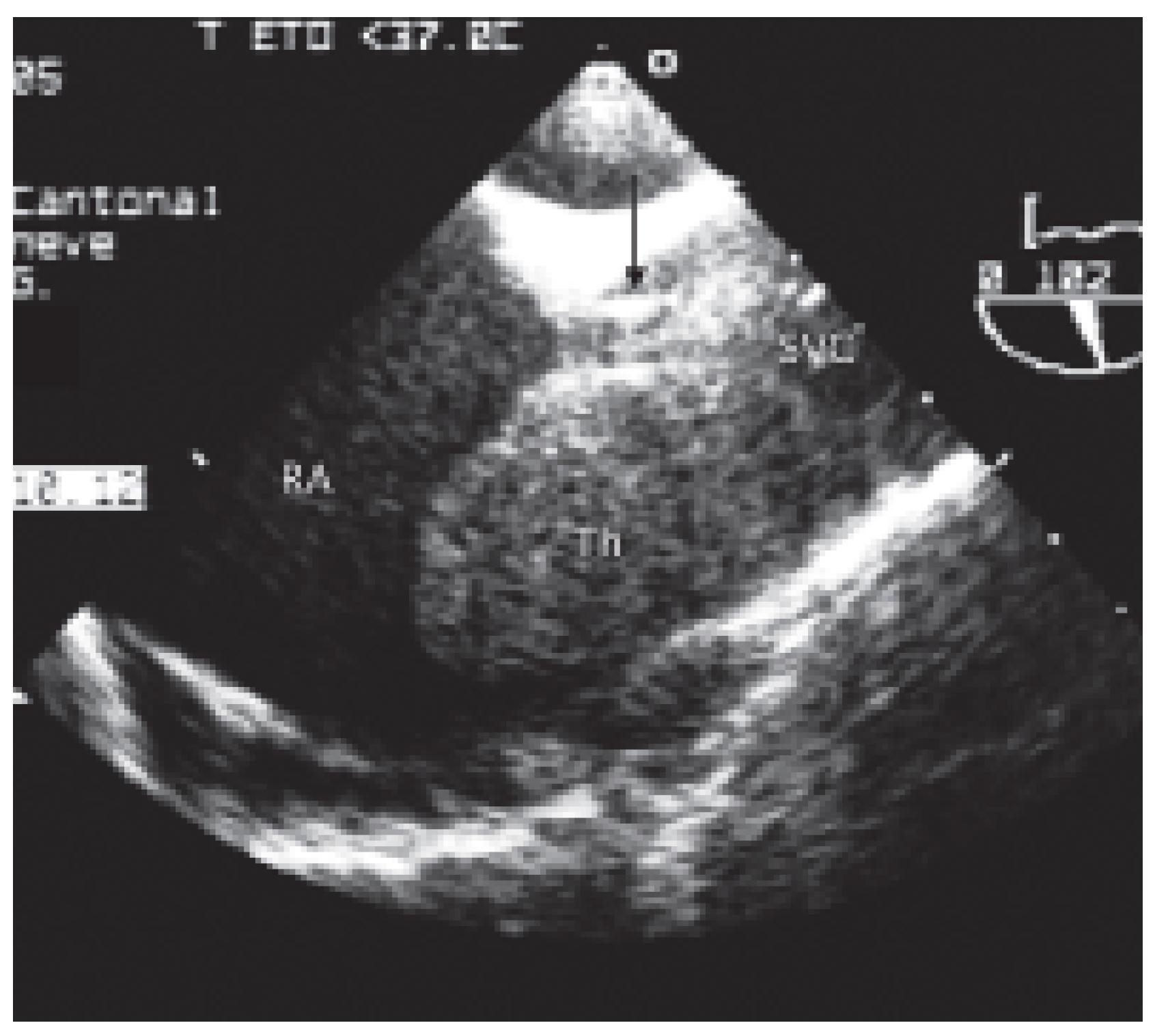
As the haemodynamic conditions became unstable the patient was taken to the operating theatre. Transoesophageal echocardiography showed a severely dilated superior vena cava (Figure 5) filled with thrombus and barely leaving place for the canula (Figure 6). The surgeons removed the clots and implanted a new bioprosthetic valve with a satisfactory result. The immediate right ventricular global systolic function was mildly decreased and the first 24 hours were uneventful. However, in the following hours right ventricular systolic function deteriorated rapidly, and did not recover in spite of a mechanical assist device. The patient died before heart transplantation could be performed.

Discussion

Replacement of the tricuspid valve (TVR) is currently reserved for those rare occasions where the valve is no longer amenable to repair. Despite advances in surgery and perioperative care, morbidity and mortality of TVR remain high, indicating that patients requiring TVR represent a high-risk group. Filsoufi et al. [1] have recently analysed a series of TVR performed in the current era; they showed that TVR continues to carry a high operative mortality (22%) and that long term survival is poor. In their series, ten year survival after TVR was 59% in patients with a mechanical prosthesis, and only 45% in patients who had been implanted a bioprosthesis.
The choice of the prosthesis remains a subject of debate [2]. In our case, the surgeon chose initially a bioprosthesis in spite of its shorter durability, because the patient was living in an environment where anticoagulation was difficult to monitor and in order to allow for easier management in case of pregnancy. The trade off of this choice was the degeneration of the bioprosthesis which led to calcification, severe tricuspid stenosis, right heart failure and clot formation.
Heart failure is the predominant cause of early and late deaths after TVR [1]; our case emphasizes the importance of regular followup after TVR and timely referral in case repeat surgery is needed.

References

  1. Filsoufi, F.; Anyanwu, A.C.; Salzberg, S.P.; Frankel, T.; Cohn, L.H.; Adams, D.H. Long-term outcomes of tricuspid valve replacement in the current era. Ann Thorac Surg 2005, 80, 845–850. [Google Scholar] [CrossRef] [PubMed]
  2. Rizzoli, G.; Vendramin, I.; Nesseris, G.; Bottio, T.; Guglielmi, C.; Schiavon, L. Biological or mechanical prostheses in tricuspid position? A meta-analysis of intra-institutional results. Ann Thorac Surg 2004, 77, 1607–1614. [Google Scholar] [CrossRef] [PubMed]
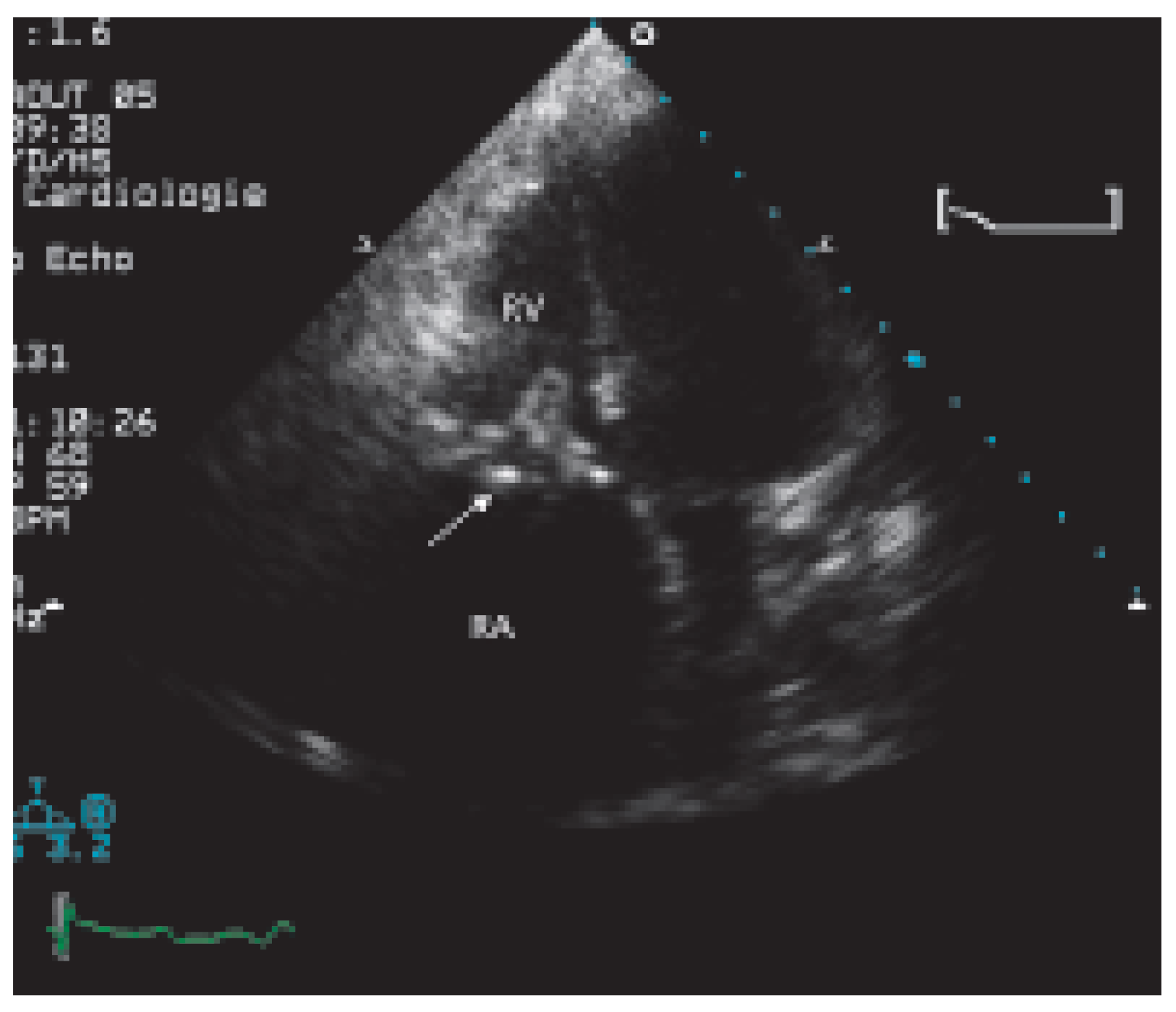
Figure 1. Transthoracic echocardiography (four-chamber view) shows a calcified bioprosthesis (arrow) in the tricuspid position and severe right atrial dilatation. RA = right atrium; RV = right ventricle.
Cardiovascmed 09 00167 g001
Figure 2. Continuous wave Doppler signal indicates an increased flow velocity through the tricuspid bioprosthesis (maximal flow velocity 2.5 m/s, peak gradient 26 mm Hg, mean gradient 20 mm Hg).
Cardiovascmed 09 00167 g002
Figure 3. Transthoracic parasternal long-axis view of the left ventricle shows marked dilatation of the coronary sinus consistent with an increased pressure in the right atrium. A heterogeneous mass is also seen in the dilated right atrium (arrow). CS = coronary sinus.
Cardiovascmed 09 00167 g003
Figure 4. A modified parasternal view of the atria depicts a large thrombus (arrow) in the right atrium. RA = right atrium; LA = left atrium.
Cardiovascmed 09 00167 g004
Figure 5. A transverse transoesophageal view shows a severely dilated superior vena cava with a large thrombus. SVC = superior vena cava.
Cardiovascmed 09 00167 g005
Figure 6. A vertical transoesophageal view of the superior vena cava. A large thrombus (Th) extends into the superior vena cava (SVC) and into the right atrium (RA) barely leaving space for the canula (arrow) inserted during surgery.
Cardiovascmed 09 00167 g006