Next Article in Journal
Risk Stratification of Pulmonary Embolism
Previous Article in Journal
Single Coronary Artery Originating from the Right Sinus Valsalva
 
 
Cardiovascular Medicine is published by MDPI from Volume 28 Issue 1 (2025). Previous articles were published by another publisher in Open Access under a CC-BY (or CC-BY-NC-ND) licence, and they are hosted by MDPI on mdpi.com as a courtesy and upon agreement with Editores Medicorum Helveticorum (EMH).
Editorial

Vena Cava Interruption and Mechanical Thrombectomy in Acute Pulmonary Embolism

Clinic of Cardiology, University Hospital Zurich, Rämistrasse 100, CH-8091 Zurich, Switzerland
Cardiovasc. Med. 2006, 9(4), 162; https://doi.org/10.4414/cvm.2006.01170
Submission received: 28 January 2006 / Revised: 28 February 2006 / Accepted: 28 March 2006 / Published: 28 April 2006

Summary

Most patients with pulmonary embolism will have an uneventful clinical course once therapeutic levels of anticoagulation are estabished. High risk patients, however, may require additional therapy to improve survival and prevent recurrent pulmonary embolism. This chapter focuses on inferior vena cava filter insertion, surgical embolectomy, and catheter interventions in pulmonary embolism.
Key words: pulmonary embolism; therapy

Zusammenfassung

Bei den meisten Patienten mit einer Lungenembolie kann ein komplikationsloser klinischer Verlauf erzielt werden, sobald ein therapeutischer Wirkstoffspiegel eines Gerinnungs hemmers eingestellt wurde. Bei Hochrisikopatienten kann jedoch eine zusätzliche Therapie erforderlich werden, um die Überlebenschancen zu verbessern und dem Wiederauftreten einer Lungenembolie vorzubeugen. Das folgende Kapitel beschäftigt sich mit der Behandlung durch Vena-cava-Filter, Embolektomie oder Katheter bei Lungenembolien.
Key words: Lungenembolie; Therapie

Vena cava interruption

Inferior vena cava filter (IVC) placement aims at preventing pulmonary embolism (PE) in high risk patients. The classical indications are (1.) failure of anticoagulation, ie, the occurrence of PE despite therapeutic levels of anticoagulation, and (2.) secondary prevention in patients with increased risk of both clotting and bleeding complications. The use of IVC filters has increased substantially in recent years, and the range of indications has broadened. Anticoagulation therapy is recommended with an IVC filter in place, if possible.
Although permanent IVC filters reduce the risk of PE, the risk of deep vein thrombosis (DVT) at one year, particularly due to filter thrombosis (Figure 1), is twice as high as in no-filter patients [1]. Newer retrievable IVC filters may be useful for patients with transiently increased risk of both clotting and bleeding complications, and many institutions now use retrievable filters in this setting. The various manufacturers of IVC filters recommend retrieval of the device within 2 weeks but retrievable filters have been removed by experienced interventionalists up to one year after placement.

Mechanical thrombectomy

In patients with massive PE, systemic thrombolysis in addition to anticoagulation is standard treatment, facilitating rapid reversal of right ventricular failure and cardiogenic shock [2]. However, approximately one third of the patients with massive PE are not eligible for thrombolysis because of major contraindications, such as recent surgery, trauma, stroke, or advanced cancer. PE thrombolysis is accompanied by a particularly high risk of bleeding complications. Among 304 patients from the International Cooperative Pulmonary Embolism registry (ICOPER) who received PE thrombolysis, 66 (21.7%) suffered major bleeding and 9 (3.0%) had intracranial bleeding [3].
The only alternative to thrombolysis for reversing PE-related right heart failure and cardiogenic shock are surgical embolectomy orpercutaneous catheter thrombectomy [4].

Surgical embolectomy

Few tertiary care centers offer surgical embolectomy with round-the-clock availability for patients with massive PE and contraindications to thrombolysis [5]. This operation mandates a median sternotomy, cardiopulmonary bypass, and incision of the main pulmonary artery (Figure 2). Cardioplegic circulatory arrest should be avoided to prevent further damage of the failing right ventricle. In the two largest PE registries, surgical embolectomy was used in only 1% of patients with massive PE and cardiogenic shock [3,6].
Surgical embolectomy in the setting of massive PE is especially useful when other cardiac surgery is required, such as removal of free-floating right heart thrombi or closure of an atrial septal defect or patent foramen ovale in the setting of paradoxical systemic embolism. The early mortality rate after surgical embolectomy is approximately 30%. The clinical outcome can be improved when surgical embolectomy is performed prior to the onset of decompensated cardiogenic shock. In a recent case series of 29 embolectomy patients, survival was 89% [5].

Percutaneous catheter thrombectomy

Catheter thrombectomy may be particularly useful if contraindications to thrombolysis are present or if surgical embolectomy is not feasible or not available.
The following three criteria should be fullfilled to consider catheter thrombectomy in a patient with acute PE [2]:
  • Pulmonary embolism and cardiogenic shock, defined as a systolic arterial pressure 90 mm Hg, a drop in systolic arterial pressure 40 mm Hg for 15 minutes, or ongoing administration of catecholamines for systemic arterial hypotension
  • Subtotal or total filling defect in the left and/or right main pulmonary artery by chest computed tomography or by conventional pulmonary angiography
  • Presence of at least one of the following contraindiations to PE thrombolysis:
    Active bleeding
    History of intracranial bleeding, or head injury, or ischaemic stroke, or brain tumor, or neurosurgery
    Surgery, delivery, organ biopsy, puncture of a non-compressible vessel within 10 days
    Gastrointestinal bleeding within 15 days
    Major trauma within 15 days
    Active cancer with known haemorrhagic risk
    Platelets <50 000 or INR >2.0
    Pregnancy

Percutaneous pulmonary embolism catheter devices

Greenfield embolectomy catheter

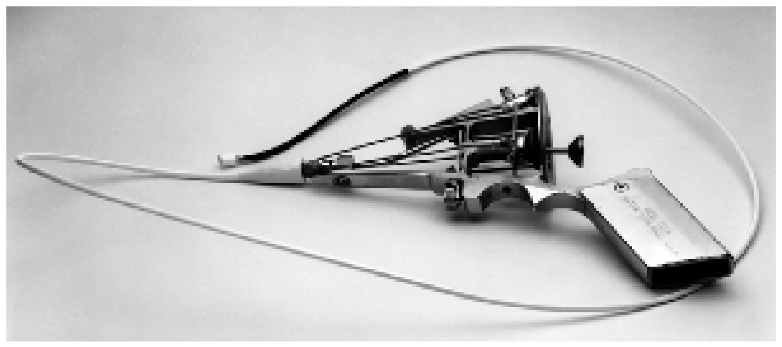
The Greenfield embolectomy device (Boston Scientific/Meditech, USA) is a 10-F, steerable catheter with a 5- or 7-mm plastic suction cup at the tip (Figure 3). This device was the first catheter that was designed for the treatment of massive pulmonary embolism and has been available for more than three decades. The major disadvantage is that it has to be inserted through a venotomy via femoral or jugular vein, and that there is no guide wire to advance the bulky device into the pulmonary circulation. The device removes the centrally located fresh embolus by manual suction with a large syringe, and requires retrieval of the device and the thrombus as a unit through the venotomy. In the hands of Dr. Greenfield, the device has been successful in extracting pulmonary thrombus in 76% of the patients, with significant improvement in haemodynamics [7,8]. The 30-day mortality rate was 30%.

Balloon angioplasty

Balloon angioplasty of obstructing emboli has been used for many years in an attempt to restore pulmonary blood flow and improve haemodynamics. Balloon angioplasty using balloon sizes of 6 to 16 mm results in compression of the embolus to the vessel wall but also to partial fragmentation of the thrombus with distal embolisation. Most of the patients who have been treated with balloon angioplasty also received local thrombolysis, with a decrease in pulmonary artery pressure over time. Therefore, it is unknown whether balloon angioplasty without concomitant thrombolysis is effective. Self-expanding wallstents [9] and self-expandable Gianturco Z stents [10] were used in patients with massive PE and failed thrombolysis or failed thrombus fragmentation.

Pigtail rotational catheter

The rotatable pigtail catheter (Cook Europe, The Netherlands) is a modified 5-F pigtail catheter with a radiopaque tip, with 10 side holes for contrast material injection. An oval side hole in the outer aspect of the pigtail loop allows direct passage of a 0.035-inch guidewire through the hole to act as a central axis around which the catheter rotates. The catheter is rotated bimanually and disrupts the clot in multiple smaller fragments, which embolise distally in the pulmonary circulation. In 20 patients with massive PE, catheter intervention with the pigtail rotational catheter showed a 33% recanalisation rate by fragmentation alone, but the catheter was more effective with adjuvant thrombolytic therapy with rt-PA[11]. Mortality in this series was 20%. One disadvantage with this catheter device is the risk of macroembolisation. Macroembolisation may cause further deterioration of haemodynamics when a large centrally located non-obstructive thrombus breaks and embolises into a previously non-obstructed lobar branch.

Hydrodynamic thrombectomy catheter devices

The AngioJet Xpeedior (Possis, Minneapolis, MN) is a 6-F, 120 cm over-the-wire catheter and is probably the most efficacious catheter among the hydrodynamic devices. A reversed high-pressure saline injection within the device creates negative pressure at the catheter tip (Venturi effect) and facilitates thrombus aspiration. However, since AngioJet was not designed to treat larger vessels of greater than 12 mm in dimension, it is also of limited effectiveness in the therapy of massive PE [12,13,14]. However, minor improvement in pulmonary perfusion often is sufficient to improve haemodynamics and clinical outcome in patients with massive PE.

Aspirex pulmonary embolism catheter

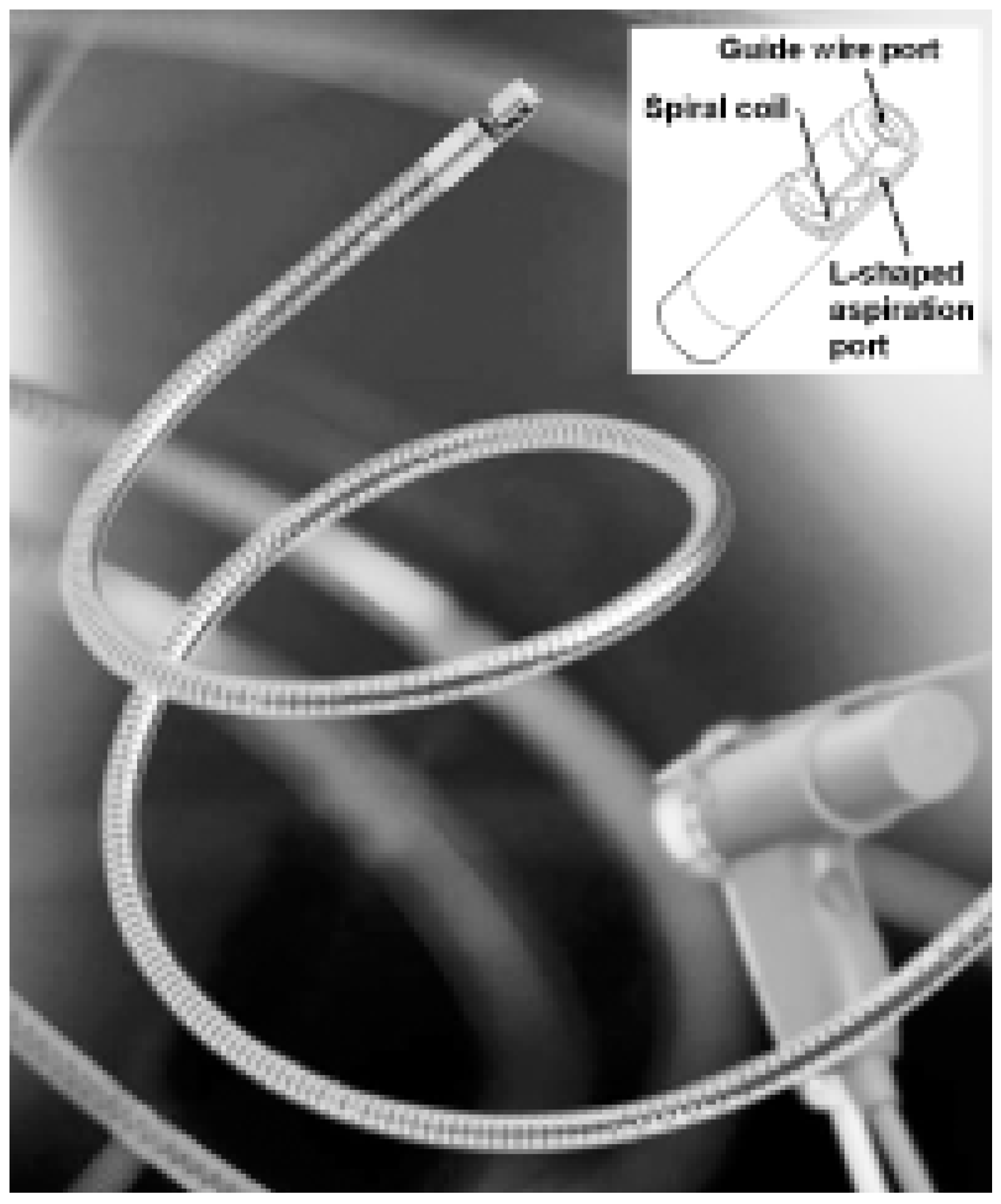
The Aspirex catheter thrombectomy device (Straub Medical, Switzerland) was specifically designed and developed for percutaneous interventional treatment of PE in pulmonary arteries, ranging from 6–14 mm in caliber. The central part of the catheter system is a highspeed rotational coil within the catheter body that (1.) creates negative pressure through an L-shaped aspiration port at the catheter tip, (2.) macerates aspirated thrombus, and (3.) removes macerated thrombus (Figure 4). The aspiration capacity of the Aspirex device was adjusted to remove thrombus from obstructed major pulmonary arteries and to minimise the risk of vascular collapse and vessel wall engagement. The Aspirex catheter is not associated with distal thrombus embolisation or haemolysis [15]. A large cohort study is currently being performed in Europe to investigate effectiviness and safety of the Aspirex device in patients with massive PE and contraindication to thrombolysis.

Catheter-directed thrombolysis

Catheter-directed thrombolytic therapy with intrapulmonary administration of a fibrinolytic drug has been used by several authors [16,17,18]. It aims to accelerate clot lysis and achieve rapid reperfusion of the pulmonary arteris. The technique requires positioning of a infusion catheter within the embolus, with injection of a bolus of thrombolytic drug followed by a continuous infusion. The following intrapulmonary thrombolytic regimens have been used in combination with a therapeutic infusion of unfractionated heparin in patients with massive PE: Urokinase 250 000 IU/h over 2 hours, followed by 100 000 IU/h of urokinase for 12–24 hours; alteplase bolus of 10 mg followed by 20 mg/h over 2 hours, or 100 mg over 7 hours. Short-acting, newer generation fibrinolytic drugs, such as alteplase (10–20 mg), reteplase (2.5–5 Units), or tenecteplase (5–10 mg) may be used.

Complications of catheter interventions

Rare but serious PE catheter thrombectomy complications include pericardial tamponade and pulmonary haemorrhage. The most serious complication is the perforation or dissection of a major pulmonary arterial branch that may cause massive pulmonary haemorrhage and immediate death. The myocardium of the right ventricle, particularly the right ventricular outflow tract, is thin and fragile, and caution is warranted to advance any device into the pulmonary arteries. The interventionalist must be able to perform an emergent pericardiocentesis in case of a perforation and should be familiar with measures to achieve rapid reversal of anticoagulation. To minimise the risk of perforation or dissection, thrombectomy should be performed only in the main and lobar pulmonary arteries, not in the segmental pulmonary arteries. Device-related complications also include blood loss and mechanical haemolysis, or arrhythmia from catheter passage through the right heart. Other complications include bleeding from heparin anticoagulation or local thrombolysis, contrast-induced nephropathy, anaphylactic reaction to iodine contrast, and vascular access complications, such as haematoma, pseudoaneurysm, or AV fistula.

References

  1. Decousus, H.; Leizorovics, A.; Parent, F. et al. A clinical trial of vena caval filters in the prevention of pulmonary embolism in patients with proximal deep-vein thrombosis. N Engl J Med 1998, 338, 409–415. [Google Scholar] [CrossRef] [PubMed]
  2. Kucher, N.; Goldhaber, S.Z. Management of massive pulmonary embolism. Circulation 2005, 112, e28–e32. [Google Scholar] [CrossRef] [PubMed]
  3. Goldhaber, S.Z.; Visani, L.; De Rosa, M. Acute pulmonary embolism: Clinical outcomes in the International Cooperative Pulmonary Embolism Registry (ICOPER). Lancet 1999, 353, 1386–1389. [Google Scholar] [CrossRef] [PubMed]
  4. Uflacker, R. Interventional therapy for pulmonary embolism. J Vasc Interv Radiol. 2001, 12, 147–164. [Google Scholar] [CrossRef] [PubMed]
  5. Aklog, L.; Williams, C.S.; Byrne, J.G. et al. Acute pulmonary embolectomy: A contemporary approach. Circulation 2002, 105, 1416–1419. [Google Scholar] [CrossRef] [PubMed]
  6. Kasper, W.; Konstantinides, S.; Geibel, A. et al. Management strategies and determinants of outcome in acute major pulmonary embolism: Results of a multicenter registry. J Am Coll Cardiol 1997, 30, 1165–1171. [Google Scholar] [CrossRef] [PubMed]
  7. Greenfield, L.J.; Proctor, M.C.; Williams, D.M.; Wakefield, T.W. Long-term experience with transvenous catheter pulmonary embolectomy. J Vasc Surg 1993, 18, 450–458. [Google Scholar] [CrossRef] [PubMed][Green Version]
  8. Greenfield, L.J.; Kimmell, G.O.; McCurdy, W.C. , 3rd. Transvenous removal of pulmonary emboli by vacuum-cup catheter technique. J Surg Res 1969, 9, 347–352. [Google Scholar] [CrossRef] [PubMed]
  9. Haskal, Z.J.; Soulen, M.C.; Huetti, E.A.; Palevsky, H.I.; Cope, C. Life-threatening pulmonary emboli and cor pulmonale: Treatment with percutaneous pulmonary artery stent placement. Radiology 1994, 191, 473–475. [Google Scholar] [CrossRef] [PubMed]
  10. Koizumi, J.; Kusano, S.; Akima, T. et al. Emergent Z stent placement for treatment of cor pulmonale due to pulmonary emboli after failed lytic treatment: Technical considerations. Cardiovasc Intervent Radiol 1998, 21, 254–255. [Google Scholar] [CrossRef] [PubMed]
  11. Schmitz-Rode, T.; Janssens, U.; Duda, S.H. et al. Massive pulmonary embolism: Percutaneous emergency treatment by pigtail rotation catheter. J Am Coll Cardiol 2000, 36, 375–380. [Google Scholar] [CrossRef] [PubMed]
  12. Muller-Hulsbeck, S.; Grimm, J.; Leidt, J.; Heller, M. In vitro effectiveness of mechanical thrombectomy devices for large vessel diameter and low-pressure fluid dynamic applications. J Vasc Interv Radiol 2002, 13, 831–839. [Google Scholar] [CrossRef] [PubMed]
  13. Koning, R.; Cribier, A.; Gerber, L. et al. A new treatment for severe pulmonary embolism. Circulation 1997, 96, 2498–2500. [Google Scholar] [CrossRef] [PubMed]
  14. Voigtlander, T.; Rupprecht, H.J.; Nowak, B. et al. Clinical application of a new rheolytic thrombectomy catheter system for massive pulmonary embolism. Cathet Cardiovasc Intervent 1999, 47, 91–96. [Google Scholar]
  15. Kucher, N.; Windecker, S.; Banz, Y.; Mettler, D.; Schmitz-Rode, T.; Meier, B. et al. Percutaneous catheter thrombectomy device for acute pulmonary embolism. Radiology 2005, 236, 852–858. [Google Scholar] [CrossRef] [PubMed]
  16. Gonzales-Juanatey, J.R.; Valdes, L.; Amaro, A. et al. Treatment of massive pulmonary thromboembolism with low intrapulmonary dosages of urokinase: Short term angiographic and hemodynamic evolution. Chest 1992, 102, 341–346. [Google Scholar] [CrossRef] [PubMed]
  17. Vujic, I.; Young, J.W.R.; Gobien, R.P. et al. Massive pulmonary embolism treatment with full heparinization and topical low-dose streptokinase. Radiology 1983, 148, 671–675. [Google Scholar] [CrossRef] [PubMed]
  18. Verstraete, M.; Miller, G.A.H.; Bounameaux, H. et al. Intravenous and intrapulmonary recombinant tissue-type plasminogen activator in the treatment of acute massive pulmonary embolism. Circulation 1988, 77, 353–360. [Google Scholar] [CrossRef] [PubMed]
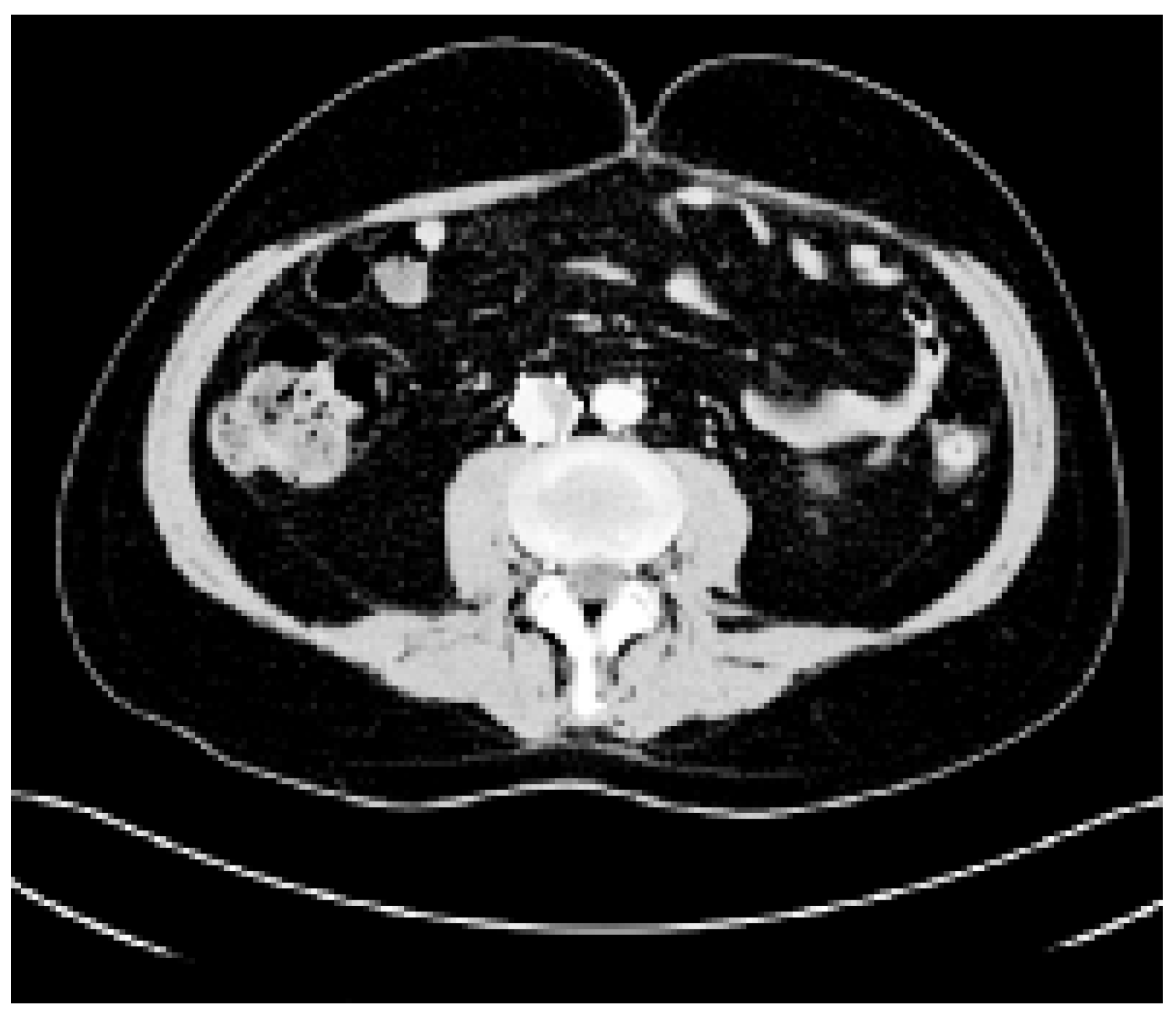
Figure 1. Inferior vena cava filter thrombosis in a patient with massive lower extremity deep vein thrombosis.
Cardiovascmed 09 00162 g001
Figure 2. Specimen from a surviving patient with massive PE who underwent emergent surgical embolectomy.
Cardiovascmed 09 00162 g002
Figure 3. The steerable 10-F Greenfield pulmonary embolectomy catheter. Figure kindly provided by Lazar Grienfield, M.D., Professor of Surgery and Chair Emeritus University of Michigan, MI.
Cardiovascmed 09 00162 g003
Figure 4. The 11-F Aspirex pulmonary embolism catheter is a promising percutaneous thrombectomy device. Figure kindly provided by the manufacturer, Straub Medical, Wangs, Switzerland.
Cardiovascmed 09 00162 g004