Next Article in Journal
Facharztprüfung für Die Erlangung des Facharzttitels Kardiologie / Examen de spécialiste en vue de l’obtention du titre en Cardiologie
Previous Article in Journal
Thrombolysis for Acute Pulmonary Embolism
 
 
Cardiovascular Medicine is published by MDPI from Volume 28 Issue 1 (2025). Previous articles were published by another publisher in Open Access under a CC-BY (or CC-BY-NC-ND) licence, and they are hosted by MDPI on mdpi.com as a courtesy and upon agreement with Editores Medicorum Helveticorum (EMH).
Editorial

Diagnosing Pulmonary Embolism

Division of General Internal Medicine, Department of Internal Medicine, Geneva University Hospital and Geneva Faculty of Medicine, Geneva, Switzerland
Cardiovasc. Med. 2006, 9(4), 136; https://doi.org/10.4414/cvm.2006.01167
Submission received: 28 January 2006 / Revised: 28 February 2006 / Accepted: 28 March 2006 / Published: 28 April 2006

Abstract

No single non invasive test has sufficient diagnostic accuracy to be used alone for diagnosing or ruling out pulmonary embolism. Therefore, modern diagnostic strategies for pulmonary embolism rely on combinations of non invasive tests such as plasma D-dimer measurement, lower limb venous compression ultrasonography, ventilation-perfusion lung scan and/or spiral CT, the results of which should be interpreted according to the clinical likelihood of pulmonary embolism. Pulmonary angiography is rarely necessary. Clinical probability of pulmonary embolism can be assessed with fair accuracy, either implicitly or by clinical prediction rules. Management studies in which patients deemed not to have pulmonary embolism are left untreated and followed up to assess their 3-month thromboembolic risk have become the benchmark for the validation of diagnostic algorithms. Haemodynamically unstable patients should be managed by quick strategies including echocardiography and ventilation-perfusion scintigraphy or spiral CT.

Résumé

Les tests non invasifs ne sont pas suffisamment précis pour permettre de diagnostiquerou d’exclure l’embolie pulmonaire lorsqu’ils sont employés isolément. C’est pourquoi les stratégies diagnostiques modernes reposent sur une combinaison de tests tels le dosage des D-dimères plasmatiques, l’échographie de compression veineuse des membres inférieurs, la scintigraphie de ventilation-perfusion et/ou le scanner spiralé. Les résultats de ces tests doivent être interprétés en tenant compte de la probabilité clinique d’embolie pulmonaire, qui peut être déterminée par le jugement clinique implicite ou par des règles de prédiction explicites. L’angiographie pulmonaire n’est plus qu’exceptionnellement nécessaire. Les études pragmatiques avec suivi des patients classés sans embolie pulmonaire et non traités par anticoagulants pour évaluer leur risque thromboembolique à 3 mois sont devenues la référence pour la validation de toute stratégie diagnostique de l’embolie pulmonaire. Les patients instables sur le plan hémodynamique doivent être investigués par des schémas rapides reposant sur l’échocardiographie et la scintigraphie de ventilation-perfusion ou le CT spiralé.
Mot-clefs: embolie pulmonaire; thrombose veineuse profonde; D-dimères; échographie veineuse; scintigraphie de ventilation-perfusion; scanner spirale; angiographie pulmonaire; évaluation clinique; échocardiographie

Introduction

Over the last fifteen years, an increasing number of tests have become available to diagnose pulmonary embolism. The North American Prospective Investigation On Pulmonary Embolism Diagnosis (PIOPED) study has definitively established the criteria for interpreting the results of ventilation-perfusion scintigraphy [1]. Plasma D-dimer measurement is now a validated and widely accepted first-line test for ruling out pulmonary embolism, provided it is used in combination with clinical probability and in the appropriate clinical setting [2,3,4]. Lower limb venous compression ultrasonography is a useful adjunct because of its high specificity for proximal deep venous thrombosis, the most frequent source of pulmonary embolism [5]. Finally, the advent of spiral computed tomography (CT) is without question a revolution in this diagnostic armamentarium [6,7]. This has, paradoxically, complicated the workup for suspected pulmonary embolism, due to the variety and complexity of diagnostic algorithms proposed in the literature [3,8,9,10]. Moreover, most algorithms were developed for the haemodynamically stable patient while the approach for suspected massive pulmonary embolism should be more direct and provide a central role to echocardiography. The aim of this article is therefore to provide a guide for combining those tests in a rational and costeffective way and review validated diagnostic strategies for the haemodynamically stable and unstable patient, respectively.

Why are diagnostic strategies necessary?

Pulmonary angiography is the only test with nearly ideal sensitivity and specificity for pulmonary embolism. Therefore, it was until recently the only test able to both rule in or out pulmonary embolism as a single procedure. However, it is invasive [11] and costly [12], and because it is but rarely performed, expertise for interpreting this difficult test is rapidly decreasing except in specialised centres. Therefore, it is generally a last resort. No single non invasive test allows reaching a definite diagnosis in all patients suspected of pulmonary embolism and it is therefore necessary to combine them. Multidetector-row CT [13] may serve that purpose, but even if this is established by further studies, it will still be more cost-effective to combine D-dimer to CT in order to spare unnecessary imaging studies. Moreover, the prevalence of pulmonary embolism has decreased from around 35% in the early 1990’s [1] to 20 to 25% in Europe [14] and even 10% in some North American centers [15]. Hence, cost-effective diagnostic strategies are required to avoid submitting a number of patients to a cascade of potentially costly tests. Finally, there is not one, but several valid diagnostic algorithms for suspected pulmonary embolism and individual institutions must select a strategy suited to their particular logistics and test availability.

What is a validated diagnostic strategy for pulmonary embolism?

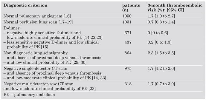
There is no absolute reference diagnostic criterion for pulmonary embolism and even pulmonary angiography is not completely accurate, due to frequent interobserver disagreement, especially at the subsegmental arterial level [1]. Furthermore, patients in whom anticoagulant treatment is withheld based on a angiogram negative for pulmonary embolism and followed-up during 3 months have a 1 to 2% rate of deep venous thrombosis or a pulmonary embolism during that period [16]. Therefore, the ultimate reference to judge the security of an exclusion criterion for pulmonary embolism is no longer the comparison with pulmonary angiography considered as the gold standard, but the results of clinical follow-up. The outcomes of follow-up should be at least as good as that associated with a negative pulmonary angiogram, ie a 1 to 2% 3-month thromboembolic risk in patients left untreated, with an upper limit of the 95% confidence interval below 4%. A rule-out criterion (whether a single test or a combination of tests) satisfying that condition does not exclude a small pulmonary embolus, but it allows ensuring the patient a safe outcome without anticoagulant treatment. Such studies are called outcome or management trials. Exclusion criteria validated in outcome studies are summarised in Table 1.

The role of ventilation-perfusion scintigraphy

This first rational diagnostic strategy for pulmonary embolism was based on the results of the PIOPED study [1]. That series which established the performance of ventilationperfusion scintigraphy in comparison with pulmonary angiography showed that a normal perfusion lung scan had an almost 100% negative predictive value for pulmonary embolism. The safety of that rule-out criterion has been verified by several outcome studies [17,18,19]. At the other end of the spectrum, a socalled “high-probability” scintigraphic pattern has a high specificity and positive predictive value for pulmonary embolism and is generally considered as an adequate rule-in criterion. Other scintigraphic results, ie low or intermediate probability according to the PIOPED interpretation criteria do not allow a definite conclusion and should be reported as non diagnostic. Therefore, performing ventilation-perfusion lung scintigraphy followed when non conclusive by an angiogram is rational. However, although well validated, that scheme is no longer widely used because of the high proportion (50 to 70%) of non diagnostic scintigraphic results.

The role of clinical probability assessment

In the PIOPED study [1], clinicians assigned each patient to one of three clinical probability categories (low, intermediate or high), based on an implicit clinical judgment incorporating the history and risk factors, physical examination and non specialised tests (chest X-ray, arterial blood gases and electrocardiogram).
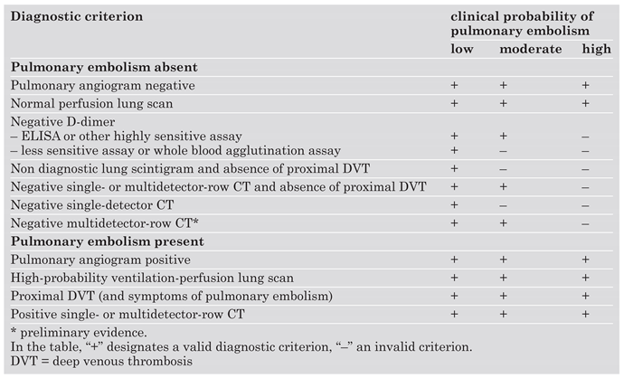
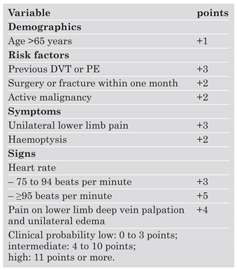
This clinical classification proved surprisingly accurate (prevalence of pulmonary embolism in the low probability group, 9%; in the intermediate probability group, 30%; and in the high probability group, 68%). Moreover, although the overall prevalence of PE was 13% in patients with a “low-probability” ventilation-perfusion scan, it was only 4% in patients with such a result and a low clinical probability of pulmonary embolism [1], low enough to rule out the disease without further testing. Hence, clinical probability is essential for interpreting the results of lung scintigraphy, but also for any diagnostic test for pulmonary embolism. The efficacy of various diagnostic criteria for ruling out pulmonary embolism according to clinical probability is shown in Table 2. Clinical probability can be validly assessed either implicitly or by prediction rules [15,20,21]. Table 3 shows the revised Geneva score [21] which attempts to combine the strengths of the Wells’ rule [15], which was based only on clinical variables, and of the former Geneva rule [20], which was entirely standardised.

The role of D-dimer

Plasma D-dimer is a product of the degradation of cross-linked fibrin and its measurement by a biological test is inexpensive. Acute venous thromboembolism provokes an elevation of plasma D-dimer in almost all patients because of the activation of coagulation and fibrinolysis. Therefore it carries a high sensitivity and is a good rule-out instrument. Its specificity is poor and an elevated D-dimer level does not rule in pulmonary embolism. Since most patients with suspected pulmonary embolism do not have the disease, it is logical to use D-dimer as a first-line test, after having assessed clinical probability of pulmonary embolism. When using a highly sensitive ELISA assay (sensitivity 95 to 100%), a negative result allows safely ruling out pulmonary embolism in patients with a low or moderate clinical probability as shown by several outcome studies [14,22,23]. Less sensitive assays (sensitivity 85 to 95%) should be restricted to patients with a low clinical probability (Table 2). D-dimer rules out pulmonary embolism in around one of every three patients suspected of pulmonary embolism in the emergency department. Due to a higher rate of false positive results, it rules out pulmonary embolism only in around one out of ten patients over 80 years, patients with cancer and inpatients [24] and performing a D-dimer test in such situations should be decided on a case-by-case basis. Whatever the diagnostic tests and sequence selected in patients with an elevated D-dimer, its use is highly cost-effective in emergency department patients, reducing costs by approximately 20% [12].

The role of lower limb venous compression ultrasonography

Most pulmonary emboli originate from clots located in the deep veins of the lower limbs, especially the proximal veins. Hence, finding a deep venous thrombosis in a patient admitted because of thoracic symptoms evocative of pulmonary embolism may be considered diagnostic, if not of pulmonary embolism, at least of acute venous thromboembolism, and warrants anticoagulant treatment. Since 40 to 50% of patients with pulmonary embolism have an ultrasound sowing a proximal deep venous thrombosis, performing ultrasonography before thoracic imaging allows a definite diagnosis in around one patient out of 10 given an overall prevalence of pulmonary embolism of 20%. The Dutch and North American teams reserve compression ultrasonography for patients with a negative CT or non diagnostic ventilation-perfusion scintigraphy. The diagnostic yield of ultrasonography in such patients is much lower. However, lower limb ultrasonography must be performed in patients with a negative single-detector CT because of the low 70% sensitivity of first-generation CT scans (see further section). The only well-validated rule-out criterion in well-designed outcome studies using single-detector CT is the combination a negative ultrasound and CT in a patient with a low or moderate clinical probability of pulmonary embolism. Ultrasonography is probably no longer necessary in patients with a negative multi-detector row CT because of that test’s high sensitivity. However, even in centers equipped with multi-detector row CT, ultrasound might still be useful to reduce costs and avoid unnecessary irradiation in patients with suspected pulmonary embolism and a deep venous thrombosis [12].

The role of echocardiography

Doppler echocardiography has several uses in suspected pulmonary embolism and it may play a role in risk stratification [25]. In a very small subset of patients with pulmonary embolism, transthoracic echocardiography allows a direct visualisation of the clot in the right heart chambers or in the right main pulmonary artery. The most frequent echocardiographic manifestations of pulmonary embolism are indirect and reflect the haemodynamic changes caused by an acute increase in pulmonary arterial resistance and pulmonary hypertension. Pulmonary arterial pressure may be estimated in most patients by the tricuspid regurgitation velocity. Signs of right ventricular strain include dilation of the right ventricle, right ventricular hypokinesis, and in severe cases, paradoxical motion of the interventricular septum. Several echocardiographic measurements have been proposed to quantify right ventricular dilation, of which the most standardised is the right ventricle over left ventricle diameter ratio. The sensitivity of these signs which are often combined, lies between 40 and 70% in clinically suspected pulmonary embolism, and their specificity is approximately 90%, provided the patient does not have another disease causing chronic pulmonary hypertension [26]. Echocardiography is the first-line test in suspected massive pulmonary embolism (see further section). Whether echocardiography should also be performed in haemodynamically stable patients with confirmed pulmonary embolism is still controversial. Patients with submassive pulmonary embolism, ie echocardiographic signs of right ventricular strain despite a normal arterial blood pressure, have a higher mortality compared with those who have a normal right ventricular function. That subset of patients can only be identified by echocardiography, but the evidence that thrombolytic treatment improves their prognosis is still scarce and relies on a single randomised trial [27].

Diagnostic strategy based on ventilation-perfusion lung scintigraphy

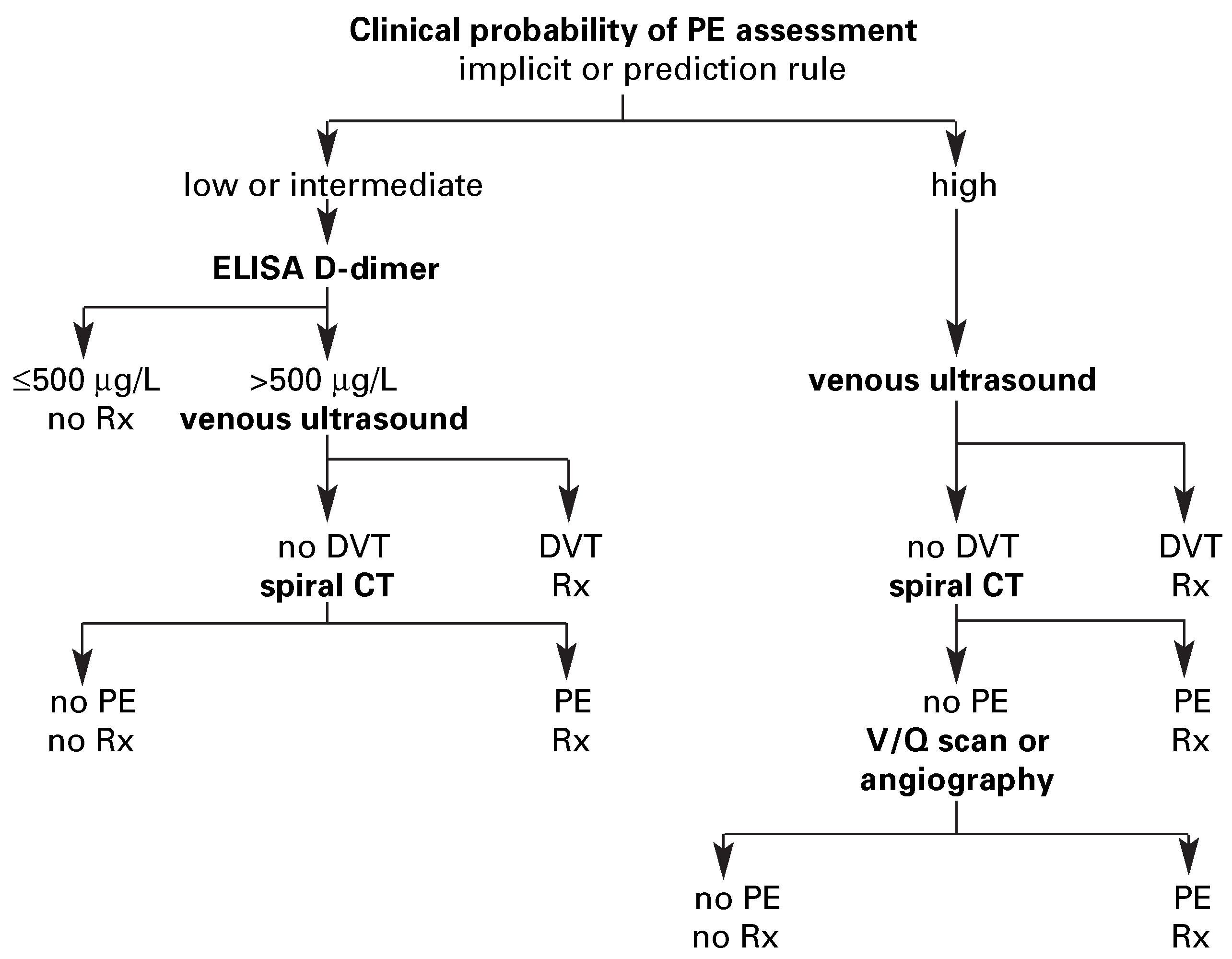
The diagnostic algorithm illustrated in Figure 1 is derived from the scintigraphy-angiography strategy described in a previous section. Measuring D-dimer as the initial test allows ruling out pulmonary embolism in around 30% of patients in the emergency department. D-dimer is negative in only 10% of patients with a high clinical probability [28]. As only 10% of all patients had a high clinical likelihood of pulmonary embolism, the overall diagnostic yield of measuring D-dimer in such patients is negligible (around 1%) and the absolute number of patients with that combination was too low to establish the safety of this rule-out criterion. Therefore, D-dimer measurement is not recommended in patients with a high clinical probability. A negative D-dimer is an adequate rule-out criterion in patients with a low-moderate clinical probability when using a highly sensitive assay, and in patients with a low clinical probability with a less sensitive assay. In this scheme, lower limb venous compression ultrasonography is the secondline test in patients with an elevated D-dimer level and the initial test in patients with a high clinical probability. It discloses a deep venous thrombosis in around 10% of patients, therefore ventilation-perfusion scintigraphy must be performed in only 50 to 60% of patients, establishing a definite diagnosis in around 15% of the entire cohort. The 3-month thromboembolic risk is less than 2% in patients who have a low clinical probability, a non diagnostic ventilation-perfusion scan and absence of proximal deep venous thrombosis [29,30]. That combination is found in around 20% of patients and is an adequate rule-out criterion. In an outcome study evaluating this algorithm, pulmonary angiography was required in only 10% of patients and the 3-month thromboembolic risk in patients left untreated based on the rule-out criteria of the strategy was around 1% [22]. According to test availability, D-dimer measurement may be forfeited without compromising patient safety although global costs and the number of necessary angiograms will be increased. In contrast, lower limb venous ultrasound is required to rule out pulmonary embolism in case of a non diagnostic ventilation-perfusion scan and a low clinical probability. Despite the growing utilisation of CT, this algorithm remains of interest in centers with easier access to scintigraphy than CT, and in patients with contraindications to CT (mainly renal failure and allergy to contrast dye).

Diagnostic strategy based on spiral computed tomography

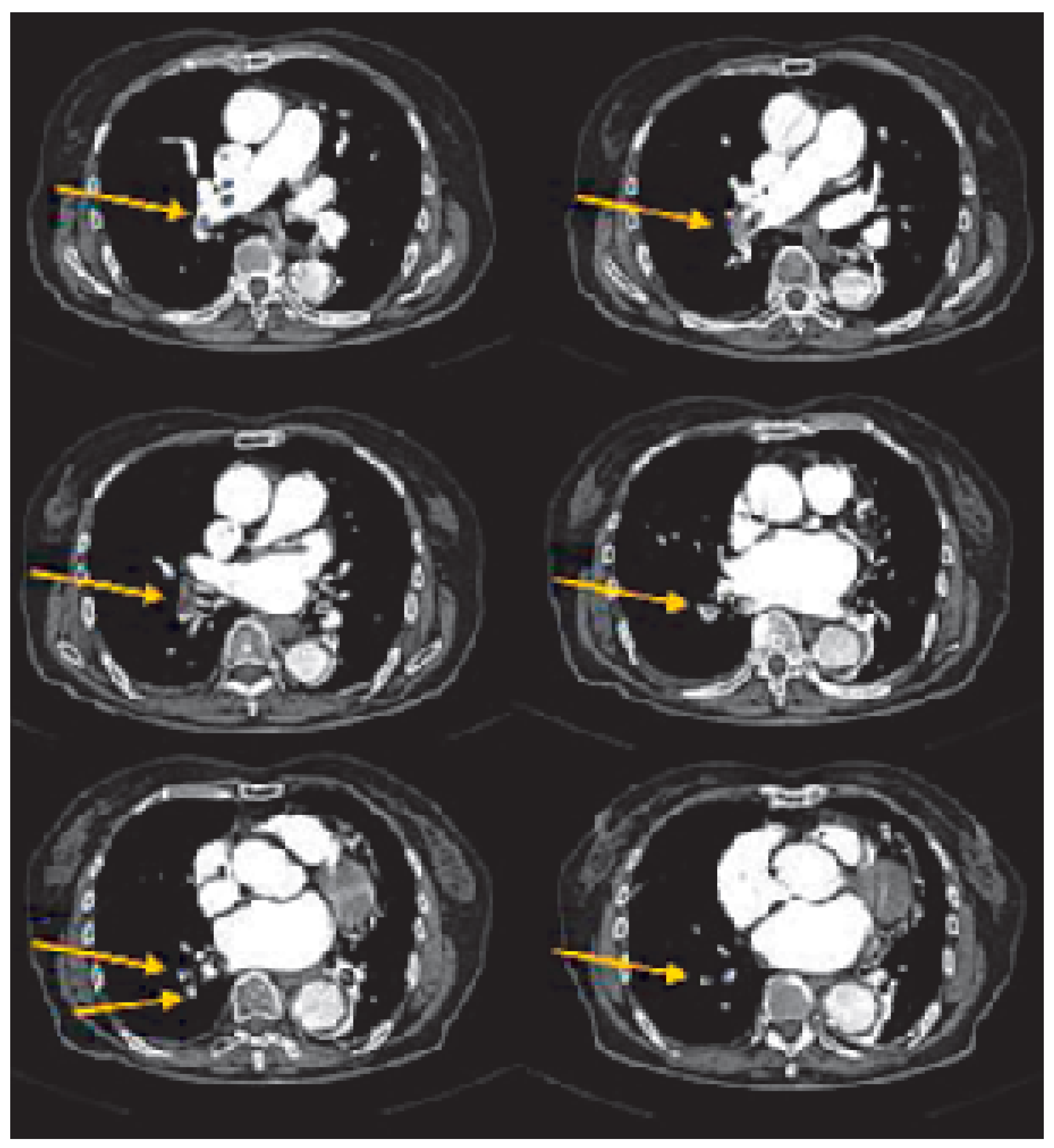
That algorithm is illustrated in Figure 2. The initial steps are the same as those of the algorithm based on ventilation-perfusion scintigraphy (Figure 1) but CT is performed instead in patients with an elevated D-dimer level and a negative lower limb venous ultrasound. CT is required in 50 to 60% of patients. Due to its low sensitivity (70%) [31,32], a negative single-detector CT does not allow ruling out pulmonary embolism, while the combination of a negative CT, a negative lower limb ultrasound and a low-moderate clinical probability safely rules out pulmonary embolism as shown by recent outcome studies [14,33,34]. For high clinical probability patients who have a negative CT and ultrasound, further testing by ventilationperfusion scintigraphy and/or pulmonary angiography is still recommended. However, this combination is rare: in the French multicentre “Evaluation du Scanner Spiralé dans l’Embolie Pulmonaire” (ESSEP) study, only 76 of the 1041 patients (7%) had such findings, of whom only 4 had a pulmonary embolism [33]. In the CTEP2 study, a three-centre outcome study by the Geneva group validating the algorithm shown in Figure 2, an angiogram was required in only 2% of patients [14]. Data on multi-detector row CT (Figure 3) are still scarce and well-designed prospective outcome studies are lacking. In the latest study from the Geneva group, all patients with elevated Ddimer levels or a high clinical probability underwent both multi-detector row CT and lower limb ultrasonography [23]. The main study hypothesis was that if multi-detector row CT has a nearly ideal sensitivity, the proportion of patients with a negative CT despite an ultrasound showing a proximal deep venous thrombosis and the 3-month thromboembolic risk in patients with a negative CT should be low. CT and ultrasonography were negative in 318 patients, of whom 3 had a definite thromboembolic event and 2 died of possible pulmonary embolism during follow-up (three-month risk of thromboembolism 1.7%; 95% CI 0.7 to 3.9). Only two patients had proximal deep venous thrombosis and a negative CT scan (0.6%; 95% CI 0.2 to 2.2). Therefore, the overall threemonth risk of thromboembolism in patients without pulmonary embolism would have been 1.5% (95% CI 0.8 to 3.0) if D-dimer and multidetector row CT had been the only tests used to rule out pulmonary embolism and ultrasonography had not been performed. This suggests that multi-detector row CT might be used as a stand-alone test for suspected pulmonary embolism, but should be confirmed in proper large-scale outcome studies, such as the CHRISTOPHER study, the results of which have just been published [35].

Diagnostic strategy for the haemodynamically unstable patient

The algorithms described in the previous sections do not apply to patients admitted with arterial hypotension and/or shock because patients with suspected massive pulmonary embolism have a very high mortality rate and require emergent thrombolytic treatment if pulmonary embolism is confirmed. Since the D-dimer result is very unlikely to be negative and a negative venous ultrasound would require further testing, those tests would only delay life-saving therapy. In patients with shock, echocardiography is extremely effective for differential diagnosis with tamponade and cardiogenic shock. Moreover, absence of pulmonary hypertension and/or right ventricular dilation and hypokinesis in that situation renders pulmonary embolism as the cause of shock unlikely. Therefore, the logical initial test in such patients is transthoracic echocardiography. An echocardiogram showing signs of acute pulmonary hypertension and right ventricular strain in a shocked patient with a normal left ventricular contractility is a very strong argument in favor of massive pulmonary embolism. In fact, most clinicians would readily begin thrombolytic treatment in such a patient without awaiting further diagnostic information, if the patient were highly unstable. On the other hand, in a patient temporarily stabilised by vasopressive drugs, confirmation may be sought by either ventilationperfusion lung scan or spiral CT, whichever is the most rapidly available. Angiography should be avoided whenever possible since it carries the highest risk in this patient population [11] and increases the risk of a major bleed at the puncture site due to thrombolytic treatment [36].

Conclusions

Numerous algorithms now exist for suspected non massive pulmonary embolism. Clinical probability assessment has become an incontrovertible first step, generally followed by plasma D-dimer measurement. Venous ultrasound may precede or follow thoracic imaging, which may consist of ventilation-perfusion scintigraphy or spiral CT. In both cases, a negative venous ultrasound is required to rule out pulmonary embolism in patients with a non diagnostic lung scintigraphy or a negative single-detector CT. Pulmonary angiography is now rarely necessary. Finally, echocardiography is very useful in haemodynamically unstable patients, but its role in stable patients is still controversial.

References

  1. The PIOPED Investigators. Value of the ventilation-perfusion scan in acute pulmonary embolism. JAMA 1990, 263, 2753–2759. [Google Scholar] [CrossRef]
  2. Stein, P.D.; Hull, R.D.; Patel, K.C.; Olson, R.E.; Ghali, W.A.; Brant, R. et al. D-Dimer for the exclusion of acute venous thrombosis and pulmonary embolism: A systematic review. Ann Intern Med 2004, 140, 589–602. [Google Scholar] [CrossRef] [PubMed]
  3. Kruip, M.J.; Leclercq, M.G.; van der Heul, C.; Prins, M.H.; Buller, H.R. Diagnostic strategies for excluding pulmonary embolism in clinical outcome studies. A systematic review. Ann Intern Med 2003, 138, 941–951. [Google Scholar] [CrossRef]
  4. Bounameaux, H.; Perrier, A. D-dimer in the diagnosis of venous thromboembolism. In Venous Thromboembolism; Dalen, J.E., Ed.; Marcel Dekker Inc: New York, NY, USA, 2003; pp. 133–148. [Google Scholar]
  5. Kearon, C.; Ginsberg, J.S.; Hirsh, J. The role of venous ultrasonography in the diagnosis of suspected deep venous thrombosis and pulmonary embolism. Ann Intern Med 1998, 129, 1044–1049. [Google Scholar] [CrossRef] [PubMed]
  6. Rathbun, S.W.; Raskob, G.E.; Whitsett, T.L. Sensitivity and specificity of helical computed tomography in the diagnosis of pulmonary embolism: A systematic review. Ann Intern Med 2000, 132, 227–232. [Google Scholar] [CrossRef] [PubMed]
  7. Mullins, M.D.; Becker, D.M.; Hagspiel, K.D.; Philbrick, J.T. The role of spiral volumetric computed tomography in the diagnosis of pulmonary embolism. Arch Intern Med 2000, 160, 293–298. [Google Scholar] [CrossRef]
  8. Fedullo, P.F.; Tapson, V.F. Clinical practice. The evaluation of suspected pulmonary embolism. N Engl J Med 2003, 349, 1247–1256. [Google Scholar] [CrossRef]
  9. Goldhaber, SZ. Pulmonary embolism. Lancet 2004, 364, 244. [Google Scholar] [CrossRef]
  10. British Thoracic Society guidelines for the management of suspected acute pulmonary embolism. Thorax 2003, 58, 470–483. [CrossRef]
  11. Stein, P.D.; Athanasoulis, C.; Alavi, A.; Greenspan, R.H.; Hales, C.A.; Saltzman, H.A. et al. Complications and validity of pulmonary angiography in acute pulmonary embolism. Circulation 1992, 85, 462–468. [Google Scholar] [CrossRef]
  12. Perrier, A.; Nendaz, M.R.; Sarasin, F.P.; Howarth, N.; Bounameaux, H. Cost-effectiveness of diagnostic strategies for suspected pulmonary embolism including helical computed tomography. Am J Respir Crit Care Med 2003, 167, 39–44. [Google Scholar] [CrossRef] [PubMed]
  13. Schoepf, U.J.; Costello, P. CT Angiography for diagnosis of pulmonary embolism: State of the art. Radiology 2004, 230, 329–337. [Google Scholar] [CrossRef]
  14. Perrier, A.; Roy, P.M.; Aujesky, D.; Chagnon, I.; Howarth, N.; Gourdier, A.L. et al. Diagnosing pulmonary embolism in outpatients with clinical assessment, D-dimer measurement, venous ultrasound, and helical computed tomography: A multicenter management study. Am J Med 2004, 116, 291–299. [Google Scholar] [CrossRef] [PubMed]
  15. Wells, P.S.; Anderson, D.R.; Rodger, M.; Stiell, I.; Dreyer, J.F.; Barnes, D. et al. Excluding pulmonary embolism at the bedside without diagnostic imaging: Management of patients with suspected pulmonary embolism presenting to the emergency department by using a simple clinical model and D-dimer. Ann Intern Med 2001, 135, 98–107. [Google Scholar] [CrossRef]
  16. Van Beek, E.J.; Brouwerst, E.M.; Song, B.; Stein, P.D.; Oudkerk, M. Clinical validity of a normal pulmonary angiogram in patients with suspected pulmonary embolism – a critical review. Clin Radiol 2001, 56, 838–842. [Google Scholar] [CrossRef]
  17. Hull, R.D.; Raskob, G.E.; Coates, G.; Panju, A.A. Clinical validity of a normal perfusion lung scan in patients with suspected pulmonary embolism. Chest 1990, 97, 23–26. [Google Scholar] [CrossRef]
  18. Kipper, M.S.; Moser, K.M.; Kortman, K.E.; Ashburn, W.L. Longterm follow-up of patients with suspected pulmonary embolism and a normal lung scan. Chest 1982, 82, 411–415. [Google Scholar] [CrossRef]
  19. Van Beek, E.J.R.; Kuyer, P.M.M.; Schenk, B.S.; Brandjes, D.P.M.; ten Cate, J.W.; Büller, H.R. A normal perfusion lung scan in patients with clinically suspected pulmonary embolism. Frequency and clinical validity. Chest 1995, 108, 170–173. [Google Scholar] [CrossRef]
  20. Wicki, J.; Perneger, T.V.; Junod, A.; Bounameaux, H.; Perrier, A. Assessing clinical probability of pulmonary embolism in the emergency ward: A simple score. Arch Intern Med 2001, 161, 92–97. [Google Scholar] [CrossRef]
  21. Le Gal, G.; Righini, M.; Roy, P.M.; Sanchez, O.; Aujesky, D.; Bounameaux, H. et al. Prediction of pulmonary embolism in emergency patients: The revised Geneva score. Ann Intern Med 2005, in press. [Google Scholar]
  22. Perrier, A.; Desmarais, S.; Miron, M.J.; de Moerloose, P.; Lepage, R.; Slosman, D. et al. Non-invasive diagnosis of venous thromboembolism in outpatients. Lancet 1999, 353, 190–195. [Google Scholar] [CrossRef]
  23. Perrier, A.; Roy, P.M.; Sanchez, O.; Le Gal, G.; Meyer, G.; Gourdier, A.L. et al. Multi-detector row computed tomography in suspected pulmonary embolism. N Engl J Med 2005, 352, 1760–1768. [Google Scholar] [CrossRef]
  24. Perrier, A. D-dimer for suspected pulmonary embolism: Whom should we test? Chest 2004, 125, 807–809. [Google Scholar] [CrossRef] [PubMed]
  25. Goldhaber, S.Z. Echocardiography in the management of pulmonary embolism. Ann Intern Med 2002, 136, 691–700. [Google Scholar] [CrossRef]
  26. Roy, P.M.; Colombet, I.; Durieux, P.; Chatellier, G.; Sors, H.; Meyer, G. Systematic review and meta-analysis of strategies for the diagnosis of suspected pulmonary embolism. BMJ 2005, 331, 259. [Google Scholar] [CrossRef] [PubMed]
  27. Konstantinides, S.; Geibel, A.; Heusel, G.; Heinrich, F.; Kasper, W. Heparin plus alteplase compared with heparin alone in patients with submassive pulmonary embolism. N Engl J Med 2002, 347, 1143–1150. [Google Scholar] [CrossRef]
  28. Righini, M.; Aujesky, D.; Roy, P.M.; Cornuz, J.; de Moerloose, P.; Bounameaux, H. et al. Clinical usefulness of D-dimer depending on clinical probability and cutoff value in outpatients with suspected pulmonary embolism. Arch Intern Med 2004, 164, 2483–2487. [Google Scholar] [CrossRef] [PubMed]
  29. Perrier, A.; Miron, M.J.; Desmarais, S.; de Moerloose, P.; Slosman, D.; Didier, D. et al. Using clinical evaluation and lung scan to rule out suspected pulmonary embolism: Is it a valid option in patients with normal results of lower-limb venous compression ultrasonography? Arch Intern Med 2000, 160, 512–516. [Google Scholar] [CrossRef][Green Version]
  30. Wells, P.S.; Ginsberg, J.S.; Anderson, D.R.; Kearon, C.; Gent, M.; Turpie, A.G. et al. Use of a clinical model for safe management of patients with suspected pulmonary embolism. Ann Intern Med 1998, 129, 997–1005. [Google Scholar] [CrossRef]
  31. Perrier, A.; Howarth, N.; Didier, D.; Loubeyre, P.; Unger, P.F.; de Moerloose, P. et al. Performances of helical computed tomography in unselected outpatients with suspected pulmonary embolism. Ann Intern Med 2001, 135, 88–97. [Google Scholar] [CrossRef]
  32. Van Strijen, M.J.; De Monye, W.; Kieft, G.J.; Pattynama, P.M.; Prins, M.H.; Huisman, M.V. Accuracy of single-detector spiral CT in the diagnosis of pulmonary embolism: A prospective multicenter cohort study of consecutive patients with abnormal perfusion scintigraphy. J Thromb Haemost 2005, 3, 17–25. [Google Scholar] [CrossRef]
  33. Musset, D.; Parent, F.; Meyer, G.; Maitre, S.; Girard, P.; Leroyer, C. et al. Diagnostic strategy for patients with suspected pulmonary embolism: A prospective multicentre outcome study. Lancet 2002, 360, 1914–1920. [Google Scholar] [CrossRef] [PubMed]
  34. Van Strijen, M.J.; De Monye, W.; Schiereck, J.; Kieft, G.J.; Prins, M.H.; Huisman, M.V. et al. Single-detector helical computed tomography as the primary diagnostic test in suspected pulmonary embolism: A multicenter clinical management study of 510 patients. Ann Intern Med 2003, 138, 307–314. [Google Scholar] [CrossRef] [PubMed]
  35. Van Belle, A.; Buller, H.R.; Huisman, M.V.; Huisman, P.M.; Kaasjager, K.; Kamphuisen, P.W. et al. Christopher Study Investigators. Effectiveness of managing suspected pulmonary embolism using an algorithm combining clinical probability, D-dimer testing, and computed tomography. JAMA 2006, 295, 172–179. [Google Scholar] [PubMed]
  36. Wan, S.; Quinlan, D.J.; Agnelli, G.; Eikelboom, J.W. Thrombolysis compared with heparin for the initial treatment of pulmonary embolism: A meta-analysis of the randomize controlled trials. Circulation 2004, 110, 744–749. [Google Scholar] [CrossRef]
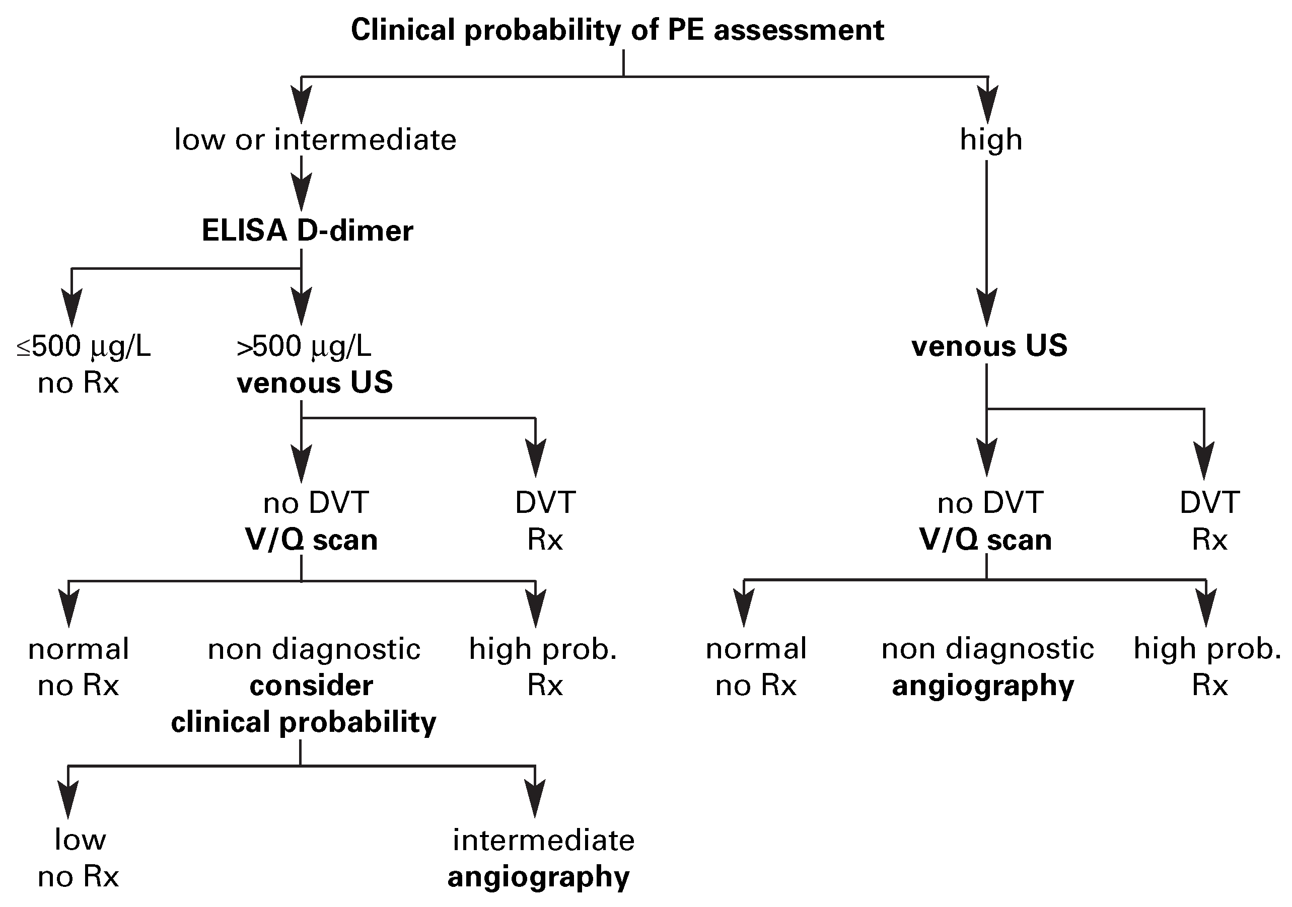
Figure 1. Validated diagnostic algorithm for suspected pulmonary embolism (PE) including spiral CT. Note that ELISA D-dimer is not useful in high-probability patients. Also, venous ultrasound must be performed when using single-detector spiral CT but it can be done either before or after CT if CT is negative. Venous ultrasound is probably unnecessary when using multidetector CT. Rx = treatment.
Cardiovascmed 09 00136 g001
Figure 2. Validated diagnostic algorithm for suspected pulmonary embolism (PE) including ventilation-perfusion (V/Q) scan. Note that ELISA D-dimer is not useful in high-probability patients. Also, venous ultrasound must be performed when using V/Q scan and the result of V/Q scan is non diagnostic, whether before or after that test. Rx = treatment.
Cardiovascmed 09 00136 g002
Figure 3. Multi-detector row CT showing a central clot in the right main pulmonary artery extending in the right lower lobar artery and its segmental branches.
Cardiovascmed 09 00136 g003
Table 1. 3-month thromboembolic risk associated with ruling out pulmonary embolism by various diagnostic criteria.
Cardiovascmed 09 00136 i001
Table 2. Appropriateness of diagnostic criteria for pulmonary embolism according to clinical probability.
Cardiovascmed 09 00136 i002
Table 3. The revised Geneva score [21].
Cardiovascmed 09 00136 i003