Next Article in Journal
Unaufhörliche Supraventrikuläre Tachykardie
Previous Article in Journal
CARTA 05, Zweiter Cardiovascular Roundtable in Weggis
 
 
Cardiovascular Medicine is published by MDPI from Volume 28 Issue 1 (2025). Previous articles were published by another publisher in Open Access under a CC-BY (or CC-BY-NC-ND) licence, and they are hosted by MDPI on mdpi.com as a courtesy and upon agreement with Editores Medicorum Helveticorum (EMH).
Interesting Images

Traumatic Tamponade and Ventricular Septal Defect

by
J. Alibegovic
1,
Y. Aggoun
1,
Cédric Vuille
1,2,* and
on behalf of the Catheterisation and Echocardiography Laboratories (U. Sigwart and R. Lerch)
1
1
Cardiology Service and Paediatric Unit, Geneva University Hospital, Geneva, Switzerland
2
Service de Cardiologie, Hôpitaux Universitaires de Genève, 24, rue Micheli-du-Crest, CH-1211 Genève 14, Switzerland
*
Author to whom correspondence should be addressed.
Cardiovasc. Med. 2006, 9(2), 83; https://doi.org/10.4414/cvm.2006.01154
Submission received: 24 November 2005 / Revised: 24 December 2005 / Accepted: 24 January 2006 / Published: 24 February 2006

Case report

A 30-year-old man sustained a knife wound in the 9th left intercostal space when he was involved in a fight as a bodyguard in a night club. In the emergency room he developed hypotension and dyspnea, with a heart rate of 110 bpm and on the ECG electrical alternans was seen. The chest X-ray showed no pneumothorax. An echocardiogram revealed an important pericardial effusion with diastolic compression of the right ventricle.
A percutaneous pericardiocentesis with drainage of fresh blood led to transient improvement. However, ten minutes later shock recurred with loss of consciousness and the patient was transferred to the operating room. Following thoracotomy a perforation of the right ventricular wall was identified one centimetre from the apex and the lesion was sutured.
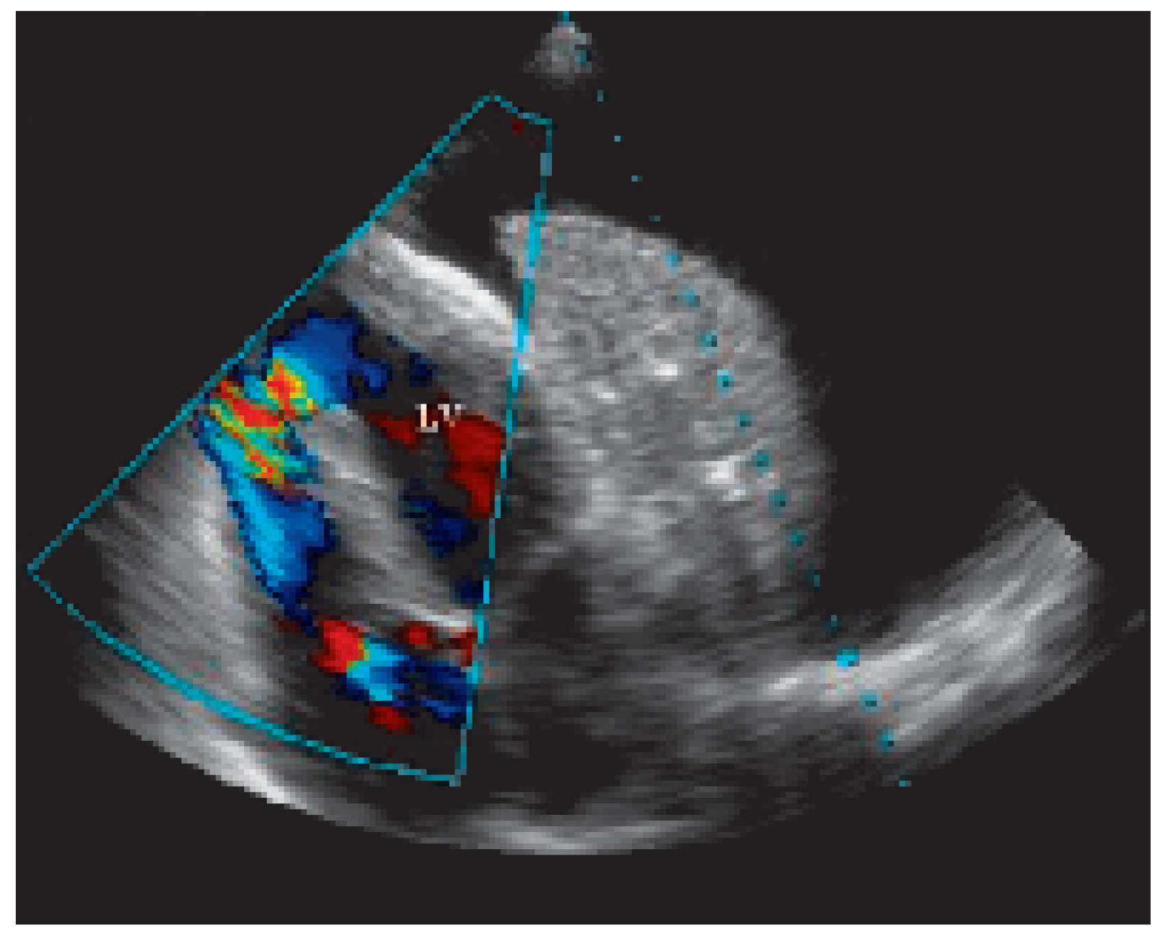
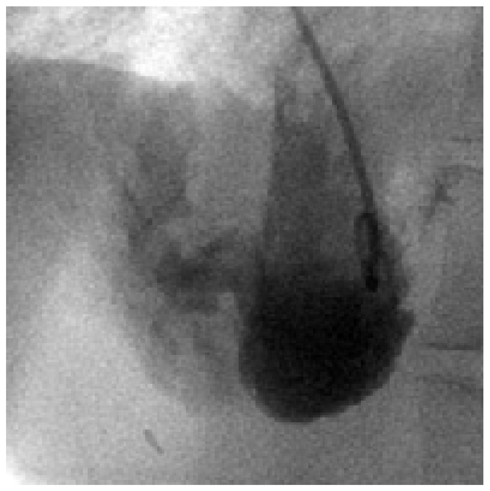
On the 2nd postoperative day a loud pansystolic murmur was heard over the precordial area. An apical ventricular septal defect (VSD) was diagnosed by echocardiography, estimated at 7 mm in size, with a left-to-right shunt (Figure 1 and Figure 2). Cardiac catheterisation confirmed the location of the VSD (Figure 3) with a step up in O2-saturations: 61% in the right atrium, 78% in the right ventricle and 68% in the pulmonary artery. The Qp/Qs-ratio was calculated at 1.35 and the pulmonary pressure was 45/14 mm Hg. A laceration of the distal left anterior descending artery and left ventricular apical akinesia were also noted. No intervention was performed and the patient was discharged from the hospital.
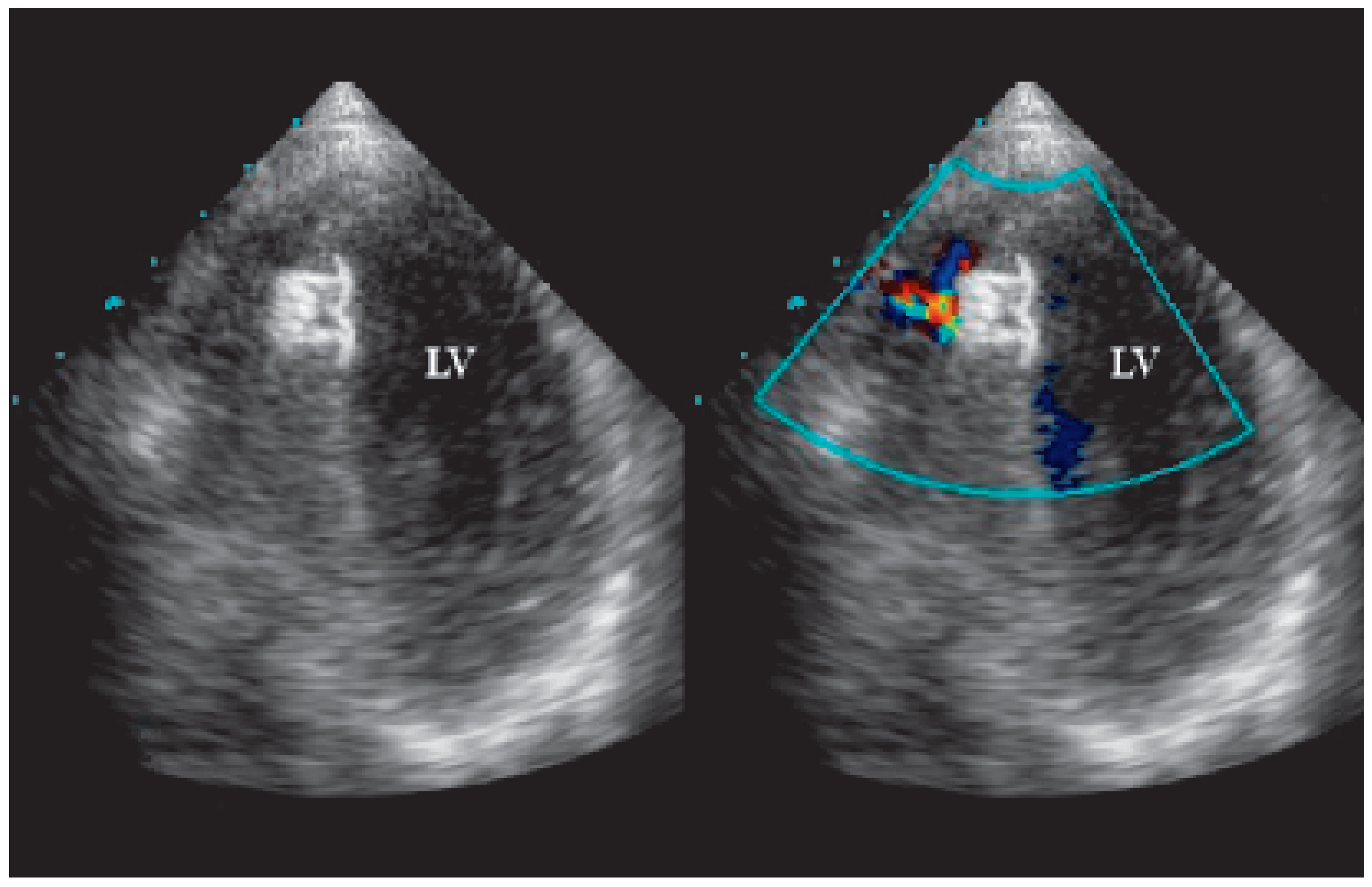
Five months later, the patient complained of exertional dyspnea and the decision to close the VSD percutaneously was made. Using a femoral approach and under local anaesthesia, a multipurpose catheter was introduced in the right heart with an exchange guidewire 0.21, and advanced through the VSD into the left ventricle and the aorta. A second catheter was introduced in the femoral artery and the descending aorta with a duplicate 0.014 guide wire forming a loop. This loop served as a lasso to snare out the exchange guide wire from the aorta through the femoral artery. Thus, a stable system with an aortovenous loop was built (Figure 4). A sizing balloon was positioned through the VSD and a stretched diameter of 24 mm was obtained. The same loop allowed the introduction and delivery of an Amplatzer Duct Occluder 9 (10–12 × 3 mm) (Figure 5). After release of the device, left ventriculography revealed only a small residual left-to-right shunt. An echocardiogram performed six months later showed the device in place, with a small persistent left-to-right shunt, no ventricular dilatation and no pulmonary hypertension (Figure 6).

Discussion

Ventricular septal defects due to penetrating chest injuries can occur either directly due to perforation of the septum or indirectly following laceration of an epicardial artery, causing local necrosis with subsequent rupture. A VSD is reported in about 1% of cases of chest trauma, the most common location being at the apex [1]. Surgical repair of a traumatic VSD implies a left ventriculotomy, with significant morbidity and mortality [2].
Percutaneous closure of VSDs has been successfully performed in children with congenital VSDs [3] and in adults with postinfarction VSDs. In most studies the use of specific Amplatzer VSD occluders are reported, but Amplatzer Duct Occluders have also been implanted [4].
In congenital defects, complete closure is achieved in 95% of patients with a muscular VSD and 92% of patients with a perimembranous VSD [3]. In a study examining post-infarction VSDs, an Amplatzer device was successfully implanted in 16 out of 18 patients, even though the 30-day-mortality amounted to 28% [5]. In a series of mixed cases the authors reported successful VSD closure in 30 out of 32 patients [6]. So far only sporadic cases of traumatic VSDs with successful percutaneous closure have been described [1,2]. The present case illustrates the usefulness of this percutaneous approach, particularly in light of the high risk of cardiac surgery in these patients.

References

  1. Bauriedel, G.; Redel, D.A.; Schmitz, C.; et al. Transcatheter closure of a posttraumatic ventricular septal defect with an Amplatzer Occluder Device. Catheter Cardiovasc Interv 2001, 53, 508–512. [Google Scholar] [CrossRef] [PubMed]
  2. Cowley, C.G.; Shaddy, R.E. Transcatheter treatment of a large traumatic ventricular septal defect. Catheter Cardiovasc Interv 2004, 61, 144–146. [Google Scholar] [CrossRef] [PubMed]
  3. Thanopoulos, B.S.; Karanassios, E.; Tsaousis, G.; et al. Catheter closure of congenital/acquired muscular VSDs and perimembranous VSDs using the Amplatzer devices. J Interven Cardiol 2003, 16, 399–407. [Google Scholar] [CrossRef] [PubMed]
  4. Fraisse, A.; Agnoletti, G.; Bonhoeffer, P.; et al. Multicentre study of percutaneous closure of interventricular muscular defects with the aid of an Amplatzer duct occluder prosthesis. Arch Mal Coeur Vaiss 2004, 97, 484–488. [Google Scholar] [PubMed]
  5. Holzer, R.; Balzer, D.; Zahid, A.; et al. Transcatheter closure of postinfarction ventricular septal defects using a new Amplatzer muscular VSD occluder: Results from the US registry. Catheter Cardiovasc Interv 2004, 61, 196–201. [Google Scholar] [CrossRef] [PubMed]
  6. Chessa, M.; Carminati, M.; Cao, Q.L.; et al. Transcatheter closure of congenital and acquired muscular ventricular septal defects using the Amplatzer devices. J Invasive Cardiol 2002, 14, 322–327. [Google Scholar] [PubMed]
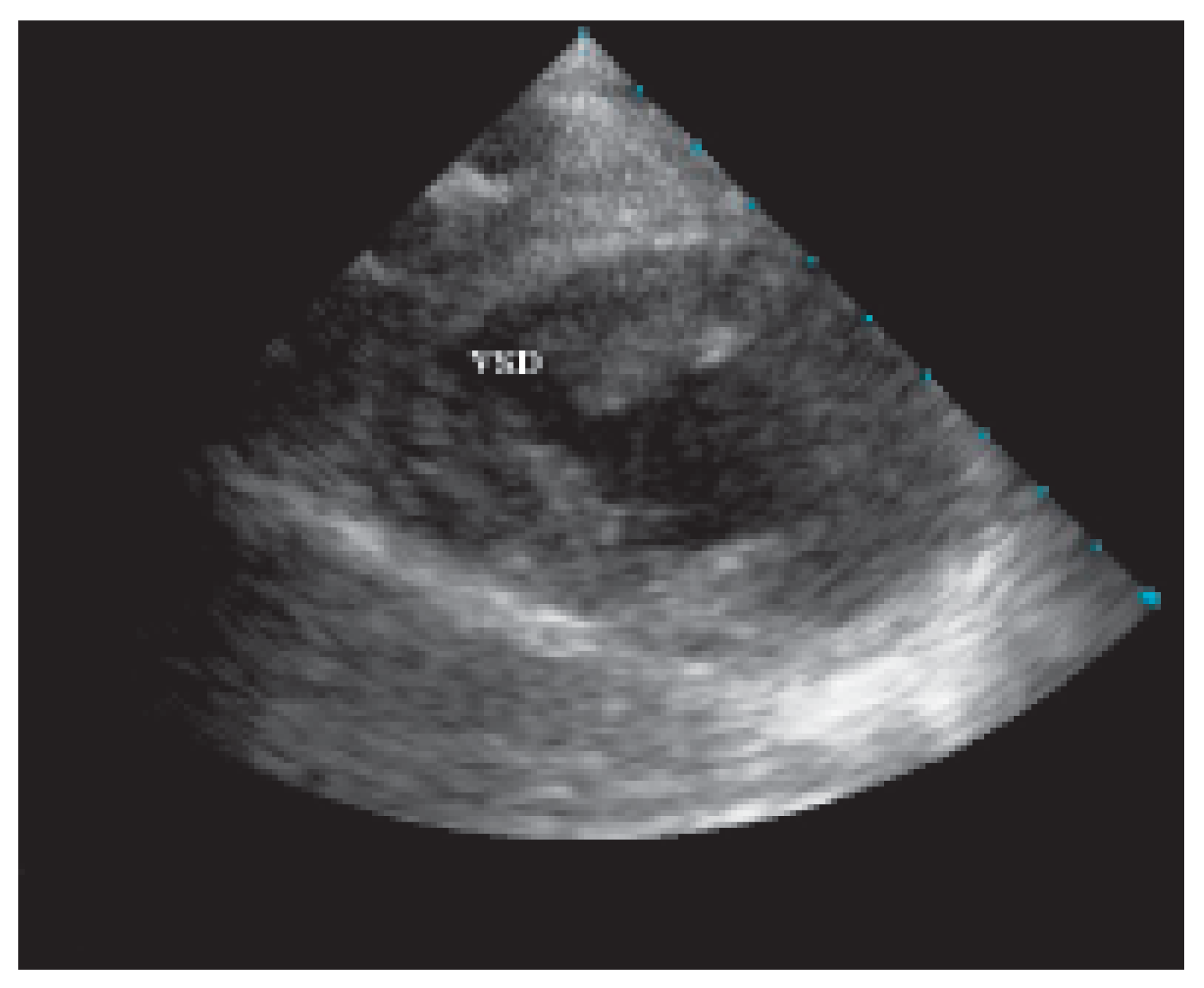
Figure 1. Transthoracic short axis view of the left ventricle with a large ventricular septal defect. VSD = ventricular septal defect.
Cardiovascmed 09 00083 g001
Figure 2. Modified apical view with color Doppler showing a left-to-right shunt through an apical VSD and a large left pleural effusion with collapsed lung. LV = left ventricle.
Cardiovascmed 09 00083 g002
Figure 3. Left ventriculography depicting a large shunt into the right ventricle through the apical VSD. VSD = ventricular septal defect.
Cardiovascmed 09 00083 g003
Figure 4. Sizing balloon placed in the VSD using an aortovenous loop created with an exchange wire travelling from the femoral vein to the right cavities, and through the VSD to the left ventricle and aorta. VSD = ventricular septal defect.
Cardiovascmed 09 00083 g004
Figure 5. Amplatzer device in the VSD before release from the catheter in the right ventricle. VSD = ventricular septal defect.
Cardiovascmed 09 00083 g005
Figure 6. Apical view showing the device implanted in the septum, with a small residual shunt detected by color Doppler. LV = left ventricle.
Cardiovascmed 09 00083 g006