Next Article in Journal
Stellenwert der Koronarrevaskularisation bei Älteren Patienten mit Chronischer Koronarer Herzkrankheit
Previous Article in Journal
Breitkomplex-Tachykardie am Monitor
 
 
Cardiovascular Medicine is published by MDPI from Volume 28 Issue 1 (2025). Previous articles were published by another publisher in Open Access under a CC-BY (or CC-BY-NC-ND) licence, and they are hosted by MDPI on mdpi.com as a courtesy and upon agreement with Editores Medicorum Helveticorum (EMH).
Interesting Images

Investigating a Continuous Heart Murmur
by
C. Roethlisberger
1,* and
M. Braunschweig
2
1
Cardiology, Spitalzentrum, Biel, Switzerland
2
Radiology, Spitalzentrum, Biel, Switzerland
*
Author to whom correspondence should be addressed.
Cardiovasc. Med. 2006, 9(1), 39; https://doi.org/10.4414/cvm.2006.01147
Submission received: 27 October 2005 / Revised: 27 November 2005 / Accepted: 27 December 2005 / Published: 27 January 2006
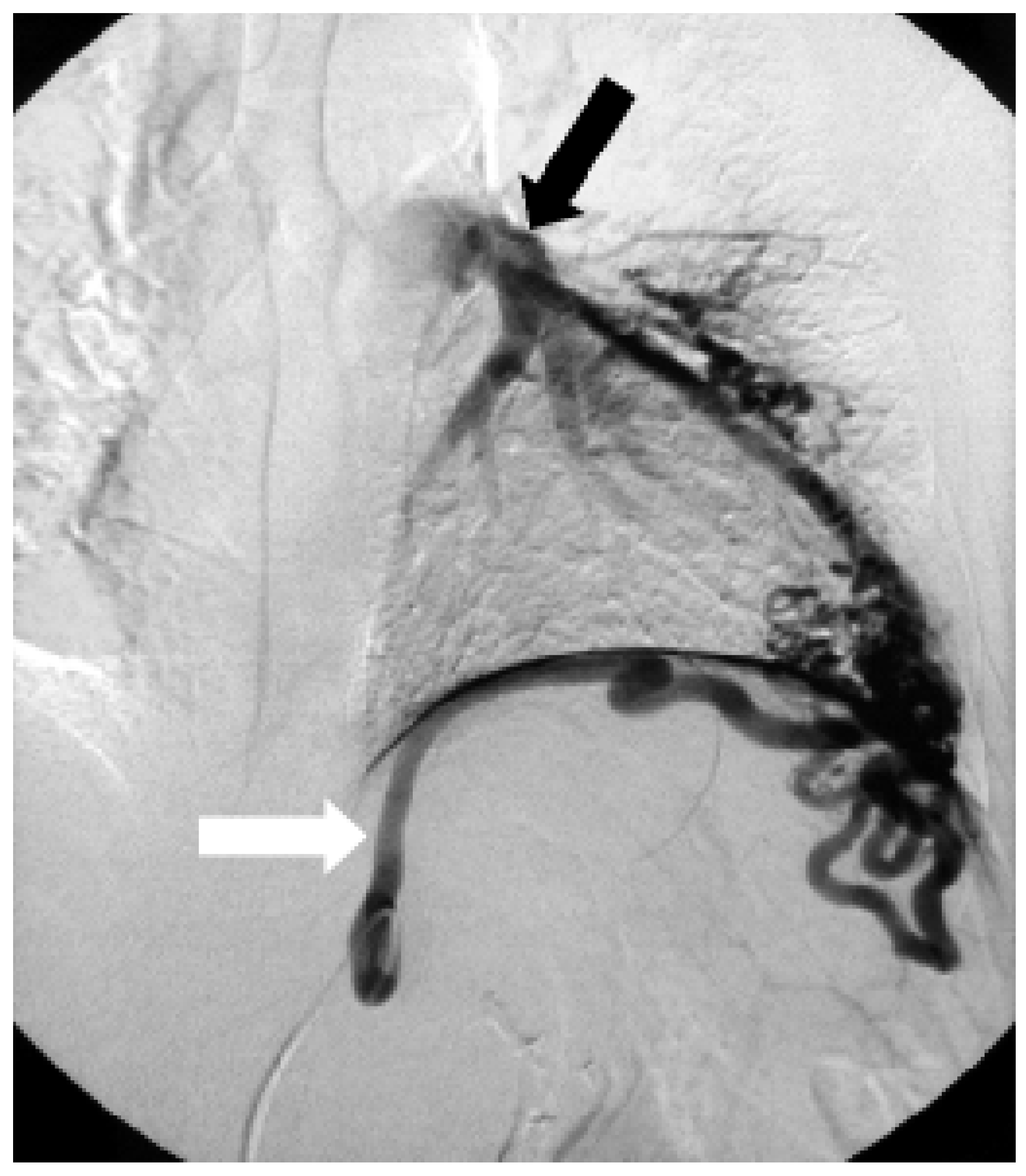
A 42-year-old male with atypical chest pain was investigated for a 2/6 continuous murmur at the apex of the heart. An electrocardiogram and a stress test were normal. Because the echocardiogram with color Doppler showed some blood flow around the apex as the only pathologic finding, a coronary fistula was suspected, but the coronary angiogram revealed completely normal coronary arteries. The computer tomogram of the chest confirmed an arterio-venous fistula around the heart’s apex which originated from the left gastric artery, but only the selective angiography (Figure 1) of the left gastric artery (white arrow) visualised the draining of the fistula into the left pulmonary artery (black arrow). As the patient was asymptomatic and signs of cardiac overload were absent, we decided to follow the patient clinically and by echocardiogram and to intervene only in case of the development of symptoms and/or left ventricular dilatation.
Figure 1. Arterio-arterial fistula around the heart’s apex originating from the left gastric artery (white arrow) and draining into the left pulmonary artery (black arrow).
Cardiovascmed 09 00039 g001