Case report
A 69-year-old woman was referred to the cardiologist for increasing shortness of breath. She had no previous cardiac examinations. The patient reported dizziness and some episodes of near-fainting in the last months. She complained of dyspnea on exertion NYHA II, but no angina and no obvious signs of heart failure. In the past, she had a history of several small surgical procedures on both hands. Her family history reveals that a brother had died suddenly a few weeks after pacemaker surgery.
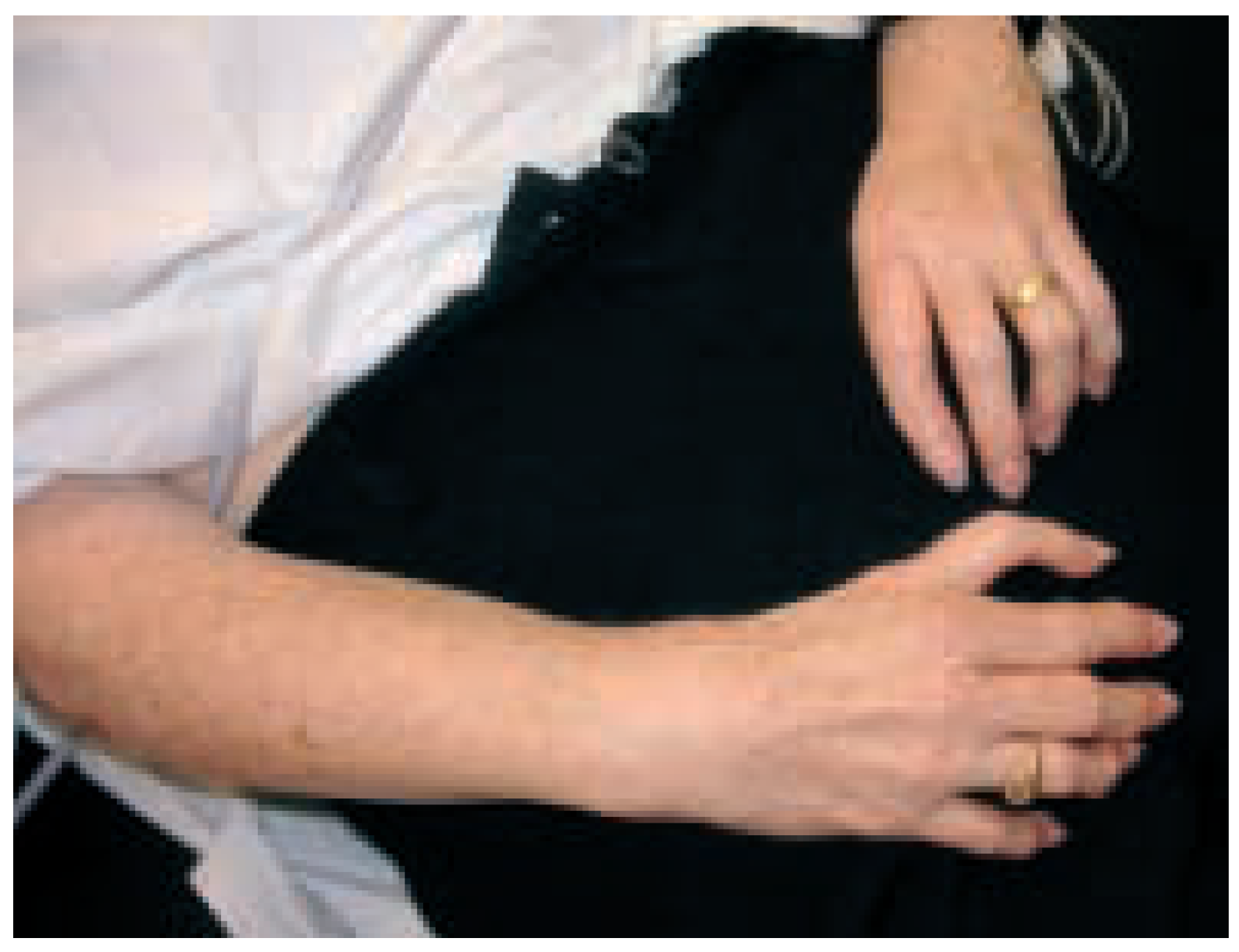
The physical exam shows obvious deformity of both forearms as shown in
Figure 1. There was neither clubbing nor cyanosis. O2 saturation at rest was normal. Heart auscultation revealed a
2/6 systolic murmur at the base and over the left second interspace. There were normal breathing sounds, no signs of biventricular heart failure and no peripheral edema. There was a digitalised triphalangeal thumb on the right side, the left sided thumb had been surgically amputated.
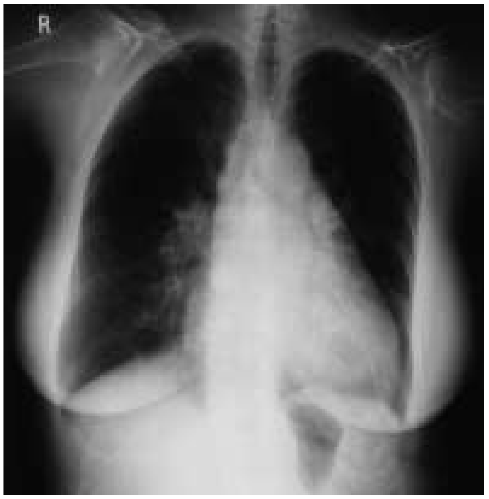
The chest-XR (
Figure 2) shows cardiomegaly, some increased pulmonary vasculature and dilatation of the main pulmonary artery on the right side.
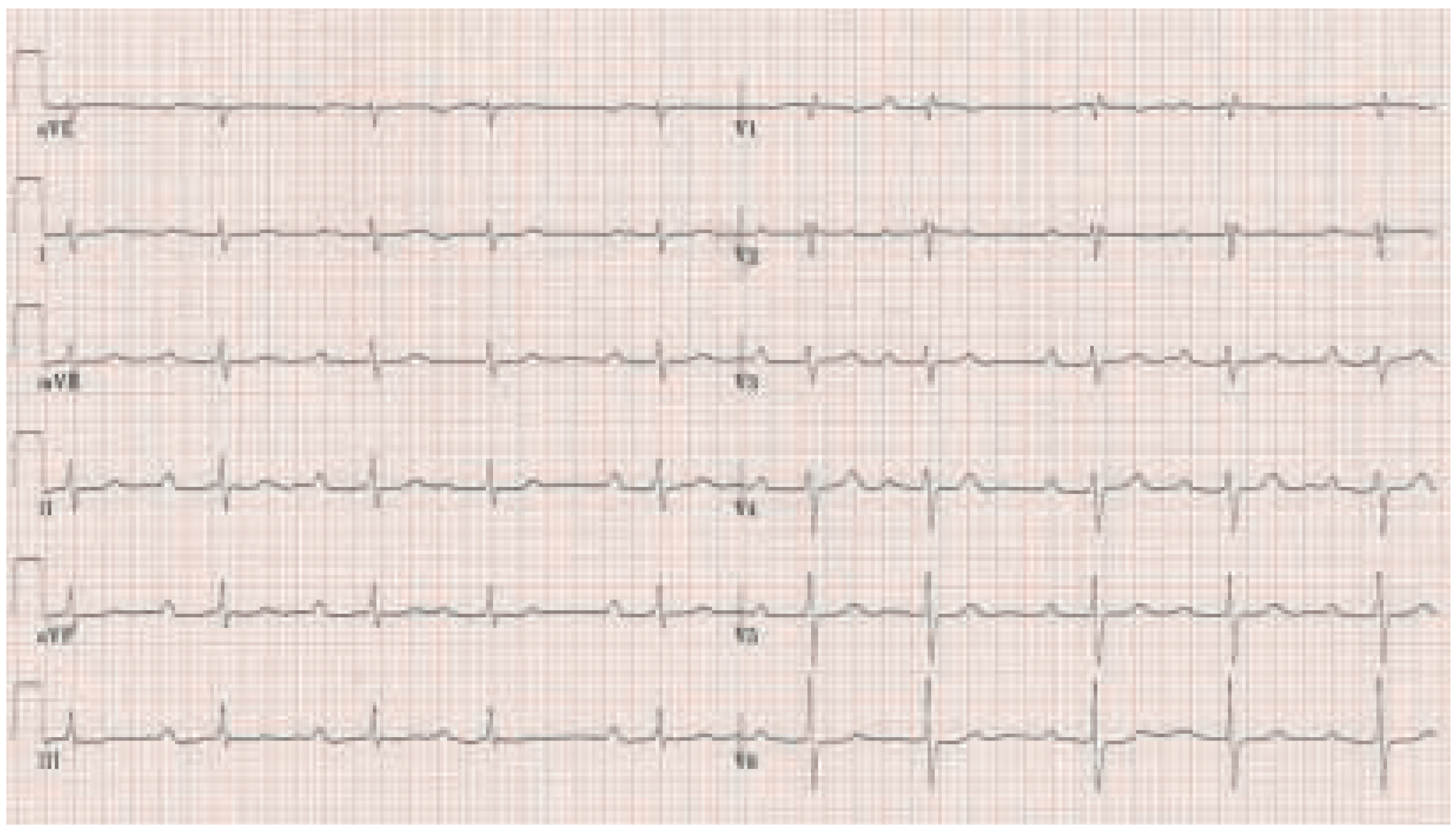
The ECG at rest is abnormal (
Figure 3): there is sinus rhythm with a very prolonged PR time of 396 ms due to first degree AV block, the QRS axis is 111 degrees, the QRS time 106 ms and QT time 426 ms. The 24-hour Holter ECG reveals intermittent second and third degree AV block.
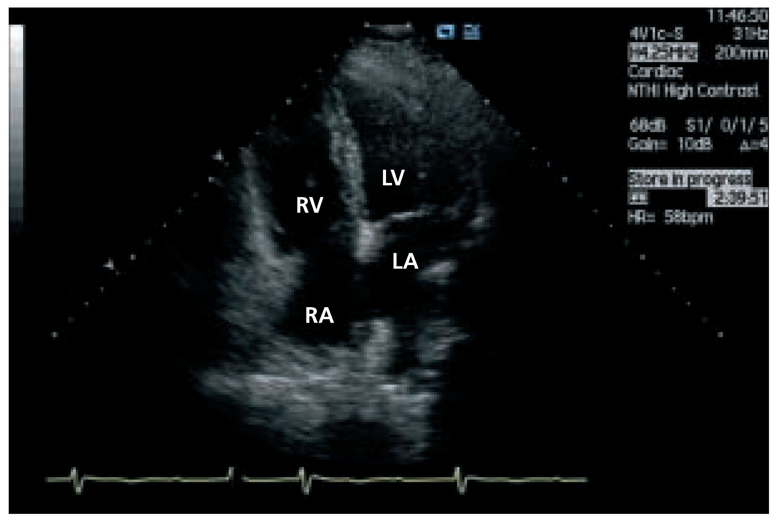
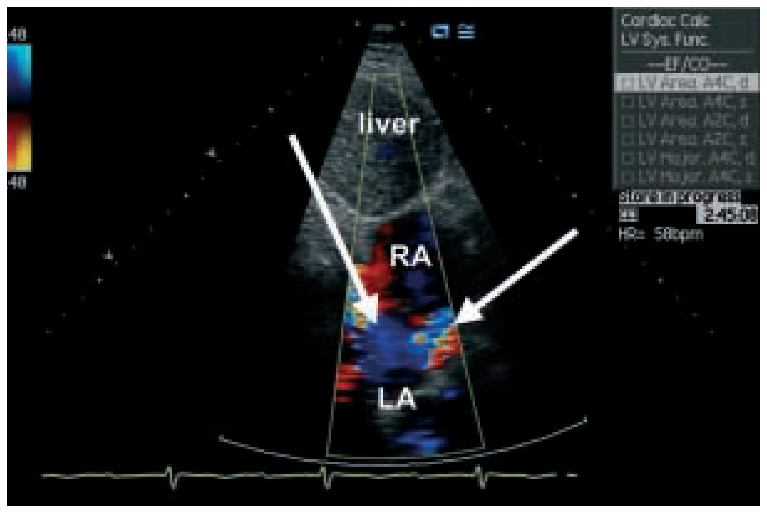
Transthoracic echocardiography (
Figure 4 and
Figure 5) is not normal and shows right heart overload with dilatation of the right sided cavities, elevated pulmonary artery pressure (estimated systolic pulmonary pressure about 56 mm Hg) and normal sized left ventricle. The reason for the dilatation of the right sided cavities was found in the presence of two secundum atrial septal defects with predominantly left-to-right shunting. Transoesophageal echocardiography was not performed.
The findings are compatible with Holt-Oram syndrome. Holt-Oram syndrome is the prototype of a heart-hand syndrome and was described in 1960 [
1]. Holt-Oram syndrome is an autosomal dominant condition accompanied by skeletal abnormalities and congenital heart disease. It occurs in one of every 100 000 life births [
2].
Cardiac defects occur in 95% of familial cases. Typical are cardiac defects of the septum including single or multiple atrial septal defects, ventricular septal defects, or atrioventricular-canal defects. Other cardiovascular defects such as hypoplastic left heart syndrome, persistent superior vena cava and mitral valve prolapse have been reported [
2]. Conduction system disease as in our patient is also common and can involve sinus bradycardia, atrioventricular block, atrial fibrillation, and sinus-node dysfunction; therefore, permanent pacemaker implantation is frequently needed [
3]. The brother of our patient who received a pacemaker had also finger deformities and thus most likely a Holt-Oram syndrome too. Finger and arm anomalies were also reported in other family members.
Bilateral forelimb deformities are common. Skeletal abnormalities include subtle carpal-bone abnormalities to triphalangeal or absent thumbs or frank upper extremity phocomelia [
3]. The radial ray is predominantly affected. The severity of skeletal deformities does not parallel the severity of cardiac malformations.
Genetically, in some families, Holt-Oram syndrome can be linked to single-gene mutations in the T-box transcription factor TBX5 [
2,
4]. Nearly 40 mutations in TBX5 have been found in Holt-Oram syndrome. TBX5 is a member of the Brachyury (T) gene family [
2,
4]. This gene defined as the TBX5 is located on the long arm of chromosome 12 (12q2). However, Holt-Oram syndrome is genetically heterogeneous. There is another autosomal dominant disorder often causing a secundum ASD and conduction system disease with no skeletal defects due to mutations in the transcription factor NKX2-5 [
5].
There can be wide variability in the expression of the phenotype within a family; however, there is 100% penetrance.
To conclude, especially in patients with an atrial septal defect, we have to consider the possibility of a heart-hand syndrome.
