Next Article in Journal
Echokardiographie bei Kardialer Resynchronisationstherapie
Previous Article in Journal
Update der Lipidmodulierenden Therapie
 
 
Cardiovascular Medicine is published by MDPI from Volume 28 Issue 1 (2025). Previous articles were published by another publisher in Open Access under a CC-BY (or CC-BY-NC-ND) licence, and they are hosted by MDPI on mdpi.com as a courtesy and upon agreement with Editores Medicorum Helveticorum (EMH).
Interesting Images

Differentiation Between Sinus Bradycardia and Second-Degree Atrioventricular Block by Left Ventricular Pressure Tracing

by
Peter Gnehm
,
Matthias E. Pfisterer
and
Piero O. Bonetti
*
Cardiology Department, University Hospital Basel, Basel, Switzerland
*
Author to whom correspondence should be addressed.
Cardiovasc. Med. 2005, 8(12), 453; https://doi.org/10.4414/cvm.2005.01137
Submission received: 30 September 2005 / Revised: 30 October 2005 / Accepted: 30 November 2005 / Published: 30 December 2005

Case Report

A66-year-old man with a subacute non-ST-elevation myocardial infarction (Creatinkinase 1540 U/l, Troponin-I 10.7 ng/l) was referred to our hospital for urgent coronary angiography. On arrival in our cardiac catheterisation laboratory, the patient was asymptomatic and the ECG showed sinus rhythm (80 bpm) with firstdegree atrioventricular (AV) block (PR interval 250 ms), but without any depolarisation or repolarisation abnormalities. Coronary angiography revealed three vessel disease with a chronically occluded right coronary artery and highly significant stenoses of the proximal segments of the left anterior descending and the left circumflex artery. Ventriculography showed normal left ventricular systolic function.
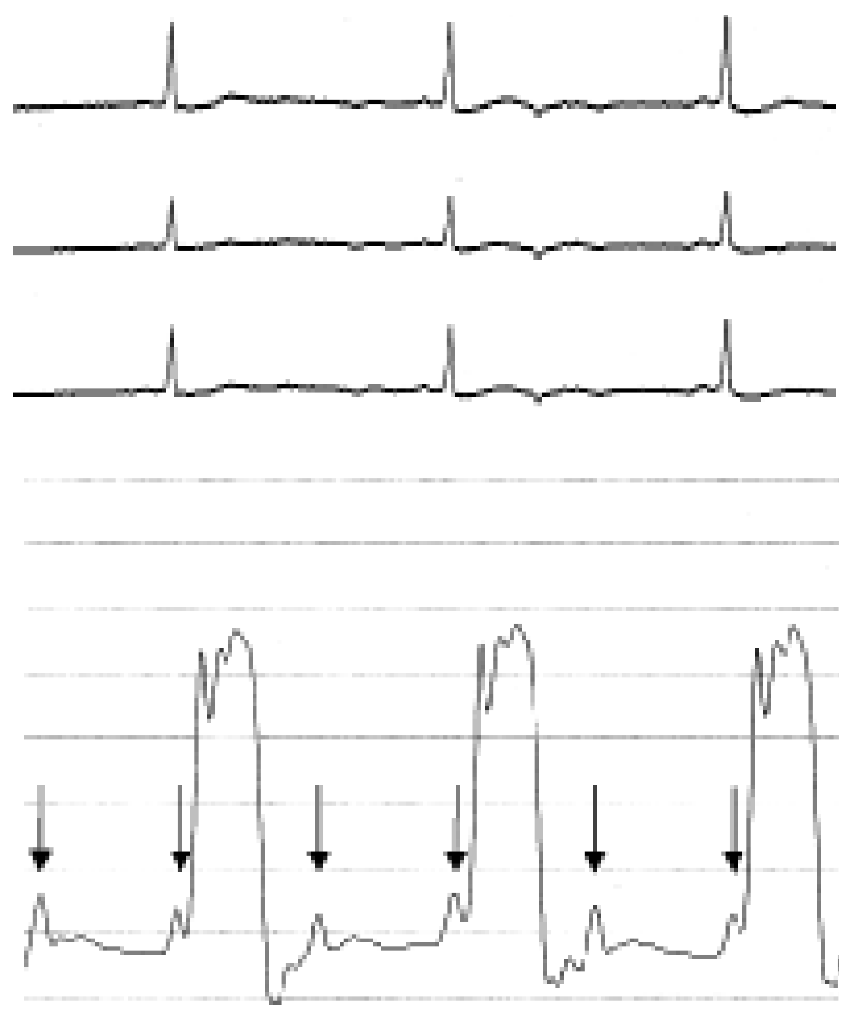
After placement of the pigtail catheter into the left ventricle (LV), the patient’s heart dropped to a regular rate of 43 bpm, whereby the patient remained haemodynamically stable and asymptomatic. The ECG showed bradycardia with a narrow QRS complex and hardly visible P-waves in front of each QRS complex (Figure 1, top) suggesting sinus bradycardia. However, the simultaneously recorded LV pressure tracing showed a pattern with two distinct regular a-waves during each diastole (Figure 1, bottom) indicating double atrial contraction and, thus, the presence of AV block with 2:1 conduction. Shortly after cardiac catheterisation, the second-degree AV block resolved spontaneously and the patient underwent successful coronary artery bypass graft surgery eleven days later.
In many centers, the patients heart rhythm is monitored by a modified 4-point ECG recording during cardiac catheterisation. Unfortunately, such simplified ECG tracings may be of insufficient quality to clearly differentiate various forms of arrhythmias. In certain cases left ventricular pressure tracing may be helpful in discriminating different forms of bradycardia such as sinus bradycardia and AV block, as is shown in the present example.
Figure 1. ECG (top) and simultaneously recorded left ventricular pressure tracing (bottom) during second-degree atrioventricular block with 2:1 conduction. Note that there are always two distinct diastolic a-waves (arrows) in the left ventricular pressure tracing suggesting double diastolic atrial contraction, although only one hardly visible p-wave can be identified during diastole in the ECG.
Cardiovascmed 08 00453 g001