Next Article in Journal
Update on Left Ventricular Hypertrophy
Previous Article in Journal
Schokolade: Genuss- Oder Heilmittel?
 
 
Cardiovascular Medicine is published by MDPI from Volume 28 Issue 1 (2025). Previous articles were published by another publisher in Open Access under a CC-BY (or CC-BY-NC-ND) licence, and they are hosted by MDPI on mdpi.com as a courtesy and upon agreement with Editores Medicorum Helveticorum (EMH).
Interesting Images

Right Bundle Branch Block or Brugada Syndrome?

by
Dagmar I. Keller
and
Stefan Osswald
*
Cardiology Department, University Hospital Basel, Basel, Switzerland
*
Author to whom correspondence should be addressed.
Cardiovasc. Med. 2005, 8(11), 410; https://doi.org/10.4414/cvm.2005.01134
Submission received: 30 August 2005 / Revised: 30 September 2005 / Accepted: 30 October 2005 / Published: 30 November 2005

Case Report

A male patient, born 1961, from Sri Lanka was admitted to hospital with two generalised seizures during sleep. The electroencephalogram was not conclusive for epileptic potentials, the MRI of the cranium was normal. Two ECG showed atypical right bundle branch block at different times (Figure 1), which normalised later during hospital stay.
What is your diagnosis?
Do you suggest further tests, and if yes, which ones?

Explanatory Answers

(1.) The history of seizures during sleep and the ECG on admission are highly suggestive for the Brugada syndrome, despite the fact that later ECG were within the normal range. Based on this evidence, an implantable cardioverter defibrillator was prophylactically inserted.
The Brugada syndrome has a high incidence in south east Asia and is known there to natives for many decades. In the Philippines for example it is known as “bangungut”, which translates as “screams and dies”, and in Thailand as “lai tai”, which means “death during sleep”. The ECG characteristics of the Brugada syndrome are a right bundle branch block pattern and descending ST segment elevation in leads V1 to V3 [1]. Clinically, the syndrome manifests by syncope and sudden cardiac death from fast polymorphic ventricular tachycardia and ventricular fibrillation typically in young male patients.
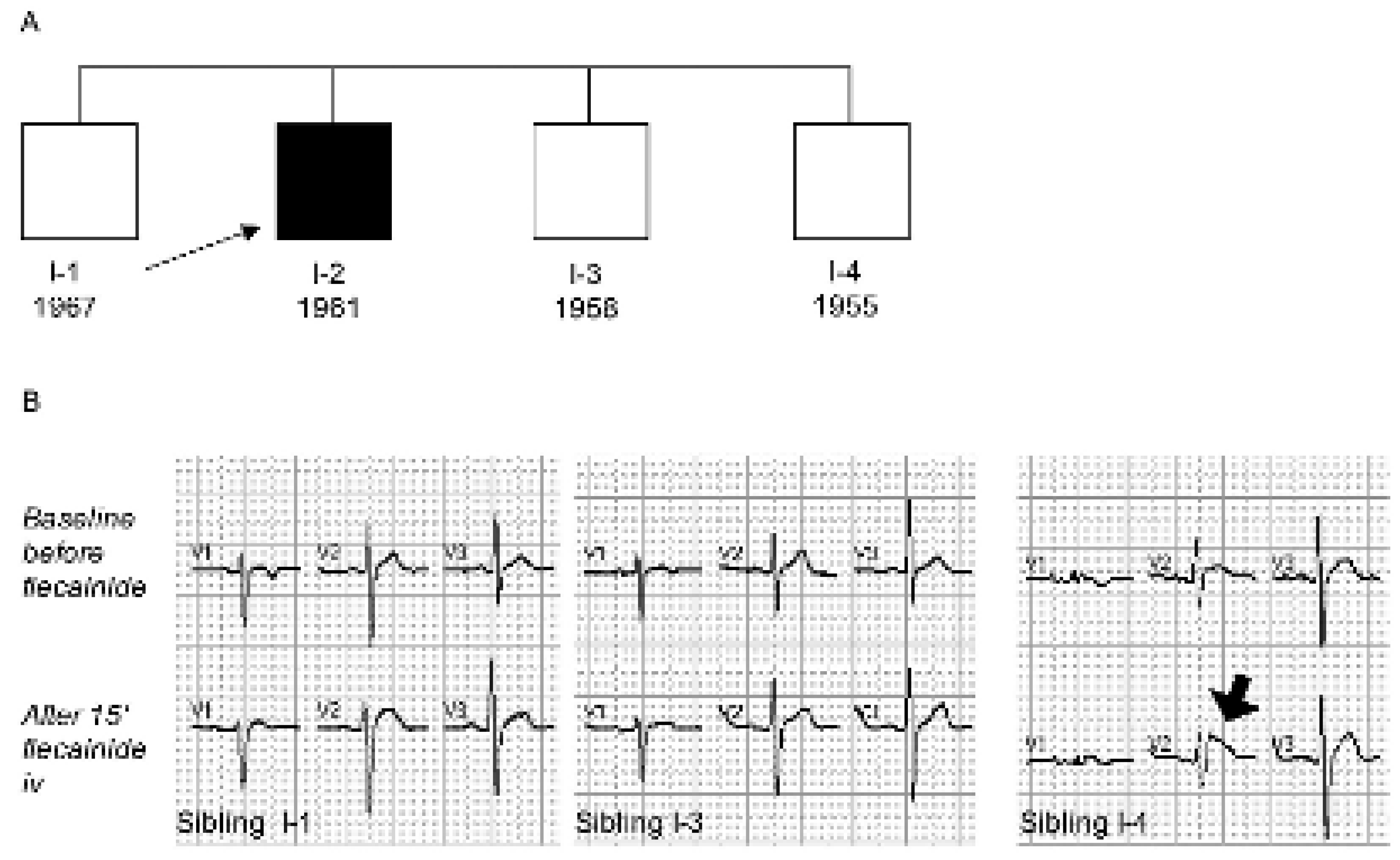
(2.) Family history is one of the cornerstone in diagnosing the Brugada syndrome, which is an autosomic dominant inherited disorder due to mutations in the SCN5A gene coding for the cardiac sodium channel (same as long QT syndrome type 3 and other congenital rhythm disorders). The diagnosis is based on the typical ECG findings that may be blunted and have to be unmasked by the administration of a class 1 anti-arrhythmic drug. Given the intermittent nature of the ECG findings, the diagnosis may be difficult in the interval. In this family, the syndrome was confirmed in the index patient and one of three siblings with normal ECG (sibling I-4) by the intravenous administration of flecainide (2 mg/kg body weight), which unmasked the typical ST-T configuration with a gradually descending ST-segment and a J wave amplitude ≥2 mm in lead V2 [2] (Figure 2A and B). In addition to class 1 anti-arrhythmic drugs fever has also been shown to unmask or trigger the Brugada syndrome phenotype [3].

References

  1. Brugada, P.; Brugada, J. Right bundle branch block, persistent ST segment elevation and sudden cardiac death: A distinct clinical and electrocardiographic syndrome. J Am Coll Cardiol 1992, 20, 1391–1396. [Google Scholar] [CrossRef] [PubMed]
  2. Wilde, A.A.M.; Antzelevitch, C.; Borggrefe, M.; Brugada, J.; Brugada, R.; Brugada, P.; et al. Proposed diagnostic criteria for the Brugada syndrome: Consensus report. Circulation 2002, 106, 2514–2519. [Google Scholar] [CrossRef] [PubMed]
  3. Keller, D.I.; Rougier, J.S.; Kucera, J.P.; Bennamar, N.; Fressart, V.; Guicheney, P.; et al. Brugada syndrome and fever: Genetic and molecular characterization of patients carrying SCN5A mutations. Cardiovasc Res 2005, 67, 510–519. [Google Scholar] [CrossRef] [PubMed]
Figure 1. Baseline ECG of index patient I-2 at different times.
Cardiovascmed 08 00410 g001
Figure 2. A Index patient I-2 and siblings. B Flecainide testing in siblings.
Cardiovascmed 08 00410 g002