Next Article in Journal
Astronaut Gets Angioplasty---Akut-PCI Unter Helmbeatmung
Previous Article in Journal
Akute Dyspnoe bei chronischem Vorhofflimmern
 
 
Cardiovascular Medicine is published by MDPI from Volume 28 Issue 1 (2025). Previous articles were published by another publisher in Open Access under a CC-BY (or CC-BY-NC-ND) licence, and they are hosted by MDPI on mdpi.com as a courtesy and upon agreement with Editores Medicorum Helveticorum (EMH).
Case Report

Percutaneous Left Ventricular Assist Device to Support PCI of Unprotected Left Main Coronary Artery Disease

by
Stéphane Cook
,
Bernhard Meier
* and
Stephan Windecker
Swiss Cardiovascular Center, Bern University Hospital, CH-3010 Bern, Switzerland
*
Author to whom correspondence should be addressed.
Cardiovasc. Med. 2004, 7(6), 258; https://doi.org/10.4414/cvm.2004.01030
Submission received: 30 March 2004 / Revised: 30 April 2004 / Accepted: 30 May 2004 / Published: 30 June 2004

Introduction

Although percutaneous coronary intervention (PCI) has long surpassed coronary artery bypass grafting (CABG) as the most common revascularization strategy in patients with coronary artery disease [1], left main coronary artery disease (LMCD) is regarded a relative contraindication to PCI [2]. This lack of enthusiasm was related to studies showing that although balloon angioplasty was effective in 94% of such high-risk procedure, long-term survival rate was dismal (36% after 3 years) in patients with unprotected LMCA [3], reinforcing CABG as therapy of choice in these patients [4].
However, recent improvements in stent technology and delivery systems, including drug-eluting stents and use of effective antiplatelet agents, have expanded the frontiers of PCI [1]. Thus, recent reports demonstrated satisfactory acute and long-term outcome after percutaneous management of protected, as well as unprotected LMCD [5,6].
Nonetheless, the risk of ischaemic complications during PCI in unprotected LMCD is of major concern and potentially responsible for the increase in mortality in this special subgroup of patients. Parallel to the breakthrough in stent technology, supportive techniques have been developed for limiting ischaemia during high-risk angioplasty. Of these, percutaneous left ventricular assist devices (pVAD) have been demonstrated to support the circulation during high-risk procedures [7].
With the advent of these recent techniques, we considered PCI in a patient with a severe lesion in the distal portion of the left main coronary artery (LMCA) who refused surgery. The patient underwent successful PCI of the left main and the left anterior descending coronary arteries after implantation of the pVAD for circulatory support during the procedure.

Case report

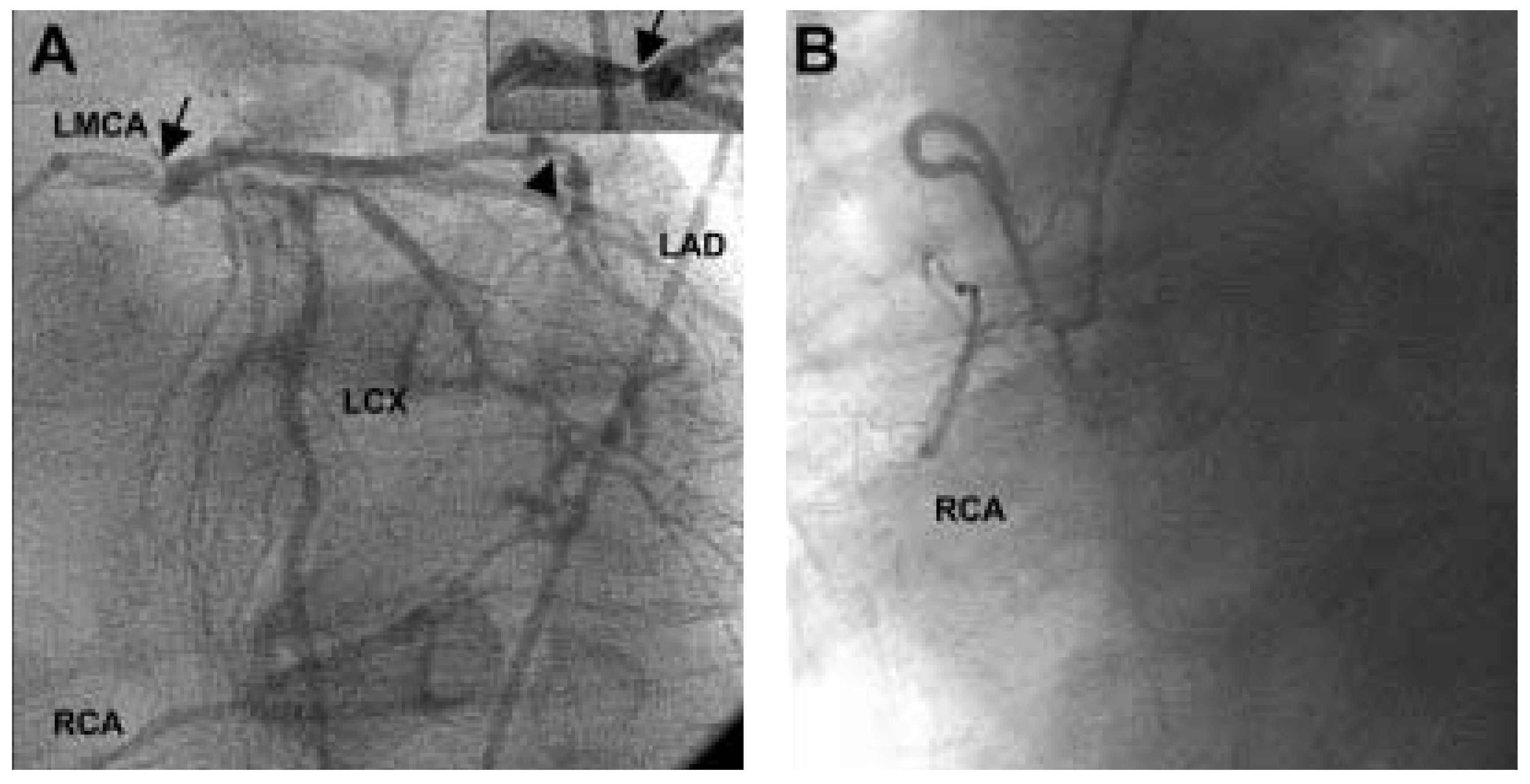
A 83-year-old man with hypertension and renal failure due to renal artery stenosis developed unstable angina pectoris and heart failure three hours after renal artery angioplasty and was transferred to our facility. Cardiac catheterization was performed and revealed occlusion of the right coronary artery with leftto-right collateral formation. The left main coronary artery had high grade disease in its distal portion and significant disease was also noted in the mid left anterior descending coronary artery as well as in the ramus intermedius (Figure 1A, 1B). Left ventricular ejection fraction was normal. A surgical myocardial revascularization was recommended, but due to the formal opposition of the patient, PCI was planned.
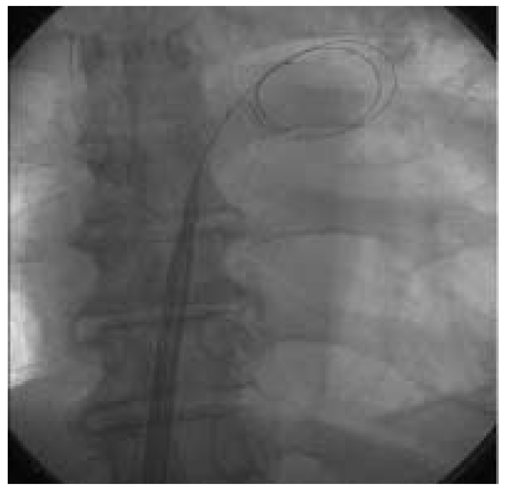
Before PCI, a percutaneous left ventricular assist device (Tandem Heart, Cardiac Assist Technologies, Inc., Ithaca, New York) was inserted. After transseptal puncture, a 21 French inflow cannula (Mullins sheath) was advanced into the left atrium (Figure 2) and the oxygenated blood harvested through this cannula was pumped back into the femoral artery through a 15 French catheter by a centrifugal continuous flow pump, providing a systemic output of up to 4 L/min. The patient received 10 000 units of heparin during the procedure. He was pretreated with acetylsalicylic acid and clopidogrel. A 6 French Q4 guiding catheter (Boston Scientific Scimed, Inc.) was placed in the ostium of the left main coronary.
Figure 1. Coronary angiograms in RAO views of the left (A) and right (B) coronary arteries with Judkins 5 French diagnostic catheters revealed a high grade lesion of the distal end of the left main coronary artery (LMCA, arrow), a significant stenosis in the mid left anterior descending artery (LAD, arrowhead) and a complete occlusion of the right coronary artery (RCA) with leftto-right collaterals. The insert shows the region of the left main stem. LCX = left circumflex artery.
Cardiovascmed 07 00258 g001
Figure 2. After transseptal puncture, an 21 French inflow cannula (Mullins sheath) was advanced into the left atrium.
Cardiovascmed 07 00258 g002
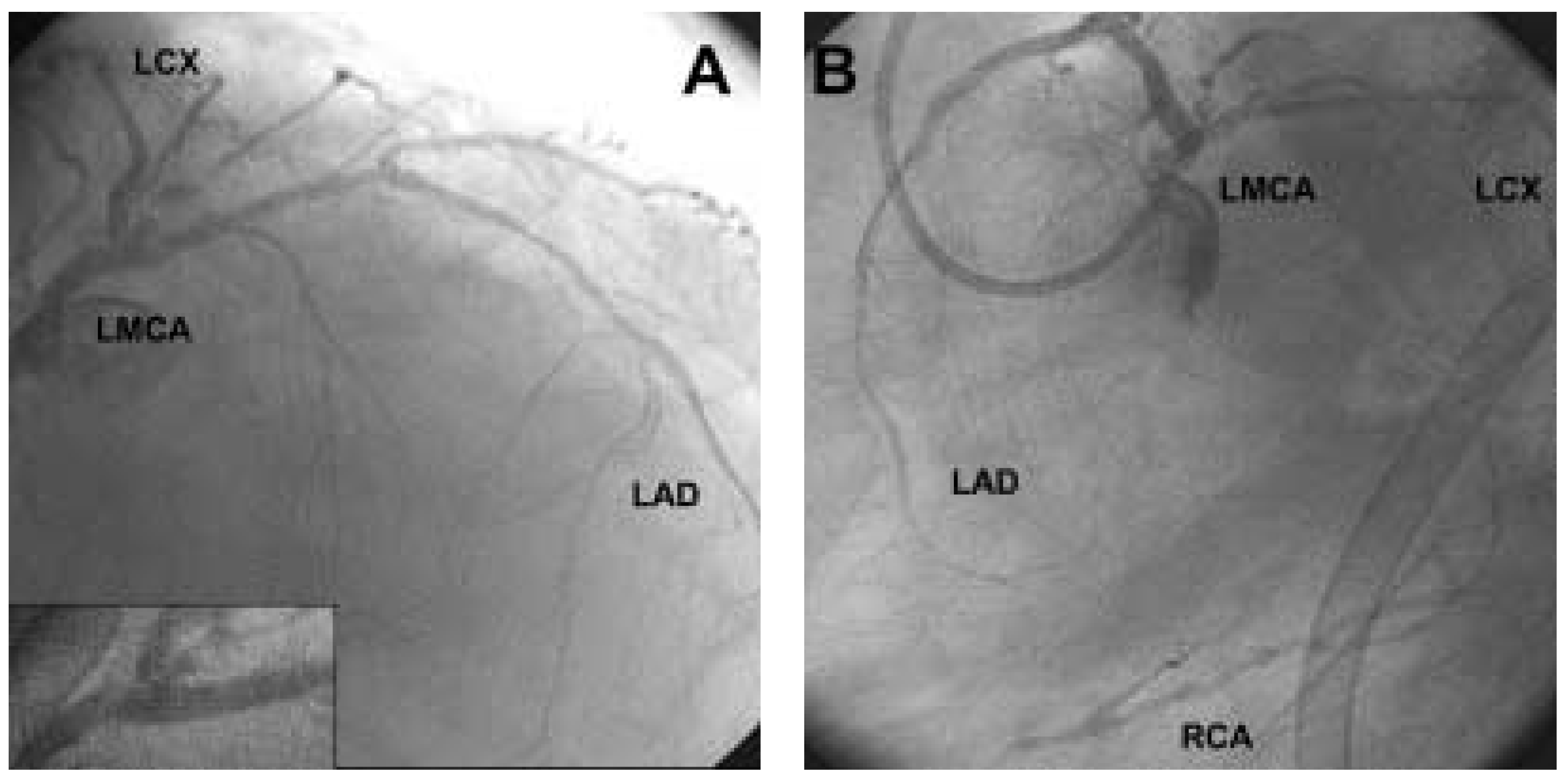
A 0.014’’ Forte guide wire (Boston Scientific Scimed, Inc.) was placed through the left main coronary artery into the LAD and the LMCA was predilated with a 3.5/20 mm balloon (Maverick, Boston Scientific Scimed, Inc., 14 atm, 30 seconds) before implantation of a 3.5/18 mm sirolimus-eluting Cypher stent (Cordis, Johnson & Johnson). After successive balloon angioplasty, a 2.5/28 mm Cypher stent was placed in the LAD and the ramus intermedius was finally treated by 1.5/20 mm Maverick balloon angioplasty. During balloon inflation, the pressure curve became a straight line at about 110 mm Hg mean pressure and the aortic valve remained closed (documented by angiography). After balloon deflation the pressure curve slowly resumed a phasic shape and the aortic valve reopened. The final coronary angiograms (Figure 3A, 3B) demonstrated a satisfactory result. Because of the uneventful course of the procedure, pVAD could be removed under manual groin compression without incident at the end of the procedure. Troponin-I levels rose to peak at 6.7 µg/l with no change in creatine kinase plasma levels or electrocardiographic evidence of infarction seen.

Discussion

Although it is accepted that PCI of LMCD is a potential alternative to CABG, the following points should be taken into consideration.
In the past decade, treatment of coronary artery disease has been advanced by the advent of stent technology, which diminished the problem of elastic recoil and abrupt closure in the acute setting. The risk of stent thrombosis has been virtually overcome by the introduction of dual antiplatelet therapy with acetylsalicylic and ticlopidine or clopidogrel.
Recent studies using stents in patients with LMCD are intriguing. Brueren and coworkers [5] reviewed the current literature and analysed the data of 71 patients who underwent PCI of unprotected LMCA in their institute. The total one year survival rate was 97%, whereas after an average follow up of 43 months survival was 90%. Most of the deaths were attributed to non-cardiac etiologies. They noted that in the 10 recently published major studies the average early mortality was 8.8%, which was a strong argument in favour of CABG in patients with LMCA stenosis. They therefore cautioned that only patients with severe concomitant disease, in whom the risk of bypass surgery was considered unacceptably high, underwent PCI, thereby introducing selection bias in the cited studies. This explains the increased mortality in past.
Figure 3. Left coronary angiograms after PCI with 6 French Q4 guiding catheter. A Forte guide wire is placed through the left main artery (LMCA) into the left anterior descending artery (LAD). The insert shows the region of the left main stem. LCX = left circumflex artery; RCA = right coronary artery.
Cardiovascmed 07 00258 g003
Recently, a single center observational study of sirolimus eluting stents in 31 patients with left main disease found no death, myocardial infarction or target lesion revascularization after a follow-up period of 5 months [8].
The major concern is related to the large myocardial area at risk, which corresponded to the entire heart in this case-report. Such patients with multivessel involvement may be less tolerant to the somewhat prolonged occlusions of the LMCA during a technically difficult PCI. Percutaneous left ventricular assist devices have shown to preserve haemodynamic stability during high-risk PCI [7] and help in patients undergoing LMCA-PCI [9], allowing the procedure to be done thoroughly taking the necessary time in the face of a comfortable patient.

References

  1. Windecker, S.; Roffi, M.; Meier, B. Sirolimus eluting stent: a new era in interventional cardiology? Curr. Pharm. Des. 2003, 9, 1077–1094. [Google Scholar] [CrossRef] [PubMed]
  2. Smith SCJr Dove, J.T.; Jacobs, A.K.; et al. ACC/AHA guidelines of percutaneous coronary interventions (revision of the 1993 PTCA guidelines) — executive summary. A report of the American College of Cardiology/American Heart Association Task Force on Practice Guidelines (committee to revise the 1993 guidelines for percutaneous transluminal coronary angioplasty). J. Am. Coll. Cardiol. 2001, 37, 2215–2239. [Google Scholar]
  3. O’Keefe JHJr Hartzler, G.O.; Rutherford, B.D.; et al. Left main coronary angioplasty: early and late results of 127 acute and elective procedures. Am. J. Cardiol. 1989, 64, 144–147. [Google Scholar] [CrossRef] [PubMed]
  4. Eagle, K.A.; Guyton, R.A.; Davidoff, R.; et al. ACC/AHA guidelines for coronary artery bypass graft surgery: executive summary and recommendations: a report of the American College of Cardiology/American Heart Association Task Force on Practice Guidelines (Committee to revise the 1991 guidelines for coronary artery bypass graft surgery). Circulation 1999, 100, 1464–1480. [Google Scholar] [PubMed]
  5. Brueren, B.R.; Ernst, J.M.; Suttorp, M.J.; et al. Long-term followup after elective percutaneous coronary intervention for unprotected non-bifurcational left main stenosis: is it time to change the guidelines? Heart 2003, 89, 1336–1339. [Google Scholar] [CrossRef] [PubMed]
  6. Kelley, M.P.; Klugherz, B.D.; Hashemi, S.M.; et al. One-year clinical outcomes of protected and unprotected left main coronary artery stenting. Eur. Heart J. 2003, 24, 1554–1559. [Google Scholar] [CrossRef] [PubMed]
  7. Vranckx, P.; Foley, D.P.; de Feijter, P.J.; Vos, J.; Smits, P.; Serruys, P.W. Clinical introduction of the Tandemheart, a percutaneous left ventricular assist device, for circulatory support during high-risk percutaneous coronary intervention. Int. J. Cardiovasc Intervent 2003, 5, 35–39. [Google Scholar] [CrossRef] [PubMed]
  8. Arampatzis, C.A.; Lemos, P.A.; Tanabe, K.; et al. Effectiveness of sirolimus-eluting stent for treatment of left main coronary artery disease. Am. J. Cardiol. 2003, 92, 327–329. [Google Scholar] [CrossRef] [PubMed]
  9. Butman, S.M.; Jamison, K.; Slepian, M.; Edling, N.; Arabia, F.; Copeland, J.G. Percutaneous intervention for unprotected left main disease prior to explantation of a left ventricular assist device. Catheter. Cardiovasc. Interv. 2003, 59, 471–474. [Google Scholar] [CrossRef] [PubMed]