Case report
An 80-year-old woman with symptomatic rheumatic mitral valve disease and chronic atrial fibrillation was admitted for mitral valvuloplasty and interventional occlusion of the left atrial appendage. Mitral commissurotomy had been performed 30 years earlier. Because of unstable INR with warfarin therapy, she was at increased risk for bleeding and thromboembolism. The left atrium was grotesquely dilated (10.3 × 4.6 cm) and showed spontaneous echo contrast. Severe calcification of the mitral valve and mitral annulus resulted in significant stenosis (mitral valve area 1.0 cm2, mean pressure gradient 11 mm Hg) and mild mitral regurgitation. The coronary arteries were normal and mitral valvuloplasty was performed resulting in only moderate improvement with a valve area of 1.3 cm2. After fluoroscopic identification of the atypically located left atrial appendage by contrast medium injection, a 10 mm Amplatzer septal occluder was inserted (fig. 1A). However, after its detachment from the catheter, the device dislodged from the left atrial appendage and was found floating in the left atrium (fig. 1B, C). In light of the residual mitral stenosis with moderate regurgitation, no attempt was made to retrieve the dislodged device percutaneously. The patient was left with bed rest and heparin. The next day the device was still in the left atrium (fig. 2) and mitral valve replacement was performed. The occluder was removed from the left atrium and found clean from thrombosis (fig. 3). An atrial septal tear resulting from the transseptal puncture was repaired, and the left atrial appendage was obliterated. Postoperative recovery was uneventful.

Figure 1.
A. Atypically located left atrial appendage outlined with contrast medium (arrows) with the 10 mm Amplatzer septal occluder inserted (arrowheads). B, C. Occluder (arrowheads) dislodges into left atrium after detachment from the catheter.
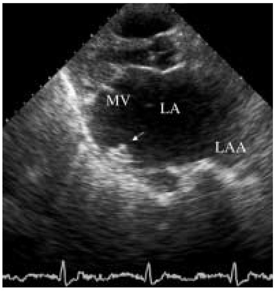
Figure 2.
Transthoracic echocardiogram. Amplatzer occluder (arrow) floating in the left atrium (LA). LAA = left atrial appendage; MV = mitral valve.
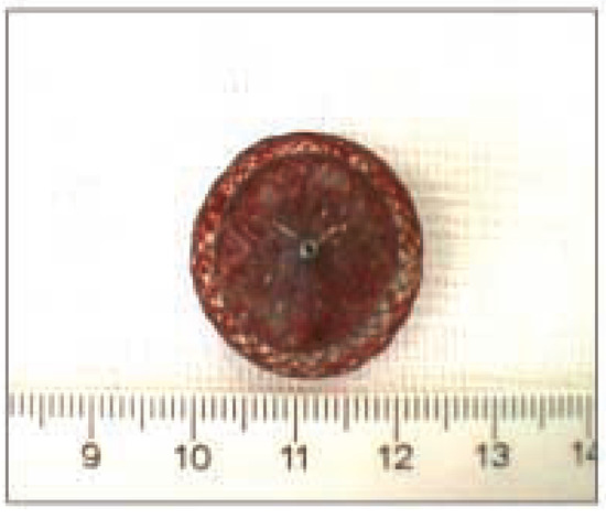
Figure 3.
Removed device showing some fibrinous coating but not clot.


© 2004 by the author. Attribution - Non-Commercial - NoDerivatives 4.0.