Next Article in Journal
Hypertension in Frail Patients
Previous Article in Journal
Balloon-Protection of the Left Phrenic Nerve During Epicardial Catheter Ablation of Ventricular Tachycardia
 
 
Cardiovascular Medicine is published by MDPI from Volume 28 Issue 1 (2025). Previous articles were published by another publisher in Open Access under a CC-BY (or CC-BY-NC-ND) licence, and they are hosted by MDPI on mdpi.com as a courtesy and upon agreement with Editores Medicorum Helveticorum (EMH).
Case Report

Flecainide Induced ST Segment Changes Can Mimic Pathological ECG Changes in Patients Undergoing Exercise Testing: A Case Report

by
Matthias Kruzik
*,
Maria Padrutt
,
Julia Stehli
,
Corinna Brunckhorst
,
Christian Schmied
and
David Niederseer
University Heart Centre, University Hospital Zurich, Zurich, Switzerland
*
Author to whom correspondence should be addressed.
Cardiovasc. Med. 2021, 24(4), 184; https://doi.org/10.4414/cvm.2021.02178
Submission received: 1 April 2021 / Revised: 1 May 2021 / Accepted: 1 June 2021 / Published: 1 July 2021

Abstract

BACKGROUND: Flecainide, a class IC antiarrhythmic drug, is used frequently in patients with supraventricular arrhythmias, mostly atrial fibrillation, and less commonly for ventricular arrhythmias. With its strong affinity to sodium channels in the cardiac cells it affects phase 0 of the action potential leading to conduction slowing, which is reflected in widening of the QRS, and it can also prolong the PR interval. These unspecific electrocardiogram (ECG) changes may mimic myocardial ischaemia in patients undergoing myocardial stress testing. The current case report reflects the diagnostic difficulties arising from this phenomenon. CASE SUMMARY: A 66-year-old patient who had been treated with flecainide for a decade for atrial fibrillation showed significant QRS widening and ST segment depression during exercise testing. The patient did not exhibit symptoms of myocardial ischaemia. A subsequent single photon emission computed tomography (SPECT) scan revealed a 10–15% cardiac ischaemic area. Invasive coronary angiography showed a chronic, complete obstruction of the left circumflex artery, which was percutaneously revascularised. DISCUSSION: This case demonstrates how pharmacological effects of flecainide and other class IC antiarrhythmic drugs may lead to suspicion of cardiac ischaemia due to QRS widening and unspecific ST-segment changes. In these patients, accurate ECG interpretation is important to avoid unnecessary invasive procedures such as coronary angiography. On the other hand, significant underlying coronary artery disease may nonetheless be present, warranting invasive diagnostic testing. Noninvasive cardiac imaging may facilitate the process of distinguishing real ischaemia from flecainide-induced ECG changes.

Introduction

Class IC antiarrhythmic drugs such as flecainide may cause QRS widening and PR prolongation, due to their effect on cardiac sodium channels [1]. Acting on activated fast-inward sodium channels, it prolongs depolarisation and slows conduction. It also blocks potassium rectifier current (IKr) channels, resulting in a prolonged action potential. Lastly, by blocking the ryanodine receptors it increases after-depolarisation [2,3]. These effects may lead to QRS widening in patients undergoing exercise testing [4] and thus provoke unwarranted invasive testing for cardiac ischaemia. It is recommended to regularly perform exercise testing in patients taking flecainide to assess for exercise-induced QRS widening, especially when increasing the dosage of the drug. If QRS widening of more than 25% of the baseline value is present, a dose reduction or discontinuation of flecainide is indicated in order to reduce the risk for possible arrhythmia [5]. Class IC antiarrhythmic drugs are contraindicated in the presence of structural or functional heart disease, especially after myocardial infarction [6]. Therefore, patients are routinely screened for structural heart disease by means of echocardiography or other imaging techniques before the drug is initiated. Although the QRSwidening and proarrhythmic effects of flecainide and other class IC anti-arrhythmic drugs are well described [7], literature regarding patients on class IC antiarrhythmic drugs with suspected coronary artery disease and pathological exercise testing remains scarce. We therefore describe this case of a 66-year-old patient on flecainide for atrial fibrillation with a pathological exercise test, illustrating this clinical dilemma.

Case presentation

A 66-year-old male Caucasian patient was referred to our outpatient cardiology clinic for the first time to assess changing his anticoagulation from rivaroxaban to apixaban becuase of recurrent but nonsignificant lower gastrointestinal bleeding. The bleeding episodes occurred at an anastomosis site of a rectal resection the patient underwent in 2016 because of adenocarcinoma of the rectum. At this visit the patient was switched to apixaban. After the diagnosis of atrial fibrillation in 2011 the patient underwent successful electrical cardioversions, once in 2011 and twice in 2012. He was anticoagulated initially with phenprocoumon and later switched to rivaroxaban in 2016. His current medication comprised rivaroxaban 20 mg once daily, flecainide 100 mg twice daily, bisoprolol 2.5 mg once daily, lisinopril 5 mg once daily, metformin twice daily and macrogol once daily. On examination, a blood pressure of 140/80 mm Hg and a heart rate of 60 bpm were noted, his weight was 100 kg, and his height was 183 cm (body mass index 29.9 kg/m2). The patient was euvolaemic. He denied dyspnoea or chest pain. Cardiovascular risk factors included arterial hypertension, current smoking (20 pack years), and diabetes mellitus type 2. An electrocardiogram (ECG) at rest showed sinus rhythm at 56 bpm, a normal QRS axis, no pathological T-wave inversions and non-significant ST depression in V4–V5 (fig. 1). Laboratory work-up showed normal electrolytes, normal renal function, a normal haemogram, low-density lipoprotein (LDL) cholesterol of 3 mmol/l and glycated haemoglobin (HbA1c) of 5.2%. Transthoracic echocardiography showed a normal ventricle size with a normal ejection fraction (left ventricular ejection fraction biplane = 64%), hypertrophy of the sub-aortic septum (12 mm) and no regional wall motion abnormalities. The left atrium appeared dilated (left atrial volume index 50 ml/m2). There was no pulmonary hypertension evident on echocardiography. An exercise test (bicycle) showed adequate performance of 158 watts, corresponding to 91% of the agepredicted normal value. The patient did not report symptoms of angina during the exercise test and there were no arrhythmias observed. The test was terminated because of exhaustion of the patient. There was significant QRS widening at maximum exertion, increasing from 80 ms at rest to 115 ms (+35 ms, +44 %) and ST depression in leads II, III and aVF as well as V4–V6 during peak performance (maximum 0.2 mV) (Figure 1). Flecainide was discontinued because of possible coronary artery disease. The patient was started on rosuvastatin and, as requested by the referring physician, apixaban was substituted for rivaroxaban.
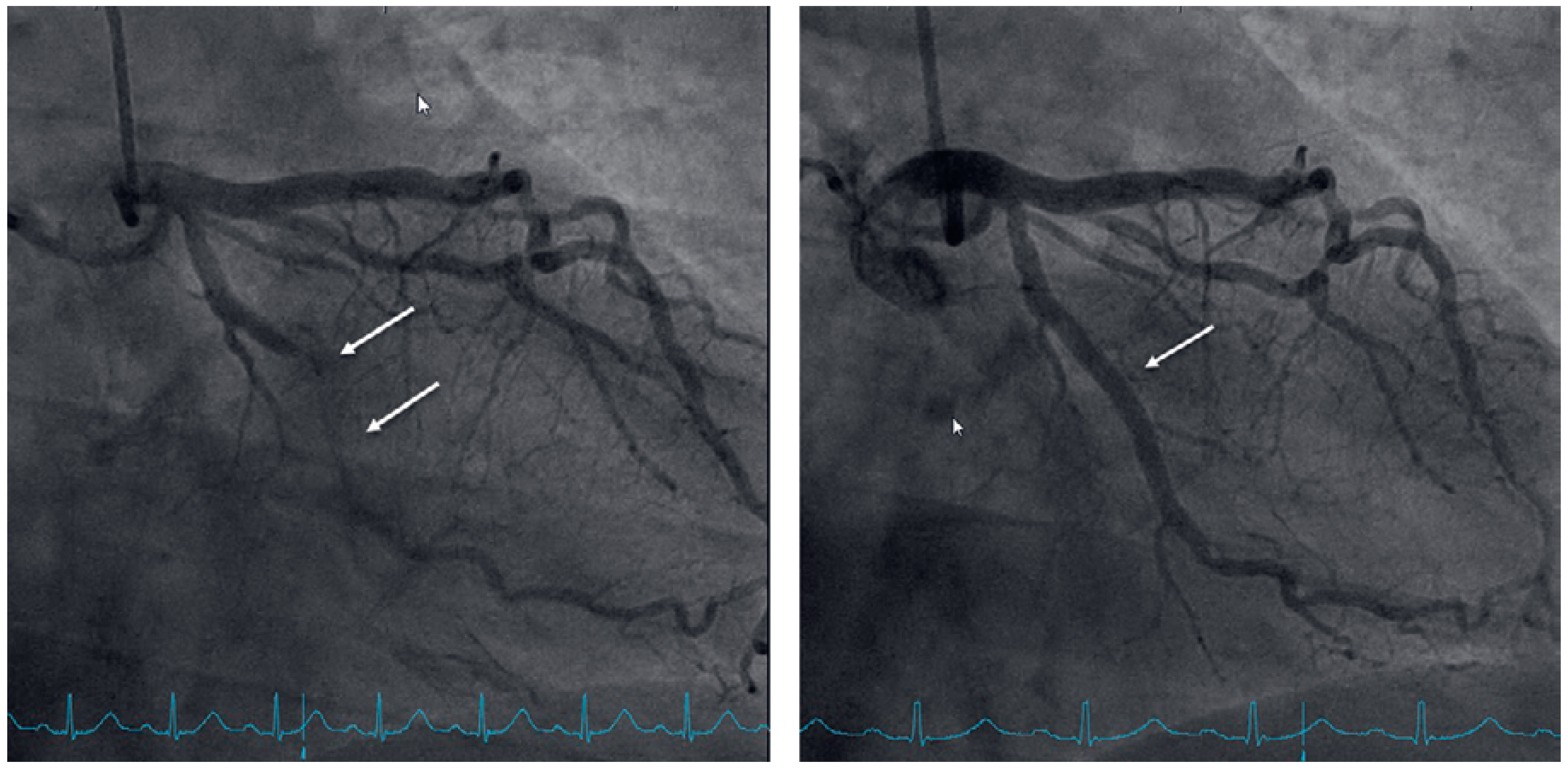
To exclude coronary artery disease the patient was referred for a cardiac single photon emission computed tomography (SPECT) scan using technetium-99m, which revealed a normal radionuclide tissue distribution at rest, but after physical stress using a bicycle ergometer significant left ventricular inferior-lateral ischaemia of 10–15% was observed (Figure 2). Elective coronary angiography confirmed coronary artery disease with chronic total obstruction of the left circumflex artery. One drug-eluting stent was placed successfully (Figure 3).

Discussion

We report on a 66-year-old Caucasian patient on flecainide for paroxysmal atrial fibrillation with significant QRS widening and progressive ST-segment depression during exercise testing. The diagnosis of coronary artery disease had not been previously established despite significant concomitant cardiovascular risk factors (hypertension, diabetes, smoking and overweight). There are two potential explanations for the pathological changes during exercise testing (widening of the QRS duration and progressive ST-segment depression): pharmacological side effects of flecainide or coronary artery disease. Progressive QRS widening during exercise testing of >25% is defined as the cut-off for discontinuation of flecainide [8]. However, ST-segment elevation, rather than depression, has previously been described in flecainide-induced QRS widening [9]. Additionally, flecainide has been shown to have proarrhythmic properties, which results in higher mortality rates in patients with coronary artery disease [10]. The long period of 9 years since the initial rule out of structural heart disease and the presence of multiple risk factors additionally raised suspicion for coronary artery disease. We therefore discontinued flecainide in this patient with a high cardiovascular risk profile, QRS widening of >25% and ST-segment depression during exercise testing. The patient then underwent a noninvasive test for myocardial ischaemia, which revealed left ventricular inferior-lateral ischaemia of 10–15%. A subsequent coronary angiogram showed a chronic total occlusion of the left circumflex artery. One drugeluting stent was successfully deployed. The patient was switched from rivaroxaban to apixaban, because of the lower gastrointestinal bleeding risk with apixaban as compared with rivaroxaban, after he had previously experienced recurrent lower gastrointestinal bleeds [11]. After stenting, one day of triple therapy with aspirin, clopidogrel and apixaban was followed by dual therapy with clopidogrel and apixaban for 3 months, according to current recommendations [12]. Flecainide, a class IC antiarrhythmic medication, is an effective agent for conversion and stabilisation of supraventricular and ventricular arrhythmias [1]. In this particular case, it was successfully used for maintenance of sinus rhythm in paroxysmal atrial fibrillation for 9 years. Major side effects of flecainide include proarrhythmic effects, conduction abnormalities, neurological side effects such as dizziness, metallic taste, blurred vision and headaches, renal dysfunction and interactions with other medications [1]. Its proarrhythmic properties where assessed in the CAST trial. The initial hypothesis was that pharmacological suppression of ventricular ectopy after myocardial infarction would reduce mortality. Contrary to that hypothesis, the authors showed an increased mortality due to arrhythmias in patients who received the drug after myocardial infarction [10]. Therefore, guidelines recommend against using flecainide in patients with coronary artery disease or other structural abnormalities of the heart (defined as ischaemic, congenital, valvular or significant myocardial heart disease, including left ventricular hypertrophy) [6]. The affinity of flecainide to sodium channels leads to slower conduction, hence potentially leading to QRS widening and an increased PR duration [3,13]. Flecainide in particular is known to prolong the QRS complex in patients undergoing exercise testing. This is referred to as use-dependence: the substance binds to the receptor of the sodium channel in systole and unbinds in diastole. When diastole gets shortened at higher heart rates, there is insufficient time to unbind and the effect of flecainide is potentiated [4]. This particular case illustrates the challenges the diagnostic algorithm may pose in a patient with suspected coronary artery disease on class IC antiarrhythmic drugs. In patients with cardiovascular risk factors, QRS widening and subsequent ST changes should not be attributed to the aforementioned pharmacological side effects of this class IC antiarrhythmic, but rather further noninvasive ischaemia testing should be pursued and/or coronary angiography performed. It is of paramount importance not to miss the diagnosis of coronary artery disease in these patients as flecainide is contraindicated in this condition.

Learning points

  • Class IC antiarrhythmic drugs inhibit sodium channels and may therefore prolong the PR interval and lead to QRS widening.
  • QRS widening in patients on class IC antiarrhythmic drugs undergoing exercise testing may mimic cardiac ischaemia.
  • Flecainide is contraindicated in patients with coronary artery disease or structural heart disease.

Disclosure statement

No financial support and no other potential conflict of interest relevant to this article was reported.

References

  1. Giardina, E.G. Major side effects of class I antiarrhythmic drugs: Up To Date. Available from: https://www.uptodate.com/contents/ major-side-effects-of-class-i-antiarrhythmic-drugs.
  2. Alzahrani, K.; Alzahrani, T. StatPearls Flecainide; StatPearls Publishing, 2020. [Google Scholar]
  3. Vik-Mo, H.; Ohm, O.J.; Lund-Johansen, P. Electrophysiologic effects of flecainide acetate in patients with sinus nodal dysfunction. Am J Cardiol. 1982, 50, 1090–1094. [Google Scholar] [CrossRef] [PubMed]
  4. Wang, J.A.; Lau, C.P.; Tai, Y.T.; Wu, B.Z. Effects of flecainide on exercise hemodynamics and electrocardiography in patients without structural heart disease. Clin Cardiol. 1995, 18, 140–144. [Google Scholar] [CrossRef] [PubMed]
  5. Vallurupalli, S.; Pothineni, N.V.; Deshmukh, A.; Paydak, H. Utility of Routine Exercise Testing to Detect Rate-Related QRS Widening in Patients Without Structural Heart Disease on Class Ic Antiarrhythmic Agents (Flecainide and Propafenone). Am J Cardiol. 2015, 116, 730–732. [Google Scholar] [CrossRef] [PubMed]
  6. Dan, G.A.; Martinez-Rubio, A.; Agewall, S.; Boriani, G.; Borggrefe, M.; Gaita, F.; et al. Antiarrhythmic drugs-clinical use and clinical decision making: a consensus document from the European Heart Rhythm Association (EHRA) and European Society of Cardiology (ESC) Working Group on Cardiovascular Pharmacology, endorsed by the Heart Rhythm Society (HRS), Asia-Pacific Heart Rhythm Society (APHRS) and International Society of Cardiovascular Pharmacotherapy (ISCP). Europace. 2018, 20, 731–732an. [Google Scholar] [PubMed]
  7. Bordier, P.; Garrigue, S.; Bernard, V.; Haissaguerre, M.; Douard, H.; Broustet, J.P.; et al. Flecainide-induced Increase in QRS Duration and Proarrhythmia during Exercise. Clin Drug Investig. 1997, 13, 326–337. [Google Scholar] [CrossRef] [PubMed]
  8. Gao, X.; Guha, A.; Buck, B.; Patel, D.; Snider, M.J.; Boyd, M.; et al. Initiation and outcomes with Class Ic antiarrhythmic drug therapy. Indian Pacing Electrophysiol J. 2018, 18, 68–72. [Google Scholar] [CrossRef] [PubMed]
  9. Nakamura, W.; Segawa, K.; Ito, H.; Tanaka, S.; Yoshimoto, N. Class IC antiarrhythmic drugs, flecainide and pilsicainide, produce ST segment elevation simulating inferior myocardial ischemia. J Cardiovasc Electrophysiol. 1998, 9, 855–858. [Google Scholar] [CrossRef] [PubMed]
  10. Echt, D.S.; Liebson, P.R.; Mitchell, L.B.; Peters, R.W.; Obias-Manno, D.; Barker, A.H.; et al. Mortality and morbidity in patients receiving encainide, flecainide, or placebo. The Cardiac Arrhythmia Suppression Trial. N Engl J Med. 1991, 324, 781–788. [Google Scholar] [CrossRef] [PubMed]
  11. Abraham, N.S.; Noseworthy, P.A.; Yao, X.; Sangaralingham, L.R.; Shah, N.D. Gastrointestinal Safety of Direct Oral Anticoagulants: A Large Population-Based Study. Gastroenterology. 2017, 152, 1014–1022. [Google Scholar] [CrossRef] [PubMed]
  12. Steffel, J.; Verhamme, P.; Potpara, T.S.; Albaladejo, P.; Antz, M.; Desteghe, L.; et al. ESC Scientific Document Group. The 2018 European Heart Rhythm Association Practical Guide on the use of non-vitamin K antagonist oral anticoagulants in patients with atrial fibrillation. Eur Heart J. 2018, 39, 1330–1393. [Google Scholar] [CrossRef] [PubMed]
  13. Hellestrand, K.J.; Bexton, R.S.; Nathan, A.W.; Spurrell, R.A.; Camm, A.J. Acute electrophysiological effects of flecainide acetate on cardiac conduction and refractoriness in man. Br Heart J. 1982, 48, 140–148. [Google Scholar] [CrossRef] [PubMed]
Figure 1. QRS complex at rest (left) and at maximum exertion (right).
Cardiovascmed 24 00184 g001
Figure 2. Single-photon emission computed tomography (SPECT); arrows indicate ischaemia in the area supplied by the left circumflex artery.
Cardiovascmed 24 00184 g002
Figure 3. Right anterior oblique projection of the left circumflex artery before (left) and after (right) placement of one drug-eluting stent.
Cardiovascmed 24 00184 g003