Next Article in Journal
COVID-19 Serology-Based Testing in Pericarditis: A Case Report
Previous Article in Journal
Maskeraden in Der Krise
 
 
Cardiovascular Medicine is published by MDPI from Volume 28 Issue 1 (2025). Previous articles were published by another publisher in Open Access under a CC-BY (or CC-BY-NC-ND) licence, and they are hosted by MDPI on mdpi.com as a courtesy and upon agreement with Editores Medicorum Helveticorum (EMH).
Case Report

Catheter Ablation of Left Atrial Macroreentrant Tachycardia After Bilateral Lung Transplantation

by
Guan Fu
1,2,
Suna Gonca
1,
Mueller Thomas F.
3,
Inci Ilhan
4,
Rüegg Christine A.
5 and
Saguner Ardan M.
1,*
1
Department of Cardiology, University Hospital Zurich, Zurich, Switzerland
2
Department of Cardiology, Capital Medical University, Beijing Shijitan Hospital, Beijing, China
3
Department of Nephrology, University Hospital Zurich, Zurich, Switzerland
4
Department of Thoracic Surgery, University Hospital Zurich, Zurich, Switzerland
5
Department of Pulmonology, University Hospital Zurich, Zurich, Switzerland
*
Author to whom correspondence should be addressed.
Cardiovasc. Med. 2020, 23(4), w02117; https://doi.org/10.4414/cvm.2020.02117
Submission received: 29 May 2020 / Revised: 29 June 2020 / Accepted: 29 July 2020 / Published: 29 August 2020

Abstract

Literature on catheter ablation of atrial arrhythmias following lung transplantation is scarce. This case highlights the feasibility, safety and efficacy of invasive 3D electroanatomical mapping guided catheter ablation of left-sided macroreentrant atrial tachycardia related to bilateral lung transplantation.

Introduction

Atrial arrhythmias commonly occur following bilateral lung transplantation, increasing the risk of postoperative morbidity. Among the atrial arrhythmias after lung transplantation, atrial fibrillation is very common in the early postoperative phase, whereas atrial tachycardia is often observed in the long term [1]. The pathophysiology leading to atrial arrhythmias following lung transplantation is multifactorial and might be due to both surgical and non-surgical factors. Principally, current surgical techniques to perform double-lung transplantation result in pulmonary vein isolation due to suture lines between the pulmonary veins of the lung donor and the left atrium of the recipient. However, oedema, fibrosis and proliferation of myocardial sleeves may cause conduction abnormalities, atrial depolarisation disorders and pulmonary vein reconnection, leading to atrial fibrillation or tachycardia [2]. Since literature on catheter ablation of atrial arrhythmias following lung transplantation is scarce, we report this case of donor-to-recipient conduction in which we successfully used invasive three-dimensional (3D) electroanatomical mapping to guide mapping and catheter ablation of a left atrial macroreentrant tachycardia in an adult with bilateral lung transplantation.

Case description

A 63-year-old bilateral lung and kidney transplant recipient with a history of coronary artery disease was referred to our cardiology department in August 2019 with episodic palpitations correlating with regular supraventricular tachycardia in his 12-lead surface ECG. Bilateral lung transplantation had been performed in August 1999 because of chronic obstructive pulmonary disease. Kidney transplantation was performed in May 2019 and the aetiology of his end-stage renal disease was presumed to be chronic calcineurin inhibitor-mediated nephrotoxicity. After intravenous adenosine administration, which was uneventful despite the lung transplantation, he was diagnosed with atrial tachycardia (Figure 1) and was started on phenprocoumon. After thrombi in the left atrium were excluded, he underwent successful electrical cardioversion. However, his arrhythmia recurred shortly after and the patient presented with signs and symptoms of congestive heart failure and worsening of his renal transplant function, indicating the importance of perfusion pressure for a single kidney graft with impaired autoregulatory responses. His left ventricular ejection fraction was slightly reduced (LVEF 48%) with previously known regional wall motion abnormalities due to his coronary artery disease and the left atrium was severely dilated (left atrial volume index 69 ml/m2). Drug therapy for rhythm control was ruled out, amiodarone owing to potential lung toxicity after bilateral lung transplantation and other antiarrhythmic drugs owing to structural heart disease (coronary artery disease), as well as digoxin owing to reduced function of the transplant kidney (baseline glomerular filtration rate around 25–30 ml/min). The patient was started on high-dose beta-blockers but suffered from recurrent decompensated heart failure as a result of atrial tachycardia with a heart rate of around 120 bpm. His LVEF further deteriorated to 39%. In an interdisciplinary approach with cardiologists, pneumologists, nephrologists and thoracic surgeons, we opted for an electrophysiological study, invasive 3D electroanatomical mapping, and catheter ablation under peri-interventional antibiotic prophylaxis with combined piperacillin and tazobactam.

Electrophysiological study and ablation

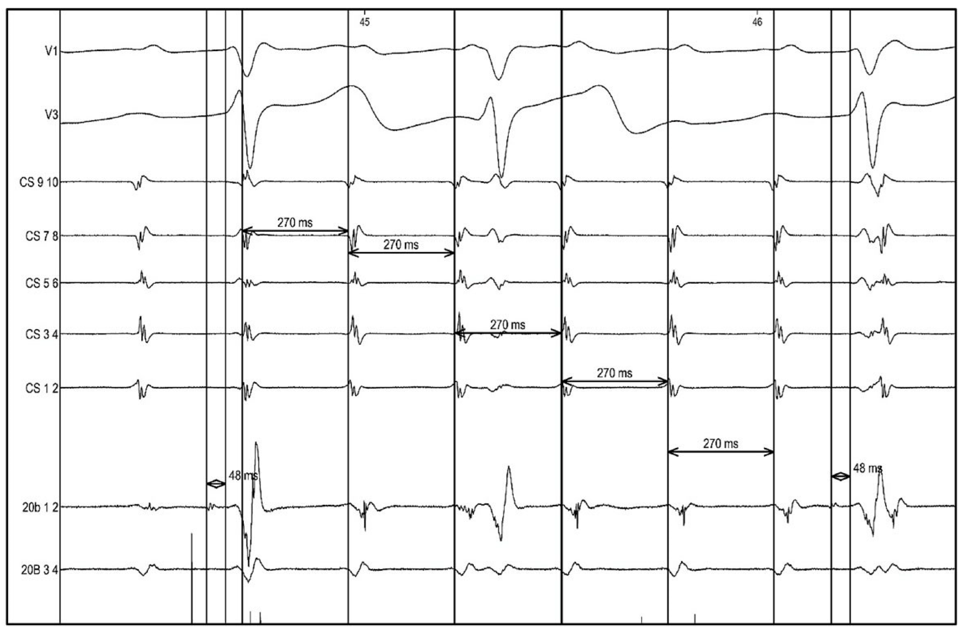
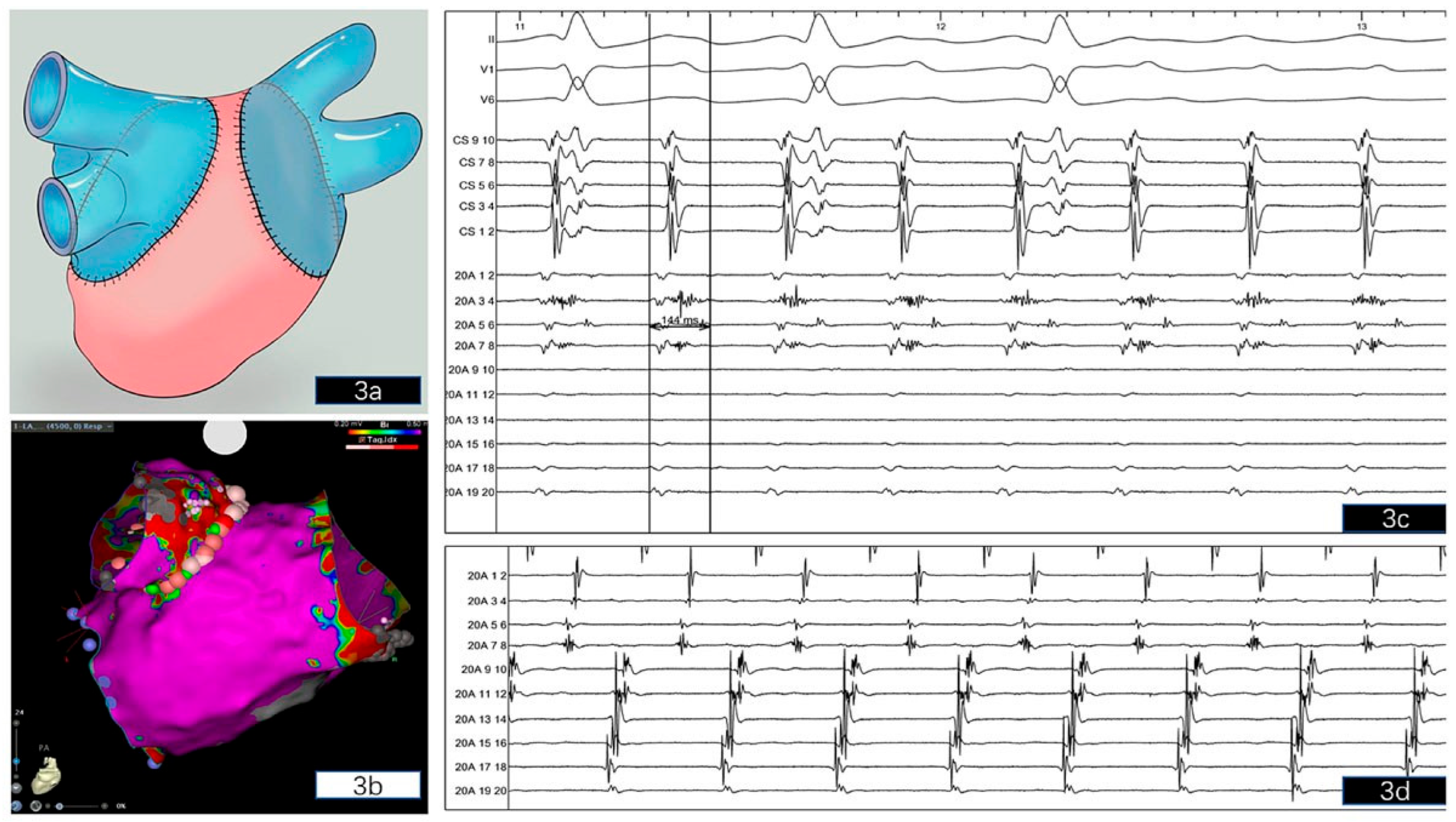
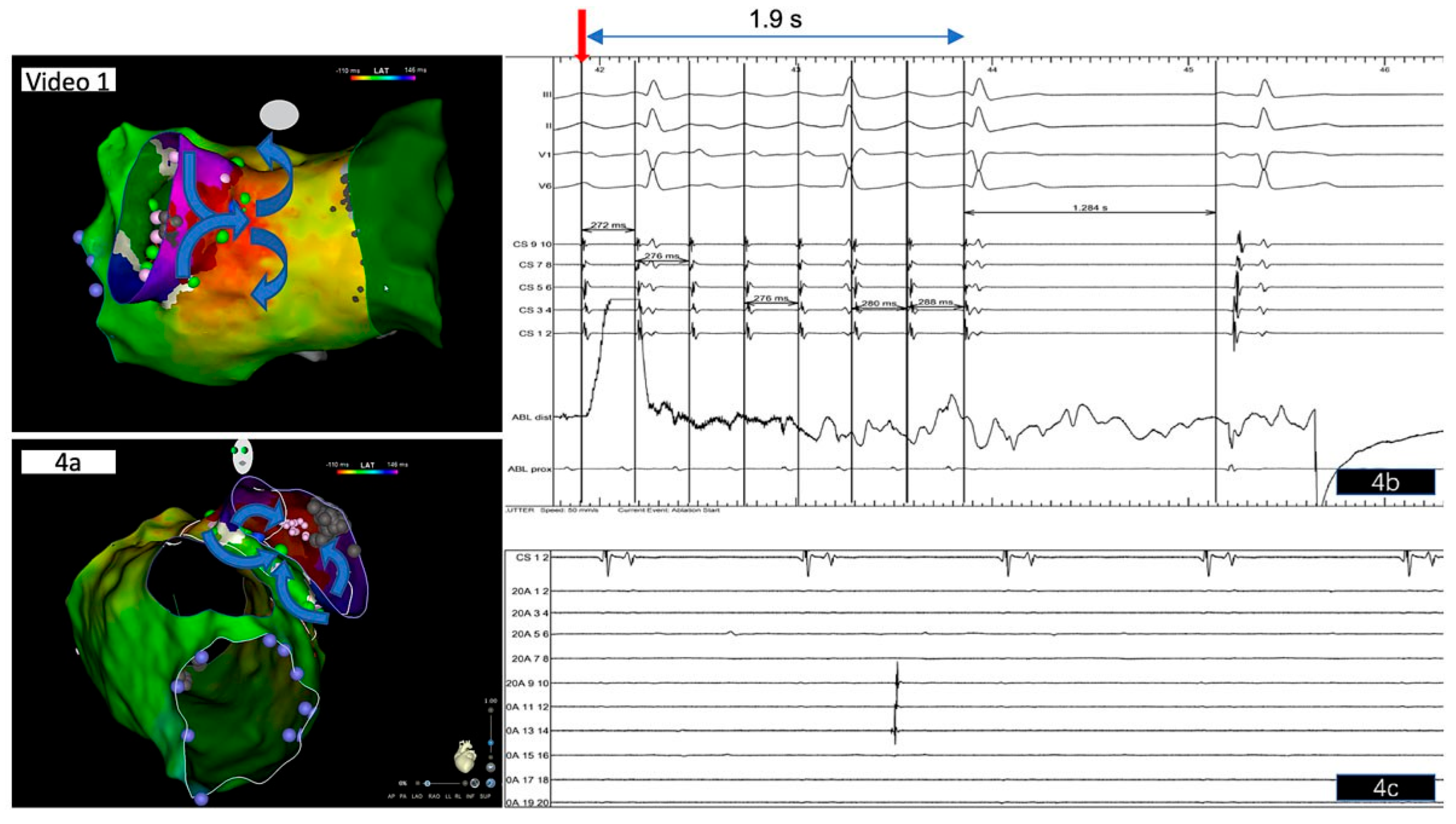
Continuous monitoring of the surface 12-lead ECG and bipolar endocardial electrograms during mapping and ablation were stored on a computer-based digital amplifier/recorder system (Bard Boston Scientific, Inc., Marlborough). Intracardiac electrograms were filtered from 30 to 500 Hz and measured at a sweep speed of 100 mm/s. A standard decapolar catheter was placed in the coronary sinus and served as the reference electrode. The eccentric coronary sinus activation (with the coronary sinus catheter in a proximal position) during atrial tachycardia suggested a left atrial origin. A quadripolar electrode was placed in the His position for baseline measurements, as well as for guiding transseptal puncture. The 12-lead surface ECG during narrow complex tachycardia constituting an atrial tachycardia with variable atrioventricular conduction showed a positive P-wave in V1 and positive P-waves in the precordial leads, as well as an inferior P wave axis, suggesting an origin in the superior part of the left atrium. The tachycardia was stable with an atrial cycle length of 270 ms (Figure 2). Single transseptal access was obtained under fluoroscopy using a 8.5 F SL-1 sheath and a BRK-1 (Abbott, Chicago, Il) transseptal needle. Next, the SL1 sheath was replaced by an Agilis steerable sheath (8.5 F, Abbott) and a multipolar high-resolution mapping catheter (PentaRay, Biosense Webster, Diamond Bar, CA) and later a 3.5-mm-tip SmartTouch ThermoCool (Biosense Webster) bidirectional ablation catheter were placed in the left atrium. A 3D endocardial high-density (Confidense module, Biosense Webster) electroanatomical activation and voltage map of the left atrium during atrial tachycardia was acquired with the PentaRay and ablation catheter covering the recipient’s left atrium and the donor pulmonary veins guided by contrast-enhanced computed tomography (CT) of the left atrium that was acquired one day prior to the procedure to exclude left atrial thrombi and to reconstruct 3D left atrial anatomy. The CT and 3D bipolar endocardial voltage mapping during atrial tachycardia confirmed that the donor bilateral pulmonary veins shared an ipsilateral common trunk (single right-sided pulmonary vein and single left-sided pulmonary vein) that was created by the thoracic surgeon during bilateral lung transplantation in order to facilitate the anastomosis of the pulmonary veins with the left atrium of the recipient. A schematic view of the donor pulmonary vein anastomosis during bilateral lung transplantation shows ipsilateral donor pulmonary veins (coloured blue) taken as common trunks with a cuff of left atrial tissue for anastomosis to the recipient left atrium (coloured pink) (Figure 3a). Theoretically, this results in complete electrical isolation of the donor pulmonary veins / atrial cuff. However, during voltage and activation mapping, the suture lines and pulmonary vein cuffs manifested as low-voltage areas, whereas the left atrium displayed no larger low voltage areas (Figure 3b). The right common trunk pulmonary vein was surgically isolated. However, positioning the PentaRay catheter across the suture line between the common trunk left pulmonary vein and left atrium recorded long fractionated low-voltage potentials and separated atrial electrograms, located at the posterosuperior and anterosuperior aspects of the left pulmonary vein-left atrium anastomosis (Figure 3c,d), indicating viable myocytes and a partial line of block in this area. The activation map confirmed a macroreentrant atrial tachycardia (donor-to-recipient conduction) involving the left donor pulmonary vein with an exit at the posterosuperior aspect of the pulmonary vein-LA junction displaying long fractionated low-voltage potentials (Figure 4a, video 1). Based on our findings, the critical isthmus of the reentrant circuit and exit of the atrial tachycardia was suspected at the posterosuperior aspect of the left pulmonary vein-LA anastomosis displaying the longest fractionated bipolar signals (Figure 3c; 20A 3-4). As previously published by other groups [3,4], the conduction gap along this anastomosis line acted as the critical isthmus of the reentry circuit, and the electrical propagation wave-front entered the donor pulmonary vein cuff at a second gap at the anterior ridge between the recipient left atrium and donor pulmonary vein, closing the circuit (Figure 4a). Radiofrequency ablation in this area terminated the tachycardia within 2 s with slowing down of the atrial tachycardia prior to termination (Figure 4b). Since a macroreentrant tachycardia incorporating the left pulmonary vein was assumed, based on 3D activation mapping, we opted for electrical isolation of the left pulmonary vein common trunk (Figure 3b and video 1), which was achieved at the end of the procedure after a waiting period of 30 min, showing isolated pulmonary vein activity with exit block in this vein (Figure 4c).
You will find the video file in the multimedia collection of Cardiovascular Medicine: https://cardiovascmed.ch/online-only-content.

Follow-up

No complications occurred, the periprocedural course was uneventful and the patient was discharged home the day after catheter ablation. At three months follow-up, he was asymptomatic with no recurrence of atrial tachycardia/fibrillation under a reduced dose of metoprolol 50 mg b.i.d. and continued oral anticoagulation.

Discussion

Since atrial arrhythmias after lung transplantation are associated with poor long-term survival [5], efforts to maintain sinus rhythm may improve survival and quality of life in these patients. As antiarrhythmic drugs are associated with increased mortality and toxicity in patients with atrial arrhythmias after lung transplantation [6], catheter ablation is a promising treatment option [3,4,7,8,9,10,11]. Frequently, double electrical connections between the donor pulmonary vein cuff and recipient left atrium, such as reported in our case, form the substrate for macroreentrant left atrial tachycardia, although other mechanisms such as enhanced automaticity can also be found. The underlying substrate can be effectively and safely targeted by catheter ablation. Compared with ablation catheter-based point-by-point mapping, multielectrode catheters with smaller electrodes and a higher resolution allow more rapid mapping in order to better understand the tachycardia mechanism and enable high success rates for ablation of substrate-based atrial tachycardia [12]. Atrial tachycardia occurring after lung transplantation often involves uncommon anatomical obstacles in which the left-sided surgical pulmonary vein-left atrium anastomosis served as the critical site (donor-to-recipient conduction). Of interest, mapping in this area in our patient demonstrated long fragmented low-voltage potentials in the posterosuperior left pulmonary vein-left atrium cuff. Considering the patient’s comorbidities, this may account for the predisposing factors leading to atrial tachycardia following bilateral lung transplantation [5]. Notably, no additional significant fibrosis or abnormal myocardium within the left atrium was found. Donor pulmonary veins are formed into single ipsilateral trunks and connected to an atrial cuff, which is sutured to the recipient’s left atrium in the double-lung transplant technique, as it was the case in our patient, shown schematically in Figure 3a. In theory, conduction block should be present along this anastomosis. Whereas anatomically the “neo pulmonary veins” are often attached to the extreme lateral ends of the posterior left atrium, the donor pulmonary veins dissected for lung transplantation usually include partial sections of donor posterior left atrium. That might lead to more potentially arrhythmogenic tissue being preserved by the procedure, and result in the posterior wall of the left atrium becoming a prominent site for initiators and drivers of atrial arrhythmias [13]. It is also possible that viable myocardium from inside the pulmonary veins, which was demonstrated in our case by documenting automaticity in the isolated left pulmonary vein, migrates towards the recipient’s left atrium and forms a myocardial bridge predisposing to atrial arrhythmias in the long term. Whether complete left-sided pulmonary vein isolation was necessary in this case can be debated. Since we identified at least two conduction gaps (one posterosuperior gap and one at the anterior ridge), as previously described by other groups [3,4], between the donor pulmonary vein cuff and the recipient left atrium forming the substrate for the clinical donor-to-recipient macroreentrant atrial tachycardia, we opted for complete unilateral pulmonary vein isolation rather than just targeting the reentrant mechanism by ablating the critical isthmus. Therefore, we first terminated the tachycardia by targeting the critical isthmus at the posterosuperior gap, and then isolated the pulmonary vein by an anatomical widearea circumferential approach. The fact that automaticity was observed in the donor pulmonary vein cuff after successful unilateral pulmonary vein isolation supported our approach, since this automaticity with residual donor-torecipient conduction could have been a trigger for future atrial arrhythmias.
In conclusion, this case highlights the feasibility, safety and efficacy of invasive 3D electroanatomical mapping guided catheter ablation of left-sided macroreentrant atrial tachycardia related to bilateral lung transplantation.

Disclosure statement

AMS received educational grants from Abbott, Biosense Webster, Biotronik, Bosto Scientific and Medtronis.

References

  1. Waldron, N.H.; Klinger, R.Y.; Hartwig, M.G.; Snyder, L.D.; Daubert, J.P.; Mathew, J.P. Adverse outcomes associated with postoperative atrial arrhythmias after lung transplantation: A meta-analysis and systematic review of the literature. Clin Transplant. 2017, 31. [Google Scholar] [CrossRef]
  2. Cui, G.; Tung, T.; Kobashigawa, J.; Laks, H.; Sen, L. Increased incidence of atrial flutter associated with the rejection of heart transplantation. Am J Cardiol. 2001, 88, 280–284. [Google Scholar] [CrossRef] [PubMed]
  3. Baykaner, T.; Cooper, J.M. Atypical flutter following lung transplantation involving recipient-to-donor tissue connections. HeartRhythm Case Rep. 2018, 4, 548–552. [Google Scholar] [CrossRef] [PubMed]
  4. Itoh, T.; Yamada, T. Left atrial and pulmonary vein flutter associated with double electrical connections after a lung transplantation. Europace. 2017, 19, 855. [Google Scholar] [CrossRef] [PubMed]
  5. Kim, B.G.; Uhm, J.S.; Yang, P.S.; Yu, H.T.; Kim, T.H.; Joung, B.; et al. Clinical significance of postoperative atrial arrhythmias in patients who underwent lung transplantation. Korean J Intern Med (Korean Assoc Intern Med). 2019. [Google Scholar] [CrossRef]
  6. Whitbeck, M.G.; Charnigo, R.J.; Khairy, P.; Ziada, K.; Bailey, A.L.; Zegarra, M.M.; et al. Increased mortality among patients taking digoxin—analysis from the AFFIRM study. Eur Heart J. 2013, 34, 1481–1488. [Google Scholar] [CrossRef] [PubMed]
  7. Nazmul, M.; Munger, T.M.; Powell, B.D. Atrial tachycardia originating from a donor pulmonary vein in a lung transplant recipient. Circulation. 2011, 124, 1288–1289. [Google Scholar] [CrossRef] [PubMed]
  8. Sacher, F.; Vest, J.; Raymond, J.M.; Stevenson, W.G. Incessant donor-to-recipient atrial tachycardia after bilateral lung transplantation. Heart Rhythm. 2008, 5, 149–151. [Google Scholar] [CrossRef] [PubMed]
  9. Sanam, K.; Holmes, D.; Shah, D.; Foster, N. Late atrial tachycardia originating from donor pulmonary vein in a double lung transplant recipient. HeartRhythm Case Rep. 2015, 1, 490–493. [Google Scholar] [CrossRef] [PubMed]
  10. See, V.Y.; Roberts-Thomson, K.C.; Stevenson, W.G.; Camp, P.C.; Koplan, B.A. Atrial arrhythmias after lung transplantation: epidemiology, mechanisms at electrophysiology study, and outcomes. Circ Arrhythm Electrophysiol. 2009, 2, 504–510. [Google Scholar] [CrossRef] [PubMed]
  11. Uhm, J.S.; Park, M.S.; Joung, B.; Pak, H.N.; Paik, H.C.; Lee, M.H. Intra-atrial reentrant tachycardia originating from the pulmonary vein cuff anastomosis in a lung transplantation patient: Ultra-high-density 3-dimensional mapping. HeartRhythm Case Rep. 2018, 4, 152–154. [Google Scholar] [CrossRef] [PubMed][Green Version]
  12. Sohns, C.; Saguner, A.M.; Lemes, C.; Santoro, F.; Mathew, S.; Heeger, C.; et al. First clinical experience using a novel high-resolution electroanatomical mapping system for left atrial ablation procedures. Clin Res Cardiol. 2016, 105, 992–1002. [Google Scholar] [CrossRef] [PubMed]
  13. Santoro, F.; Sohns, C.; Saguner, A.M.; Brunetti, N.D.; Lemes, C.; Mathew, S.; et al. Endocardial voltage mapping of pulmonary veins with an ultra-high-resolution system to evaluate atrial myocardial extensions. Clin Res Cardiol. 2017, 106, 293–299. [Google Scholar] [CrossRef] [PubMed]
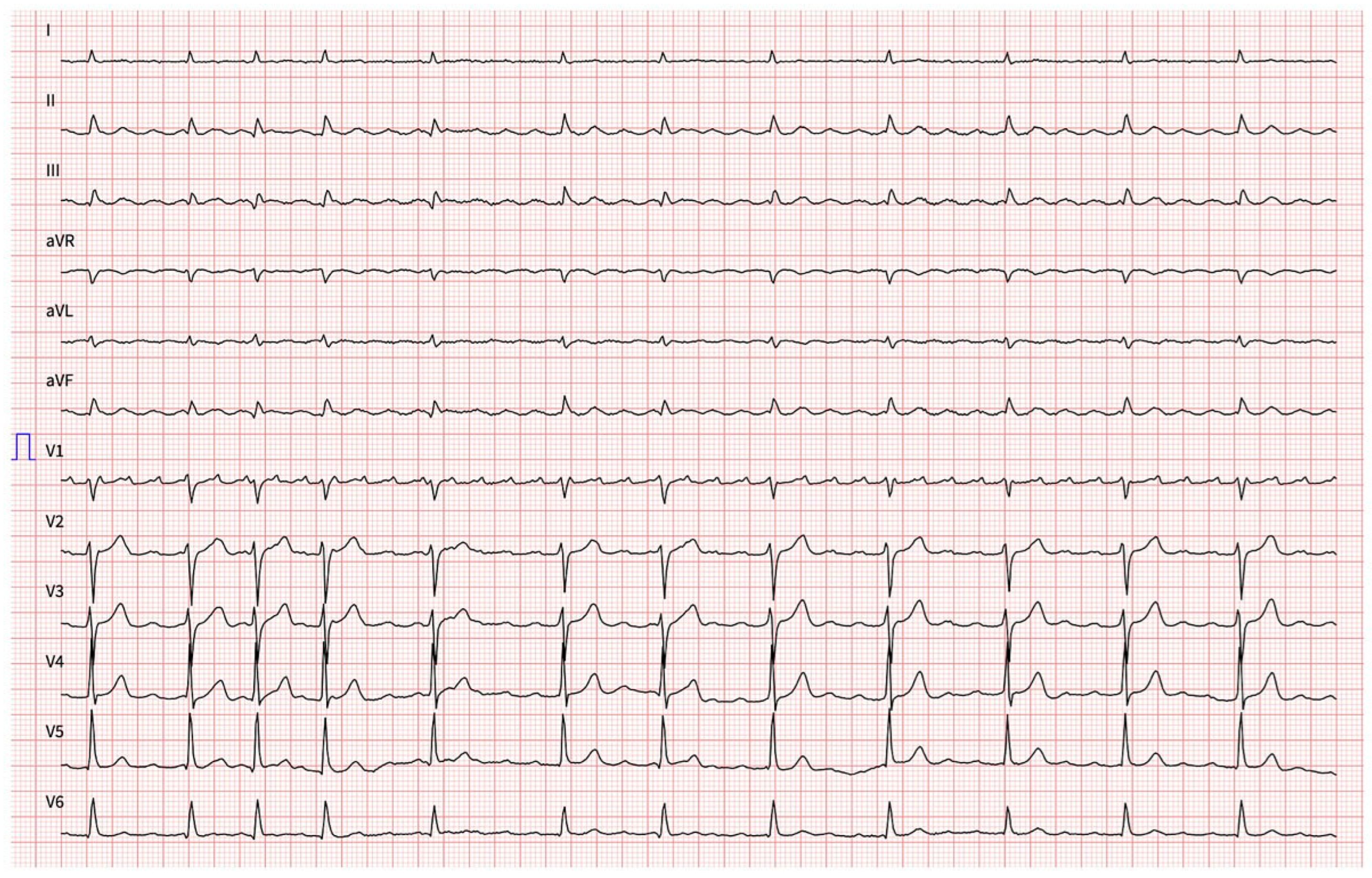
Figure 1. Surface 12-lead ECG (speed 25 mm/s) after adenosine administration constituting an atrial tachycardia with variable atrioventricular conduction; note the positive P-wave in V1 and positive P-waves in the precordial leads, as well as an inferior P-wave axis in the inferior leads, suggesting an origin in the superior part of the left atrium.
Cardiovascmed 23 w02117 g001
Figure 2. Surface electrocardiogram (V1 and V3) and intracardiac electrograms recorded during atrial tachycardia, with a sweep speed of 100 mm/s. Baseline measurements: CL of atrial tachycardia 270 ms with variable atrioventricular conduction (2:1 and 3:1), HV 48 ms. CS = decapolar catheter in the coronary sinus (distal placement).
Cardiovascmed 23 w02117 g002
Figure 3. (a) Schematic PA view of donor pulmonary vein anastomosis during double lung transplantation. Ipsilateral donor pulmonary veins (blue) are taken as common trunks with a cuff of left atrial tissue for anastomosis to the recipient left atrium (pink). In theory, this results in complete electrical isolation of the donor pulmonary vein/atrial cuff. (b) 3D electroanatomical reconstruction of the recipient left atrium and donor left-sided common trunk pulmonary vein. The common trunk right-sided pulmonary vein is not shown. Low-voltage areas (pink small points) located at the posterosuperior and anterosuperior aspects of the left pulmonary vein-left atrium anastomosis. No significant low voltage areas within the left atrium were found in this patient, the right-sided pulmonary vein trunk was electrically isolated. (c) Intracardiac electrograms on the high-resolution PentaRay mapping catheter showed long fractionated low-voltage potentials indicating pulmonary vein reconduction in this area and the critical site of atrial tachycardia (note bipolar signals from 20A 3-4) and double potentials indicating a complete surgical suture line with a “line of block” (20A 5-6), which located on the surgical posterosuperior left pulmonary vein-left atrium anastomosis. (d) Intracardiac electrograms also showed fragmented and late potentials at the anterior aspect of the left pulmonary vein-left atrium anastomosis corresponding to the donor pulmonary vein entry site of the macroreentrant circuit (note signals from 20A 7-8 to 20A 9-10, which are located on both sides of the suture line indicating a “line of block” in this area). CS = coronary sinus; 20A 1-2 to 19-20 = multipolar PentaRay catheter; PA = posteroanterior projection.
Cardiovascmed 23 w02117 g003
Figure 4. and video 1: (a) Activation map during macroreentrant atrial tachycardia depicting electrical propagation (blue arrows) entering the donor pulmonary vein at an anterior ridge donor-to-recipient conduction gap and exiting at a second posterosuperior donor-to-recipient conduction gap (critical isthmus) located at the posterior atriotomy suture between the donor left pulmonary vein trunk and recipient left atrium. Fragmented potentials and double potentials were seen most prominently at the posterosuperior ridge left pulmonary vein cuff (pink points), which could be visualised on the intracardiac electrograms shown in Figure 3c. Radiofrequency ablation at this area terminated the atrial tachycardia within 2 s (blue arrow shows the site of termination). (b) Tracing showing atrial tachycardia termination 1.9 s (blue line) after the onset of RF delivery (red arrow) with slowing down of the atrial tachycardia prior to termination. (): Dissociated pulmonary vein potentials (automaticity with exit block) recorded in the left pulmonary vein common trunk with the PentaRay catheter (20A 9-14) after successful left-sided anatomical wide-area pulmonary vein isolation (see also large rose to red ablation points encircling the left-sided pulmonary vein trunk in Figure 3b and Figure 4a).
Cardiovascmed 23 w02117 g004