Next Article in Journal
Benefit of Upstream Impella Insertion in Cardiogenic Shock
Previous Article in Journal
Patients with Syncope Remaining Unexplained After a Structured Work-Up Share the Characteristics of Patients with Neurally Mediated Syncope
 
 
Cardiovascular Medicine is published by MDPI from Volume 28 Issue 1 (2025). Previous articles were published by another publisher in Open Access under a CC-BY (or CC-BY-NC-ND) licence, and they are hosted by MDPI on mdpi.com as a courtesy and upon agreement with Editores Medicorum Helveticorum (EMH).
Case Report

Unusual Collateralisation of a Chronic Total Ccclusion of the Left Anterior Descending Artery

Hôpitaux Universitaires de Genève, Genève, Switzerland
*
Author to whom correspondence should be addressed.
Cardiovasc. Med. 2020, 23(2), w02092; https://doi.org/10.4414/cvm.2020.02092
Submission received: 20 January 2020 / Revised: 20 February 2020 / Accepted: 20 March 2020 / Published: 20 April 2020

Summary

When performing revascularisation of a chronic total occlusion, contralateral injection is essential to visualise the distal bed of the occluded vessel, adding to procedural success. Here we describe a case of antegrade recanalisation of a left anterior descending artery chronic total occlusion with retrograde injection through a well-developed conus branch.

Case Report

Following a positive stress test performed in the context of shortness of breath on exertion, coronary angiography was planned for a 67-year-old patient with a history of hypertension, dyslipidaemia and recent radiofrequency ablation of an atrial flutter. Echocardiography showed conserved left ventricle ejection fraction. Coronary angiography revealed sub-occlusion of the proximal left circumflex (LCX) artery with bridging collaterals. We could not easily differentiate the left anterior descending (LAD) artery from a large first diagonal branch on the right anterior oblique (RAO) cranial view (Figure 1A). Using the lateral view, we visualised a chronic total occlusion (CTO) of the mid segment of the LAD artery (Figure 1B). During right coronary artery angiography (Figure 1C), we unintentionally injected a well-developed branch—with a separate origin in the aorta next to the right coronary ostium—providing large collateral vessels to the distal LAD artery (Figure 1D). The LCX artery lesion was treated ad hoc with a drug eluting stent (Ultimaster 2.5 × 38 mm, Terumo Europe, Leuven, Belgium) (Figure 1E,F) and we decided to perform a noninvasive functional test to assess the presence and severity of any ischaemia in the territory of the LAD artery. The patient was discharged on acetylsalicylic acid 100 mg/day, clopidogrel 75 mg/day (for 3 months) and rivaroxaban 15 mg/day.
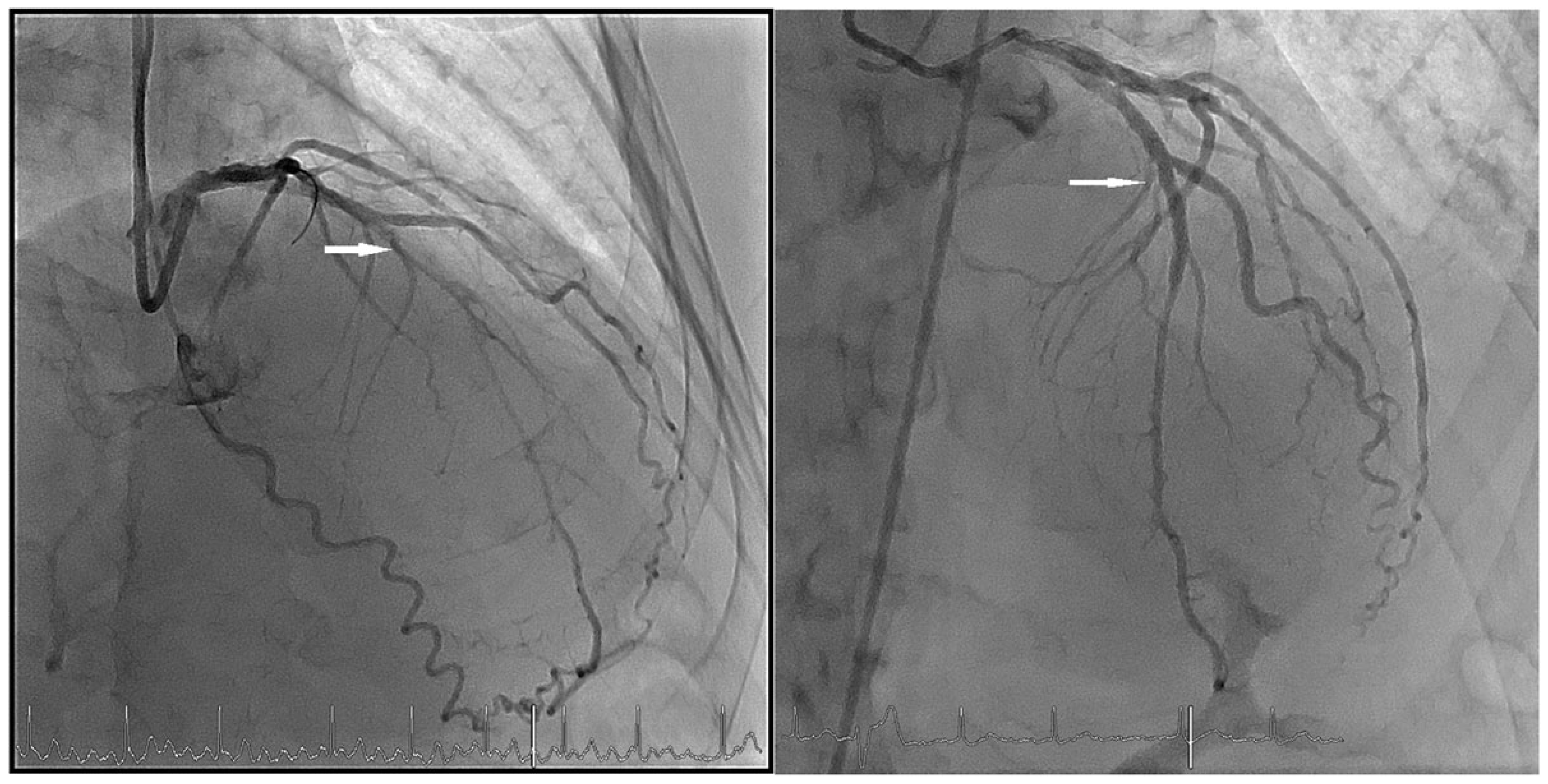
Subsequently, cardiac positron emission tomography showed a large area of ischaemia secondary to the LAD artery occlusion, with a total reversible deficit involving 19% of the left ventricle volume. Hence the patient was readmitted for revascularisation. The conus artery was selectively cannulated with a 5F left coronary bypass catheter (Cordis, Miami Lakes, Florida, USA) via the right radial artery to visualise the distal LAD artery by simultaneous injection. A 0.014 blue SION guidewire (Asahi, Aichi, Japan) was used to stabilise the diagnostic catheter. The left main coronary artery was engaged with a 7F EBU 4.0 guiding catheter (Medtronic, Minneapolis, MN, USA) via the right femoral artery (Figure 2A). The occlusion was crossed using an Asahi Fielder XT-A, subsequently exchanged for a Galeo Pro (Concert Medical, Norwell, Massachusetts, USA) wire using a Finecross micro-catheter (Terumo, Tokyo, Japan). A drug eluting stent (Ultimaster 2.5 × 33 mm, Terumo) was implanted with a good angiographic result (Figure 2B). The patient was discharged on the same triple therapy regimen.
You will find the video files in the multimedia collection of Cardiovascular Medicine: https://cardiovascmed.ch/online-only-content.

Discussion

Despite providing complementary visualisation of the mid LAD and distal LAD artery around the apex, the lateral view is no longer routinely used. The major concern about the steep left anterior oblique (LAO) tube angulation is radiation exposure for patients and staff [1]. However, as in our case, when the cranial angulations are not enough to differentiate between a well-developed diagonal artery (with LAD occlusion) or the LAD artery, lateral projection can be a valuable option.
In our case, we could argue that the source of the collaterals could be either a well-developed conus branch, considering its origin (the conus branch arises directly from the aorta in 20 to 30% as in our case,) or a right ventricular (RV) branch with an atypical origin. Indeed, the path of the collateral was compatible with an RV branch, as was its connection to the LAD on its distal portion. Interestingly, an observational study including 481 patients with 519 CTOs demonstrated that collaterals from the conus branch were less frequently encountered (5.9%) than collaterals from typical RV branches (26.8% and second most frequent source of collaterals to the LAD) in LAD CTOs. Of note, collaterals to the LAD develop most commonly from the posterior descending artery (80.6%) [2].
The importance of careful, selective cannulation of the conus branch in the presence of chronic occlusion of the LAD artery without any collateral filling, and with wellpreserved left ventricular function, has been highlighted [3]. This pattern of collaterals to the LAD artery from the conus artery is known as the arterial circle of Vieussens or Vieussens’ ring [4]. Coronary computed tomography angiography may be useful to depict this if not seen during coronary angiography. Recanalisation of a CTO in the LAD artery using contralateral injection via an isolated conus artery has been described in a case presentation [5]. A major concern during selective cannulation of the conus artery is provocation of major arrhythmias. However, it was suggested that short and severe pressure damping during selective cannulation is not dangerous if no test injection of contrast is performed and the catheter is quickly withdrawn after angiographic acquisition, especially in the event of contrast staining [3].

Conclusions

When it is suspected that a well-developed diagonal artery (with LAD occlusion) is mistaken for the LAD artery, the lateral view can help to show whether the LAD artery is present or not. Furthermore, in patients with LAD artery CTO and preserved left ventricular function, the absence of collateralisation is unlikely. In this setting, as in our case, collaterals may develop from an unusual artery and should be assessed.

Conflicts of Interest

The authors declare no conflict of interest.

References

  1. Kuon, E.; Dahm, J.B.; Empen, K.; Robinson, D.M.; Reuter, G.; Wucherer, M. Identification of less-irradiating tube angulations in invasive cardiology. J. Am. Coll. Cardiol. 2004, 44, 1420–1428. [Google Scholar] [CrossRef] [PubMed]
  2. McEntegart, M.B.; Badar, A.A.; Ahmad, F.A.; Shaukat, A.; MacPherson, M.; Irving, J.; et al. The collateral circulation of coronary chronic total occlusions. EuroIntervention 2016, 11, e1596–e1603. [Google Scholar] [CrossRef] [PubMed]
  3. Tanigawa, J.; Petrou, M.; Di Mario, C. Selective injection of the conus branch should always be attempted if no collateral filling visualises a chronically occluded left anterior descending coronary artery. Int. J. Cardiol. 2007, 115, 126–127. [Google Scholar] [CrossRef] [PubMed]
  4. Deora, S.; Shah, S.; Patel, T. “Arterial circle of Vieussens”—An important intercoronary collateral. Int. J. Cardiol. Heart Vessels. 2014, 3, 84–85. [Google Scholar] [CrossRef] [PubMed]
  5. Kawamura, A.; Jinzaki, M.; Kuribayashi, S. Percutaneous revascularization of chronic total occlusion of left anterior descending artery using contralateral injection via isolated conus artery. J. Invasive Cardiol. 2009, 21, E84–E86. [Google Scholar] [PubMed]
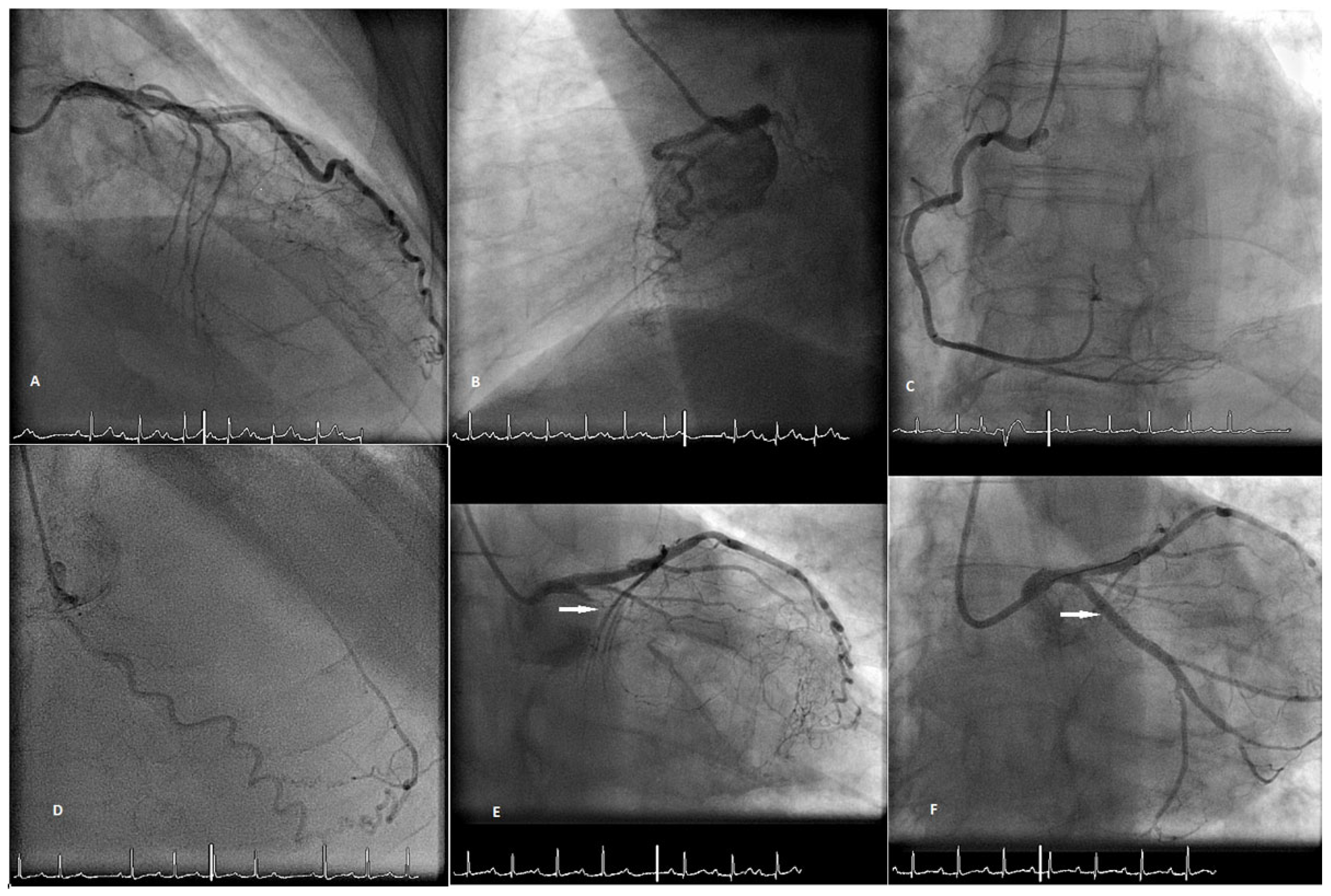
Figure 1. (A) RAO cranial view showing a sub-occlusion of the proximal LCX artery. The second vessel is either the LAD or a well-developed first diagonal branch with occlusion of the LAD artery. (B) Lateral view angiogram showing no vessel extending to the apex which implies that the LAD artery is occluded and that we are looking at a well-developed first diagonal branch. (C) Right coronary artery. (D) Vessel with an origin on the aorta next to the right coronary ostium (conus branch) and providing large collateral flow to the LAD artery. (E,F) Angiography of the LCX artery before and after angioplasty, respectively.
Cardiovascmed 23 w02092 g001
Figure 2. (A) Simultaneous injection of the LAD artery and the conus branch. (B) Angiography showing the result after LAD artery percutaneous coronary intervention.
Cardiovascmed 23 w02092 g002