Next Article in Journal
Remotely Navigated Ablation of RVOT Tachycardia
Previous Article in Journal
Severe Pulmonary Embolism: Surgical Aspects
 
 
Cardiovascular Medicine is published by MDPI from Volume 28 Issue 1 (2025). Previous articles were published by another publisher in Open Access under a CC-BY (or CC-BY-NC-ND) licence, and they are hosted by MDPI on mdpi.com as a courtesy and upon agreement with Editores Medicorum Helveticorum (EMH).
Interesting Images

Franck’s Sign

Division of Cardiology, Geneva University Hospitals, 1205 Genève, Switzerland
Cardiovasc. Med. 2017, 20(6), 162; https://doi.org/10.4414/cvm.2017.00485
Submission received: 21 March 2017 / Revised: 21 April 2017 / Accepted: 21 May 2017 / Published: 21 June 2017

Case Description

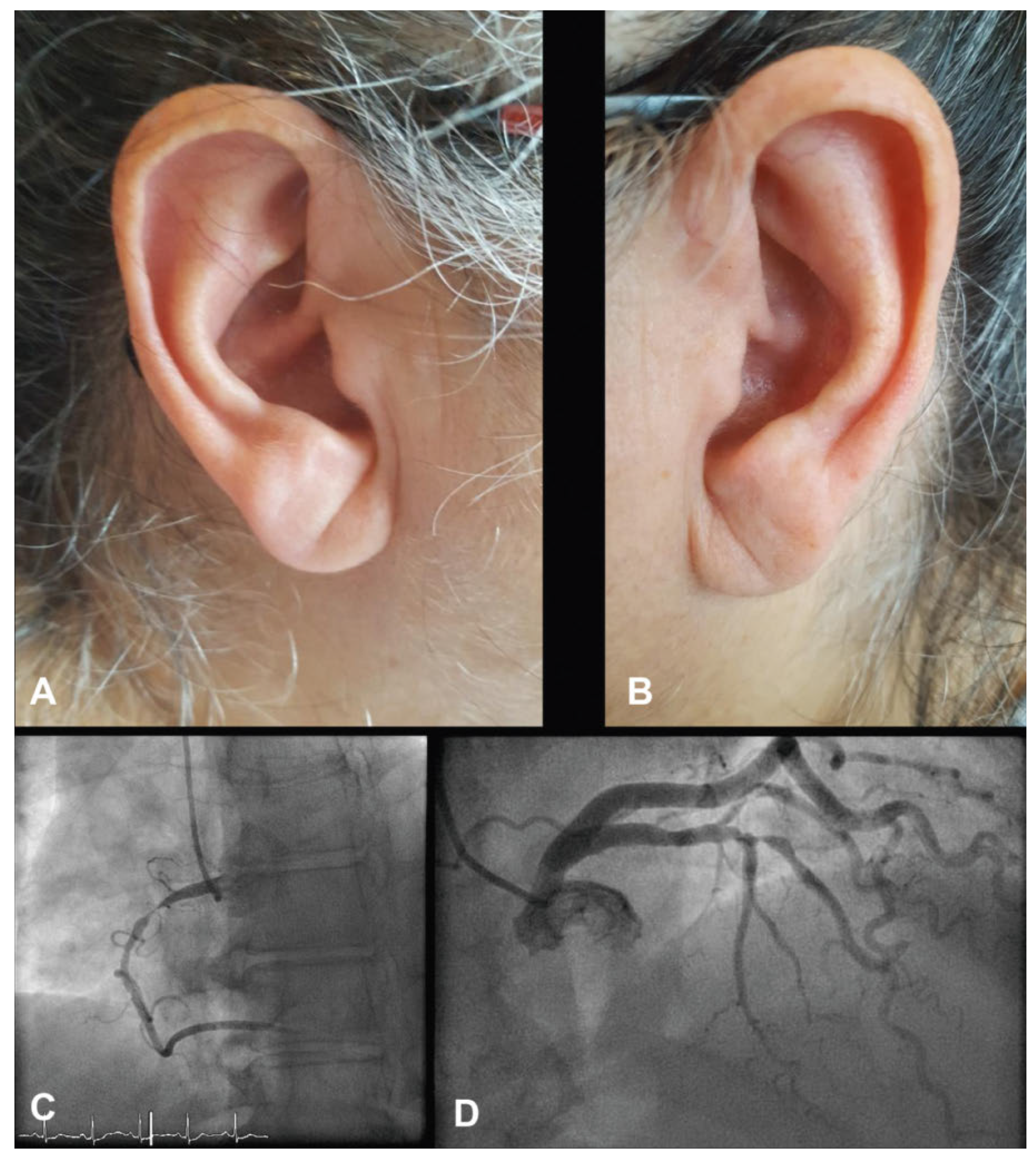
A 61-year-old obese female with Parkinson’s disease reported recurrent chest pain and exertional dyspnoea in the previous 24 hours. Clinical examination was normal, but careful inspection of both ear lobes showed bilateral oblique creases extending backwards from the intertragic notch to the edge of the lobule (Figure 1, panels A–B). The electrocardiogram was unremarkable; however, cardiac troponin was mildly elevated. A coronary angiogram showed two-vessel coronary artery disease, which was successfully treated with drug-eluting stents (Figure 1, panel C–D). The subsequent course was uneventful and the patient was discharged on dual antiplatelet therapy and a statin and counselled for intensive cardiovascular risk factor management including cardiac rehabilitation.
Ear lobe crease, commonly referred as “Franck’s sign”, was initially described more than forty years ago by Sanders T. Franck, as a cutaneous sign of coronary artery disease. Several pathophysiological explanations have been proposed, including loss of elastic fibres due to increased free radical oxidative stress and diminished blood supply in the absence of collateral vessels in the ear lobe. A substantial number of studies have evaluated the diagnostic performance of ear lobe crease as a marker of coronary artery disease. In a recent meta-analysis including more than 31 000 patients, Franck’s sign was associated with a 3.3-fold increase in the risk of coronary artery disease. The sign itself had sensitivity of 62% and specificity of 67%.
The finding of Franck’s sign should thus prompt further questioning for conventional cardiovascular risk factors and family history of coronary artery disease.

Disclosure Statement

No financial support and no other potential conflict of interest relevant to this article was reported.

Author Contributions

Conception and design: FR, UV, GG. Drahing the manuscript for important intellectual content: FR, UV, GG.
Figure 1. Panel A–B: Right and left ears with Franck’s sign or ear lobe crease. Panel C: Proximal right coronary artery subocclusion. Panel D: Mid left anterior descending artery subocclusion. The patient gave her consent for the publication of the images.
Cardiovascmed 20 00162 g001