Next Article in Journal
Gibt es Dogmen in der Medizin?
Previous Article in Journal
Franck’s Sign
 
 
Cardiovascular Medicine is published by MDPI from Volume 28 Issue 1 (2025). Previous articles were published by another publisher in Open Access under a CC-BY (or CC-BY-NC-ND) licence, and they are hosted by MDPI on mdpi.com as a courtesy and upon agreement with Editores Medicorum Helveticorum (EMH).
Case Report

Remotely Navigated Ablation of RVOT Tachycardia

Cardiology/Electrophysiology, University Hospital Basel and CRIB–Cardiovascular Research Institute Basel, University of Basel, 4001 Basel, Switzerland
Cardiovasc. Med. 2017, 20(6), 160; https://doi.org/10.4414/cvm.2017.00481
Submission received: 21 March 2017 / Revised: 21 April 2017 / Accepted: 21 May 2017 / Published: 21 June 2017

Abstract

Ablation of tachycardia and premature ventricular contractions originating from the right ventricular outflow tract can safely be performed in patients with normal anatomy via the femoral approach. We present a case of catheter ablation in the right ventricular outflow tract in a patient with a venous malformation of the inferior vena cava by means of remote magnetic navigation via the internal jugular vein.

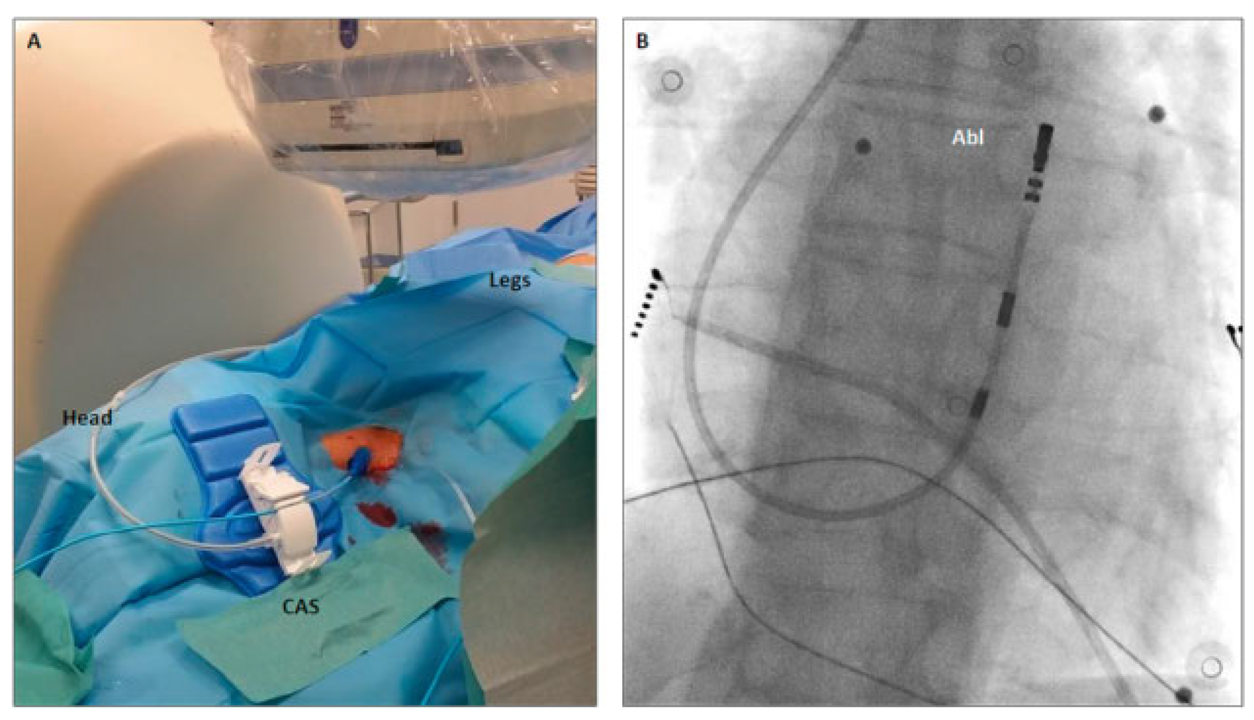
A 51-year-old female was referred for radiofrequency ablation of symptomatic frequent premature ventricular contraction (PVCs) originating from the right ventricular outflow tract (RVOT) (PVC burden 20%). Because of a congenital interruption of the inferior vena cava (IVC) between the renal and hepatic veins, the right jugular vein was chosen for access to the right ventricle via the superior vena cava. For optimal steerability of the catheter, we used the Niobe remote magnetic navigation system (Stereotaxis Inc, St Louis, MO, USA). The right jugular vein was punctured under echocardiographic sonographic guidance and a 3.5 mm open irrigated-tip ablation catheter (Navistar RMT TC, Biosense Webster) was advanced into the right atrium over an 8 Fr introducer. For remote advancement of the catheter via the tricuspid valve into the right ventricle, the catheter advancer system (Cardiodrive unit, Stereotaxis Inc, USA) was placed alongside the head of the patient (Figure 1A). A fast anatomical map was created by remote navigation with the CARTO 3 RMT system (Biosense Webster, Diamond Bar, CA, USA). Despite isoproterenol infusion, only very few PVCs were documented at the beginning of the intervention. Therefore, pace mapping was performed to localise the origin of the extrasystoles. A pace map with 12 out of 12 matching electrocardiogram leads (Figure 2A,B) was found in the anterolateral segment of the RVOT. Ablation was performed for 618 seconds and with a power of 35 watts (Figure 2C). Total procedure duration was 111 minutes with a fluoroscopy time of 0.8 minutes. After a waiting period of 20 minutes and isoproterenol infusion, no PVCs could be documented. Twenty-four-hour Holter monitoring 1 month after the ablation showed no recurrence of the PVCs.

Discussion

Vascular Access via the Internal Jugular Vein

In patients without congenital malformations, the RVOT is usually accessed via the femoral vein and the IVC. In our patient without an IVC connection between the renal and hepatic vein, this standard approach was precluded. Jugular vein access was favoured over a femoral vein puncture and then access to the right atrium via the azygos vein because the latter may be more challenging for anatomical reasons. To enable optimal performance, we used the right internal jugular vein and not the subclavian vein to allow straight access of the catheter via the catheter advancer system and the 8 Fr introducer (Figure 1A) without additional bending of the catheter. Puncture of the vein, fixation of the catheter advancer system and of the catheter (Figure 1A) could safely be performed.

Remote Magnetic Navigation

Electroanatomical mapping and ablation of PVCs originating from the RVOT with conventional ablation catheters via the internal jugular vein may be challenging because of the unfamiliar orientation for the operator. Remote magnetic navigation allows performing the procedure from the control room with maximal flexibility of catheter manoeuvrability [1]. The Niobe system also enables positioning of the catheter in small increments and high position stability due to its constant magnetic field [2,3]. Owing to the lower catheter contact force of remote magnetic navigation in comparison with conventional ablation, we increased the ablation power by 5 watt. Because of the floppy nature of the catheter, we observed, on both fluoroscopy and the Carto3 system, striking catheter movement with breathing. This pattern is reflected by the colour-coded energy distribution of the Niobe software on the static Carto3 map (Figure 2C). However, this did not have an impact on either the outcome or safety of the intervention.

Conclusion

We showed that access to the right cardiac chambers via the internal jugular vein with use of remote magnetic navigation is feasible in patients without the possibility for standard femoral access through the IVC as a result of venous malformation

Disclosure Statement

No financial support and no other potential conflict of interest relevant to this article was reported.
References

  1. Knecht, S.; Sticherling, C.; Kuhne, M. Entering through the back-door: remotely navigated ablation of leh atrial tachycardia in the presence of a large atrial septal defect occluder. Europace 2013, 15, 943. [Google Scholar] [CrossRef] [PubMed]
  2. Thornton, A.S.; Jordaens, L.J. Remote magnetic navigation for mapping and ablating right ventricular outflow tract tachycardia. Heart Rhythm. 2006, 3, 691–696. [Google Scholar] [CrossRef] [PubMed]
  3. Ernst, S.; Ouyang, F.; Linder, C.; Hertting, K.; Stahl, F.; Chun, J.; et al. Initial experience with remote catheter ablation using a novel magnetic navigation system: magnetic remote catheter ablation. Circulation 2004, 109, 1472–1475. [Google Scholar] [CrossRef] [PubMed]
Figure 1. (A) Clinical setting with the catheter advancer system (CAS) alongside the head of the patient and the access of the ablation catheter over an 8 Fr introducer via the internal jugular vein. (B) Fluoroscopic image in anterior-posterior projection of the catheter position during electroanatomical mapping of the right ventricular outflow tract.
Cardiovascmed 20 00160 g001
Figure 2. (A) Twelve-lead surface ECG of the spontaneous premature ventricular contraction and the pace-map (B) with 12 out of 12 matching leads. (C) Screenshot of the Niobe system. The left window shows the mapped right ventricular outflow tact tract geometry from the Carto3 system. The asterisk indicates the pacing site for the pace-map of panel B. The graded red area on the grey right ventricular ouflow tract (RVOT) map represents the delivered energy distribution during radiofrequency ablation on the static Carto map. The right upper window shows an overlay of the RVOT map and the fluoroscopic image in anterior-posterior projection and the lower window shows the Niobe model of the right ventricle with the RVOT encircled by the dashed line.
Cardiovascmed 20 00160 g002