Next Article in Journal
Review of the 2016 European Dyslipidaemia Guidelines
Previous Article in Journal
Chefredaktion «Cardiovascular Medicine» ab 2017
 
 
Cardiovascular Medicine is published by MDPI from Volume 28 Issue 1 (2025). Previous articles were published by another publisher in Open Access under a CC-BY (or CC-BY-NC-ND) licence, and they are hosted by MDPI on mdpi.com as a courtesy and upon agreement with Editores Medicorum Helveticorum (EMH).
Case Report

Atrioventricular Nodal Reentrant Tachycardia After Heart Transplant

Division of Cardiology, University Hospital Basel, CH-4031 Basel, Switzerland
*
Author to whom correspondence should be addressed.
Cardiovasc. Med. 2016, 19(12), 332; https://doi.org/10.4414/cvm.2016.00440
Submission received: 21 September 2016 / Revised: 21 October 2016 / Accepted: 21 November 2016 / Published: 21 December 2016

Abstract

We report the case of a 59-year old patient with a history of orthotopic heart transplantation, who was referred to us because of recurrent syncope due to atrioventricular nodal reentrant tachycardia. This case report is one of few in the literature describing an invasive treatment approach to a classical supraventricular tachycardia attributable to a pre-existing substrate of the donor’s heart.

Case Report

A 59-year-old man with a history of coronary artery disease and concomitant end-stage heart failure received an orthotopic heart transplant (OHT) with biatrial anastomosis at the age of 49 years. Overall, the procedure was tolerated well. The state of the donor heart years after OHT was good (left ventricular ejection fraction 60%, coronary sclerosis without significant stenosis). Cumulatively, there were three episodes of moderate acute cellular rejection (International Society of Heart and Lung Transplantation grade 3A). Ten years after successful transplantation the patient was admitted for unexplained syncope. During another episode of presyncope as an inpatient, the 12-lead electrocardiogram showed a regular narrow complex tachycardia at a rate of 220 bpm with suspected p-waves at the end of the QRS complex (Figure 1). Administration of 6 mg of adenosine promptly terminated the tachycardia. Because of the severity of symptoms, an electrophysiological study was performed.
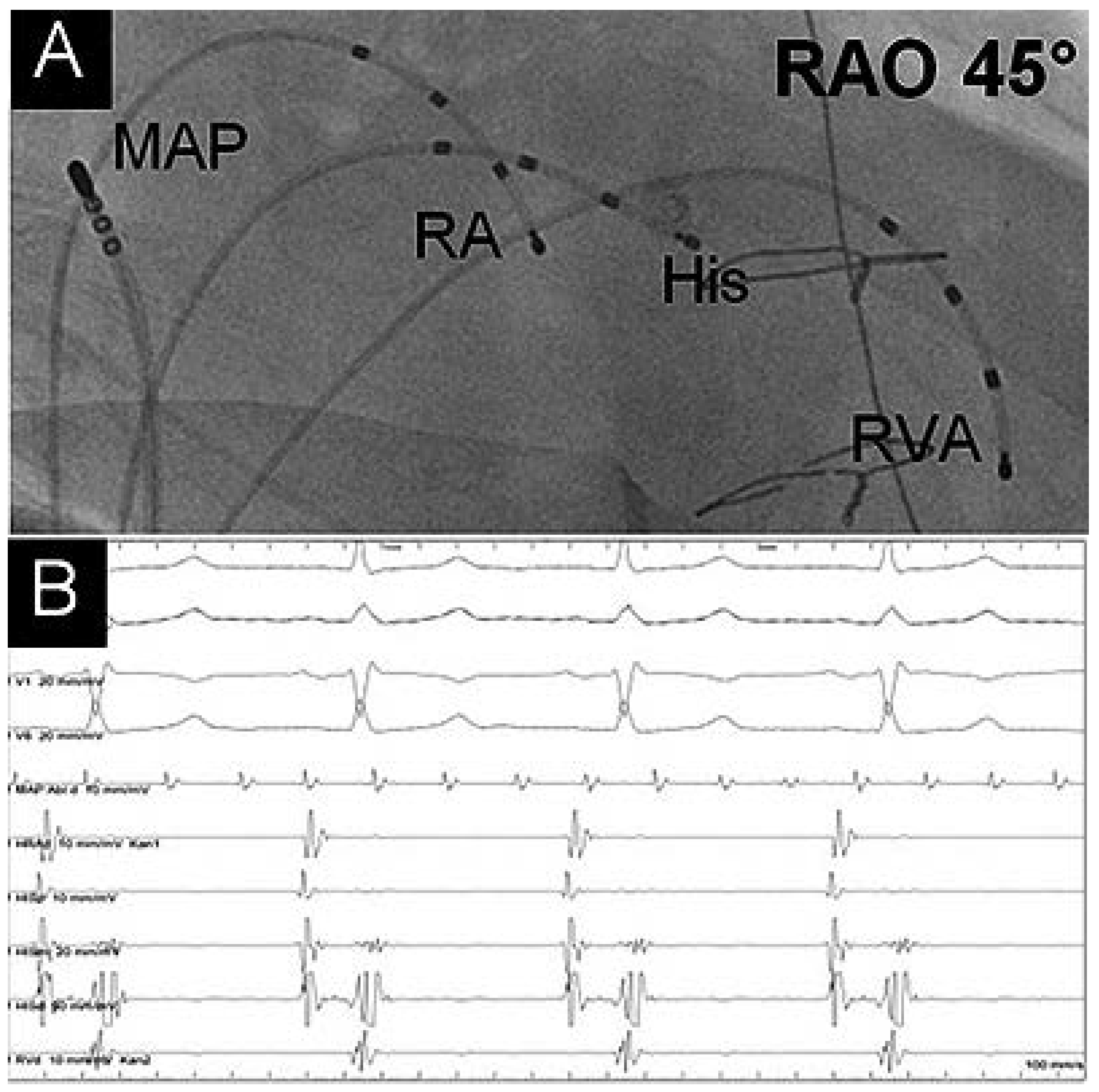
The study was performed with the heart in sinus rhythm. Atrial fibrillation was detected in the recipient’s right atrium (cycle length 164 ms, ablation catheter in Figure 2, panel B), while the donor right atrium showed sinus rhythm (cycle length 700 ms, Figure 2, panel A). With programmed atrial stimulation, typical atrioventricular nodal reentrant tachycardia (AVNRT) was easily inducible.
The slow pathway could be located in the classical posteroseptal position and was ablated successfully. During 6 months of follow-up, the patient has done well without recurrent syncope or tachycardia.
Supraventricular tachyarrhythmias (SVTs) late after OHT are rarely observed and have been reported with an incidence of approximately 7% [1]. The majority of these late SVTs in stable OHT patients are attributed to macro-reentrant tachycardias (e.g. atrial flutter and atrial scar reentry tachycardias). The occurrence of classical SVT attributable to a pre-existing substrate such as AVNRT, as observed in our patient, or of AVRT is exceedingly rare, with a reported incidence of <0.5% [1]. Even though surgical techniques in heart transplantion have evolved (e.g., from biatrial anastomosis as in our patient towards a bicaval technique recently), there is no evidence on resulting benefits regarding a reduction of reentry tachycardia [2]. In OHT patients these arrhythmias can be ablated using the standard approach [3,4]. However, if antiarrhythmic drug therapy is chosen, hypersensitivity of transplantated hearts towards adenosine has to be considered [5].

Disclosure Statement

No financial support and no other potential conflict of interest relevant to this article was reported.

References

  1. Vaseghi, M.; Boyle, N.G.; Kedia, R.; Patel, J.K.; Cesario, D.A.; Wiener, I.; et al. Supraventricular Tachycardia Aher Orthotopic Cardiac Transplantation. J. Am. Coll. Cardiol. 2008, 51, 2241–2249. [Google Scholar] [CrossRef] [PubMed]
  2. Nof, E.; Stevenson, W.G.; Epstein, L.M.; Tedrow, U.B.; Koplan, B.A. Catheter ablation of atrial arrhythmias after cardiac transplantation: Findings at EP study utility of 3-D mapping and outcomes. J. Cardiovasc. Electrophysiol. 2013, 24, 498–502. [Google Scholar] [CrossRef] [PubMed]
  3. Elsik, M.; Teh, A.; Ling, L.H.; Virdee, M.; Parameshwar, J.; Fynn, S.P.; Kistler, P.M. Supraventricular arrhythmias late after orthotopic cardiac transplantation: Electrocardiographic and electrophysiological characterization and radiofrequency ablation. Europace 2012, 14, 1498–1505. [Google Scholar] [CrossRef] [PubMed]
  4. Zhu, D.X.; Sun, H. Case Report: Radiofrequency Catheter Ablation of Atrioventricular Nodal Reentrant Tachycardia in a Patient with Heart Transplantation. J. Interv. Card. Electrophysiol. 1997, 2, 87–89. [Google Scholar] [CrossRef] [PubMed]
  5. Ellenbogen, K.A.; Thames, M.D.; DiMarco, J.P.; Sheehan, H.; Lerman, B.B. Electrophysiological e ects of adenosine in the transplanted human heart. Evidence of supersensitivity. Circulation 1990, 81, 821–828. [Google Scholar] [CrossRef] [PubMed]
Figure 1. 12-Lead ECG at admission.
Cardiovascmed 19 00332 g001
Figure 2. (A) Fluoroscopic image in right anterior oblique (RAO) 45° with diagnostic catheters placed in the right ventricular apex (RVA), the His Bundle (His) and the donors’ right atrium (RA). The ablation catheter (MAP) is placed in the recipient’s right atrium. Panel B) Intracardiac electrogram recordings. Atrial fibrillation can be seen on the ablation catheter (MAP abl), while normal sinus rhythm is seen on the surface ECG leads as well as on the diagnostic catheters in the donor’s heart.
Cardiovascmed 19 00332 g002