Next Article in Journal
A Valve-in-Valve Approach for Ebstein’s Anomaly
Previous Article in Journal
EKG bei akuten Koronarsyndromen
 
 
Cardiovascular Medicine is published by MDPI from Volume 28 Issue 1 (2025). Previous articles were published by another publisher in Open Access under a CC-BY (or CC-BY-NC-ND) licence, and they are hosted by MDPI on mdpi.com as a courtesy and upon agreement with Editores Medicorum Helveticorum (EMH).
Review

Real-Time Contact Force Measurement for Catheter Ablation of Cardiac Arrhythmias

Hôpitaux Universitaires de Genève, Service de Cardiologie, Geneva, Switzerland
Cardiovasc. Med. 2015, 18(5), 155; https://doi.org/10.4414/cvm.2015.00313 (registering DOI)
Submission received: 27 February 2015 / Revised: 27 March 2015 / Accepted: 27 April 2015 / Published: 27 May 2015

Abstract

Radiofrequency (RF) energy nowadays enjoys an indisputable value as the dominant energy source for the treatment of various arrhythmias amenable to catheter ablation, which has gradually replaced surgical techniques for the treatment of supraventricular and ventricular arrhythmias with electric, thermal, light, mechanical and chemical means. It has become obvious in recent years that the success and sustainable effect of the ablation may depend on a critical understanding of the biophysics of lesion creation and its control.

Biophysics of lesion formation

Current radiofrequency (RF) generators use an unmodulated sine-wave alternating current (AC) at a frequency of 300–750 kHz (too high to provoke rapid myocardial depolarisation and thus the induction of atrial or ventricular fibrillation) to deliver RF energy in a unipolar fashion between the electrode tip of the ablation catheter and a dispersive electrode/cutaneous patch, while body tissue in-between serves as a resistive medium. During RF energy delivery, the electrical energy is converted to thermal energy at the electrode-tissue interface by resistive or ohmic heating, and is transmitted by conduction to the myocardial tissue and into the blood pool.
The passage of current produces resistive heating within a narrow rim at the electrode-tissue interface, while deeper tissue layers are heated as a result of passive heat conduction [1,2,3]. Resistive heating is proportional to the square of the current density, which, in turn, is inversely proportional to the square of the distance from the ablation electrode. As a result, resistive heating decreases with the distance between the ablation electrode and the electrode-tissue interface to the fourth power [4]. In-vivo studies further showed that lesion size is also proportional to RF power delivery due to a greater current density at the electrode tip [5]. Therefore, increasing the heating effect at the electrode-tissue interface would also increase the temperature gradient, and thus lesion size and depth, and that monitoring levels of power delivery during RF ablation would be a convenient indicator of lesion formation. However, current flow is affected by impedance changes. The higher the impedance, the greater the voltage drop and hence the higher the amount of electrical energy dissipating as heat. Since laboratory equipment forming most of the ablation circuit has a high conductance and so minimises power loss, the major dissipation of RF energy occurs from the electrode into the circulating blood pool, resulting in a smaller lesion size [6]. Although greater tissue heating has been shown to be associated with larger lesion size, temperatures ≥100 °C result in boiling of tissue water and coagulum formation (resulting from protein denaturation) at the tip of the catheter, producing a rapid increase in impedance and loss of effective tissue heating [7]. Adequate tissue heating is reflected by an impedance drop of 5–10 ohms, whereas a significant rise may occur in the case of excessive tissue heating, with charring or vaporisation of surrounding blood and steam formation within tissue, which may result in myocardial rupture and tamponade [1,8,9,10,11,12]. In fact, larger initial impedance decreases significantly increase the risk of a subsequent significant impedance increase and adverse effects [2].
Another important determinant of lesion formation is duration of RF delivery. Steady-state temperatures are reached within seconds at the electrode-tissue interface, but deeper tissue layers require much more time for passive heat conduction. Various studies have reproducibly shown that the half-time of lesion growth is ca. 8 seconds and a maximum lesion volume is achieved when RF energy is applied for 30–40 seconds [5,13]. Finally, once myocardium is exposed to heating, loss of membrane excitability and a drop in conduction in tissue occurs as a sign of tissue injury. Whereas excitability remains almost normal at temperatures below 45 °C, reproducible irreversible injury of tissue occurs at above 50 °C. Of note, these effects have been shown to be exclusively thermally mediated and unrelated to the dissipation of highfrequency current (see below) [6,14,15,16].

Advances in ablation catheter technology

Catheter technology has significantly advanced in recent years with the common goal of optimising the size and transmurality of RF lesions while minimising collateral damage.
One of the first modifications for improved efficacy and control of tissue heating and, hence, lesion size, was the incorporation of thermocouples or thermistors within or on the tip electrode of the catheter, in order to measure temperature values during RF ablation. This allows an automatic adjustment of the applied power as a function of the target temperature (commonly 55 °C or 60 °C) based on a closed-loop control system, thus potentially preventing an impedance rise and steam/soft thrombus formation [17]. Notably, the thermocouples/thermistors do not provide accurate information on tissue temperature values, but measure the temperature only at the electrode-tissue interface, thereby often recording falsely low measurements owing to heat loss to the blood pool [18]. Moreover, RF power is significantly reduced in areas of low blood flow, which causes the electrode to reach the target temperature at lower power levels as a result of poor cooling despite low tissue temperatures, such as in a deep pouch in the subeustachian cavotricuspid isthmus or dilated and poorly contracting atria and ventricles [19]. Increasing the electrode target temperature to 65 °C or 70 °C in these areas where lesion size is adversely affected because of low local blood flow, only marginally increases delivered RF power, but also increases the risk of thrombus formation and impedance rise [20,21,22,23,24]. A larger electrode (up to 8–10 mm in length) results in an increased electrode surface area exposed to the blood pool, allowing greater cooling, better maintenance of targeted RF power and thus larger and deeper lesions in various areas [10,25]. The subsequent development of active electrode cooling by means of fluid irrigation (a closed loop system with circulating fluid within the electrode or an open irrigation system with saline flushed through openings in the electrode) has allowed greater independence from the limited electrode-tissue interface cooling produced by luminal blood flow [24,26,27].
The preceding account shows that RF power and duration, electrode size and tissue temperature are all well-known determinants for effective heating of the myocardial target site and thus for efficient lesion creation. Lesion creation is also obviously and very importantly determined by the contact between the energy delivering electrode and the target tissue, although until recently this has not been considered a quantifiable parameter. Maximising the contact area reduces the electrode surface area exposed to the low impedance shunt, i.e., the luminal circulating blood pool, thus favouring current delivery to the target tissue. Poor contact and spatial instability may lead to unnecessary heating of the blood pool, coagulum formation and failure to achieve the required myocardial temperatures regardless of the voltage and power applied. The key to effective and safe treatment with RF catheter ablation is to control lesion size, which implies controlling tissue temperature, although this cannot currently be determined in vivo. Controlling lesion size is important for ablation efficacy – particularly for substrates requiring multiple ablations, such as linear lesions making strategies such as for typical/atypical atrial flutter, atrial fibrillation and ventricular tachycardia.

The role of catheter contact: a historical perspective

The correlation of electrode-tissue contact with lesion size and thus the importance of catheter contact during RF ablation has been recognised as a major determinant of lesion formation through in-vitro experiments performed in the 1990s [13,28]. In one study, serial RF lesions were created with RF energy delivery of 90 seconds and power adjusted to maintain a constant electrode-tissue interface temperature at 80 °C on canine free right ventricular freewall preparations. The electrode was carefully positioned perpendicularly to the myocardial surface prior to energy delivery and the force of contact between the electrode and tissue was varied between 0 and 400 mN (0–40.8 g). The authors observed that lesion depth increased statistically significantly, but not dramatically, with higher contact forces (CFs) between 10 mN (~1 g) and 400 mN (~40 g) in a linear relationship, while power settings were titrated to maintain a constant electrode-tissue interface temperature. Higher CFs (i.e. >40–400 mN) did not result in significantly increased lesion depth because of downregulation of delivered power to maintain elec-trode-tissue interface temperature. In the second study, in anaesthetised dogs with hearts exposed through a median sternotomy, a catheter holder was used to position a 4-mm ablation electrode with four different levels of contact with the epicardial surface (+3 contact or +1 contact – electrode pressed 3 mm or 1 mm into epicardium, respectively; 0 contact or –5 contact – electrode lightly touching or retracted 5 mm above, respectively). The results clearly showed that an increase in electrode-tissue contact is associated with a temperature rise and impedance drop, and an increase in lesion diameter and depth. When the electrode was positioned without contact and thus with zero force 1 mm above the endocardium, the lesion size was dramatically reduced since no direct resistive heating of the myocardium occurred. One important observation was that better contact resulted in deformation of the tissue surface, allowing the ablation electrode to embed slightly and the contact to improve, thereby resulting in more efficient formation and control of the lesion. Accordingly, poorer contact was associated with significant increases of power to maintain a constant electrodetissue interface temperature (Figure 1) [13,28]. Furthermore, differences in stiffness, plasticity or elasticity of the target tissue may influence the “dose-response” of contact force and thus lesion size by altering contact area.
These observations clearly indicate that insufficient contact results in smaller lesions with regards to volume and depth, but it has taken many years to overcome the technical challenges of an accurate and reliable implementation of real-time contact sensing modalities in RF catheters, which is why until now operators have had to rely on passive tactile feedback, fluoroscopic appearance and movement of the catheter tip, electromyograms (EGMs), intracardiac ultrasound and/or a combination of the “traditional” measurements discussed above, such as local electrogram attenuation, electrode temperature and impedance drop, as surrogate measures [29]. However, new technologies now allow continuous and accurate real-time evaluation of tissue contact by means of precise measurement of real-time CF, yielding valuable insights for a better understanding of lesion creation and its control.
A recent study investigated the time-dependent evolution of CF, quantified as the force-time integral (FTI) in gram-seconds (gs) or as the area under the CF curve, with regards to lesion size while RF power and peak CF were maintained constant in an in-vitro model simulating the beating heart. An open-tip irrigated catheter incorporating a dynamic force sensor (Tacticath, see below) was attached to a movable mount. Radiofrequency energy was delivered during constant contact (20 g, simulating the pattern in the fibrillating heart chamber), variable contact (20 g peak and 10 g nadir) and intermittent contact (20 g peak and 0 g nadir) for 60 seconds with 17 cc/min of irrigation. The variable and intermittent contact protocols, modelling organised rhythms, were performed at 50 and 100 movements/min and a systole:diastole time ratio of 2. The authors observed that measured FTI was highest in the constant contact group, and was intermediate and lowest in the variable and intermittent contact groups, respectively. Furthermore, FTI correlated linearly with lesion volume in an equivalent proportion between the different contact groups. These results clearly showed that a clinical strategy of achieving a variable contact pattern (without exceeding safe peak CF values) might allow effective and predictable lesion creation. Likewise, an intermittent contact pattern should be recognised in order to avoid ineffective RF delivery and, eventually, conduction recovery. Furthermore, respiratory movements may also have repercussions on FTI and thus lesion size, which is why FTI over one to two respiratory cycles may be useful to anticipate lesion size and its control [30].

Available systems

Currently, four systems, allowing continuous and accurate real-time estimation/measurement of tissue contact and CF are available: IntelliSense® by Hansen Medical (Hansen Medical Inc., Mountain View, CA, USA). TactiCath™ Quartz by St. Jude Medical (formerly Endosense), ThermoCool® SmartTouch™ by Biosense Webster (Biosense Webster Inc., MA, USA), and EnSite Contact VeriSense™ by St. Jude Medical (St. Jude Medical, St. Paul, MN, USA). Technical refinements and adaptations of the existing systems are ongoing after recent strategic changes of the abovementioned companies, such as the takeover of Endosense by SJM (Figure 2 and Figure 3).
The earliest available technology was the robotic catheter remote contact sensor system IntelliSense® Fine Force Technology using two force sensors that grip the shaft of any ablation catheter, which protrudes from the system-inherent steerable sheath (Artisan). The ablation catheter was pulsed four times per second <1.5 mm in and out of the Artisan sheath and coaxial force data (based on a resistance measurement of the moving catheter) was acquired with each pulse thereby also eliminating static friction as a source of force noise [31,32].
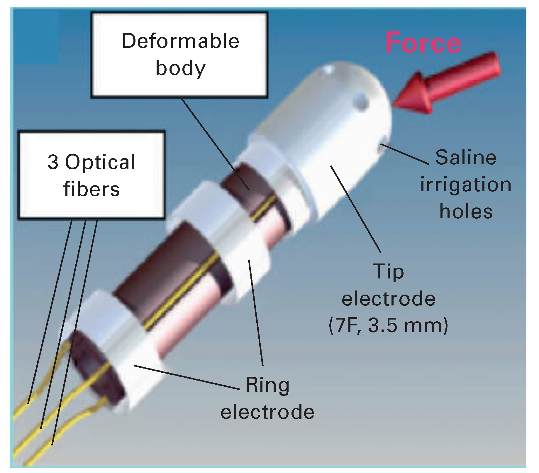
The TactiCath™ (formerly by Endosense SA, Geneva, Switzerland) is a 3.5 mm open irrigated-tip ablation catheter, with a thermocouple and a force sensor in its distal part between the second and third electrode. This force sensor consists of an elastic polymer (i.e., deformable body, elastomer) and three circumferentially aligned optical fibres allowing the calculation of CF as a vector sum (based on deformation of the elastic polymer), thus providing information on the total force and the direction of the applied force with a sensitivity of 1 g and a sampling rate of 50 Hz. Any force on the catheter tip causes corresponding microdeformation of the elastic polymer. The optical fibres transmit an infrared laser light which is reflected by fibre Bragg gratings with a changed wave-length due to the microdeformation of the elastic polymer – the changes in wavelength being proportional to the CF applied [33,34].
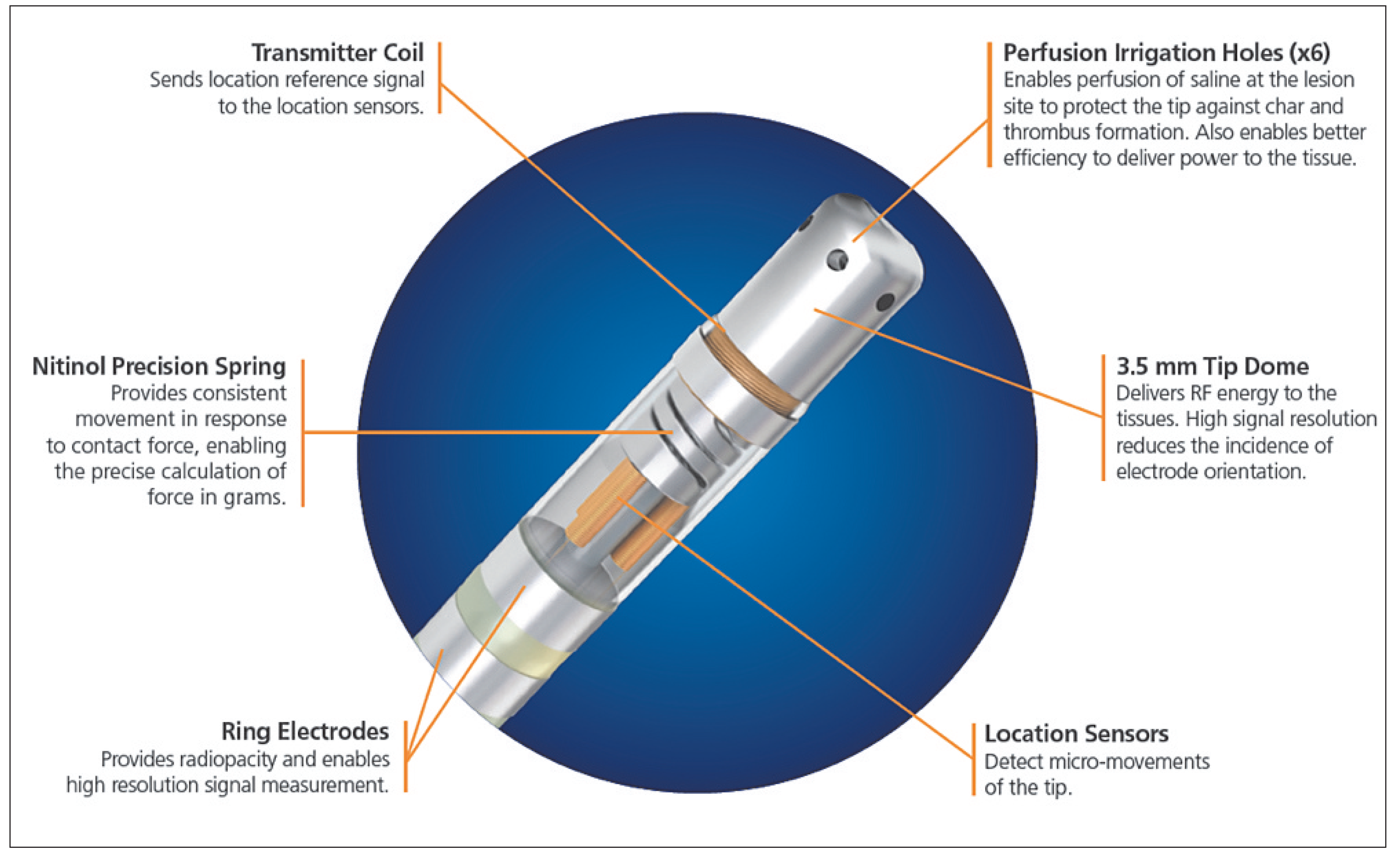
The SmartTouch™ catheter is a 3.5 mm open irrigated-tip ablation catheter. The force-sensing capability of this catheter is based on the electromagnetic location technology used in the CARTO 3D mapping System (Biosense Webster, Inc., MA, USA). The catheter tip electrode is mounted on a precision spring that permits a small amount of electrode deflection. A transmitter coil that is coupled to the tip electrode, distal to the spring, emits a location reference signal. Three magnetic location sensor coils placed at the proximal end of the spring detect micromovements of the transmitter coil, representing movement of the tip electrode on the spring. The system senses the location information of the sensor and calculates the respective force based on the known spring characteristics. The movements are sampled at 20 Hz and calibrated to produce a CF reading that is averaged over 1 second and accurate within 1 g. Of note, the catheter is fully integrated within the electroanatomical mapping system [35].
The EnSite Contact VeriSense™ System uses the principle of electrical coupling as the local measure of impedance (unlike the impedance normally used, which is a global measure between the catheter tip and a body-surface electrode) specific to the catheter tip-totissue interface through a three–terminal circuit model. The complex local coupling information is then provided as ECI (electrical coupling index) units within the three-dimensional cardiac mapping system using a real-time curve, a contact meter and an adaptive colour-coded beacon as the tip of the ablation catheter. This technology requires: scaling to the individual patient by acquisition of noncontact ECI baseline values, while the catheter rests free in the left atrium (LA) body; definition of the patient-specific noncontact/contact threshold 15 ECI units above the noncontact baseline; and acquisition of an upper safety indication while firmly pressing the catheter against the posterior LA wall. This scaling needs to be repeated every 30 minutes to adapt to shifts in the baseline values during the procedure [36].

IntelliSense® Fine Force Technology and EnSite Contact VeriSense™

Early in-vitro reports on the actual remote robotic catheter control system were able to show precise navigation and control of the remote catheter with the ability to reach a targeted endocardial site accurately and rapidly [37]. Thereafter, in-vivo studies followed and provided the basis for the above-mentioned CF system [31,38,39]. An in-vitro study examined the direct impact of catheter CF on lesion formation by using the remote robotic catheter control system [40]. The authors used intracardiac ultrasound (ICE) and fluoroscopy for validation of catheter tip / tissue contact and CF in twelve dogs undergoing irrigated-tip atrial ablation at 15 W for 30 seconds. Two different validation protocols were applied. In the first protocol, catheter tip / tissue contact as visualised by use of ICE/fluoroscopy was defined as “no contact”, “minimal contact”, “consistent contact throughout cardiac and respiratory cycles” and “tissue tenting”. Measured corresponding CF values, to which the operator was blinded, were correlated with each condition. In the second protocol, catheter tip / tissue contact was generated by <2 g, 2–10 g, 10–20 g and 20 g of CF, and catheter tip / tissue contact was subsequently graded as above. Importantly, catheter tip / tissue surface angles were considered perpendicular if the angle between the tissue surface and the catheter was >45°. In addition, the impact of catheter tip / tissue contact on 3D-electroanatomical mapping and contact-dependency of lesion size were assessed. In summary, the authors concluded that, in accordance to earlier reports, both mapping and ablation with this robotic sheath guidance system are critically dependent on generated CF and they suggested further that ablative lesion size may be optimised by the application of 10–20 g of CF, whereas mapping requires application of lower CF to avoid image distortions and increases in chamber volumes [40]. A more recently published in-vitro analysis using the same system was able to confirm these findings, observing a correlation between transmurality and CF, with an increased risk of steam pop and char formation when applied with ≥40 g, whereas a CF between 20–30 g and a power setting of 40 W appeared to be associated with transmurality by preserving safety [32].
One major limitation of this technology is that it does not provide feedback on different directional forces applied to the catheter (i.e. angle-dependent CF values), which is why visual fluoroscopy information remains crucial. As the authors point out, a nonperpendicular catheter orientation and force application might go along with contact loss and impair the measurement of the generated CF. Furthermore, parasitic frictional forces have to be overcome during every catheter placement [40].
Feasibility in human validation and use of the ECI using the EnSite Contact™ system have been assessed in two main studies with patients suffering from atrial fibrillation (AF). While the operators had to place the ablation catheter in the LA in different areas of unambiguous “qualities” of contact as determined by fluoroscopy, tactile feedback, unipolar and bipolar EGM recordings measurement and validation of the CF surrogate ECI were performed [41]. The same group recently described a prospective randomised pilot study, which demonstrated an added value of ECI for lesion creation as measured as higher rates of pulmonary vein isolation (PVI) after anatomical encircling [36]. However, ECI and the nature of relative changes during tissue contact are not fully understood and remain more complicated than established measures for CF. Furthermore, it lacks the precise distinction between different levels of contact as well as short instantaneous changes, e.g., during cardiac cycle and respiratory movements [41].

TactiCath® and SmartTouch™

An ex-vivo porcine model to determine the importance of CF during irrigated-tip RF ablation has been described [33,42,43]. Basically, it could be demonstrated that CF is a main determinant of tissue temperature and lesion size, when controlled for power and duration of open-irrigated RF energy delivery. Largest lesions were obtained using a high fixed power and CF (30 W / 60 g; 12 mm wide and 8.1 mm deep). The results showed that higher CF was associated with a higher incidence of thrombus formation and/or steam pops. Of note, in contrast to experiences gathered from closed-irrigation ablation systems, this study found that the impedance drop during RF energy delivery was not predictive of the degree of CF, whereas other works showed that the initial impedance at the start of the application, as well as the impedance drop in the first 5 seconds, correlate well with CF, suggesting the potential use of these parameters when direct CF measurements are not available [43,44,45,46].
The first clinical study investigating device and procedure-related safety (12 months of follow-up) with the TactiCath® system for the RF ablation of rightsided supraventricular tachycardias (SVTs) and AF was the TOCCATA study [47]. Two patient groups with various atrial arrhythmias were enrolled: a rightsided SVT group (n = 43) including patients with a confirmed diagnosis of atrioventricular-nodal reentry tachycardia, Wolff-Parkinson-White syndrome, atrial tachycardia and cavo-tricuspid isthmus-dependent atrial flutter and a left-sided AF group (n = 34) including patients with confirmed paroxysmal AF. The TOCCATA-study protocol required the CF values to be concealed from the investigators (all experienced operators) during mapping in order to minimise bias on the force being applied, whereas CF data were available during the ablation phase. Based on earlier experiences, high CF values in the AF group were defined as increases to >100 g for over 200 ms and, since the study was specifically designed for the assessment of safety, prespecified safety rates were derived from the literature (estimated incidence of serious adverse events for right-sided SVT at 11.4%, for patients with AF at 16.8%) [48]. The authors concluded that the safety profile was comparable to conventional irrigated-tip RF catheters and the incidence of serious adverse events in both groups clearly below the prespecified rates (2% and 12%, respectively). The study further highlighted a marked inter- and intraoperator variability during the assessment of CF values and that high CF values naturally may occur at any moment during catheter manipulation, regardless of whether one ablates or not. As a matter of fact, the one perforation event in the TOCCATA-study was shown to be preceded by a very high transient force during catheter manipulation and not during ablation. The high force of 137 g was then followed by a sudden decrease in force. Such a CF pattern typically occurs at and immediately after a perforation, as has been described in a recent analysis of forces required to perforate mechanically and transmurally the walls of the four cardiac chambers of 50–60 kg pigs [48]. Further important observations by the same authors were: (a) perforation forces are significantly lower in the right atrium and right ventricle (RV) (301 ± 117 g, 297 ± 82 g, respectively) as compared with the left atrium (LA) and left ventricle (LV) (417 ± 167 g, 457 ± 204 g, respectively) without any differences between the respective ventricles and atria; (b) perforation forces are significantly lower through transmural RA free wall RF lesion than through healthy, unablated RA tissue (172 ± 79 g vs 301 ± 117 g, p <0.0002); and (c) cardiac perforation with a catheter within a sheath is more rapid and easier than without the use of a sheath, the latter preventing the distal catheter shaft from buckling and dissipating the CF delivered by the operator proximally. Of note, the minimum CF for perforation in healthy tissue without a sheath was 131 g and lowest in the RA, while 159 g in the LA, 168 g in the RV and 227 g in the LV [48].
Such observations clearly suggest that the avoidance of CF values exceeding 100 g at any time during the procedure is crucial. Moreover, CF should be kept at low levels in the vicinity of recently ablated sites, where the tissue is structurally weakened [47,48]. Finally, TOCCATA was the first clinical study to con-firm that knowledge of the CF during the entire procedure may clearly improve safety aspects and increases the operators’ awareness of high-risk situations. The clinical outcome of the AF group of the TOCCATA study population was investigated further by assessing the relationship between CF and clinical recurrences during the 12-months’ follow-up. Acute pulmonary vein isolation was achieved in 100% of the patients, yet all patients treated with an average CF of <10 g (five of five patients) experienced AF recurrences, whereas 80% of the patients treated with an average CF of >20 g (8 of 10 patients) had none [49]. Of note, fluoroscopy and total RF times were higher in the patient group with AF recurrences, possibly suggesting difficulties with stable catheter positioning as a reason for low CF, all the more so as the operators were not blinded to CF in the ablation part and thus actually would have wanted to achieve higher CFs but had difficulties doing so. Apart from the absolute and average CF value during RF application, time-dependent evolution of CF quantified as the force-time integral (FTI or area under the CF curve, in gram-seconds, gs) proved to correlate linearly with lesion size [30,49]. The findings of this onearm prospective study underlined once more the importance of a real-time measurement of CF during RF, which allows the operator to base his/her ablation strategy on causally interrelated data, and thus compensate for low CF due to anatomical and/or technical factors by varying either RF power and/or duration or repositioning the catheter to improve contact. In this context, important further evidence about the relationship between CF measurement during RF application (EFFICAS I study) and the incidence of isolation gaps in the pulmonary vein (PV), and about areas of intrinsically good, poor, and excessive contact at the PV antrum, has been gathered through recently published studies [34,35,45,50,51,52]. The findings of the EFFICAS I study, where invasive electrophysiological assessment of conduction gaps at PVI ablation sites was performed 3 months after the index ablation procedure, showed a strong correlation between the minimum CF and minimum FTI values and the subsequent gap formation. These and other authors suggested that CF stability is required before ablation in order to minimise the risk of unstable contact and ineffective lesion formation, particularly in the left anterior segment, and suggested a target CF of 20 g, albeit with an absolute minimum CF of 10 g and an absolute minimum FTI of 400 gs [34,53]. Last but not least, safety and efficacy results from the SMART-AF trial, a prospective, nonrandomised study of 172 enrolled patients with paroxysmal AF, have recently been presented. Comparable to previous studies assessing safety, the authors reported a 12-month success rate of 72%. Furthermore, increased percent of time with physician-targeted CF correlated with increased freedom from arrhythmia recurrence, with 84.4% of subjects being arrhythmia-free at 12 months when the CF was within the targeted range >82% of the time [54].
Regardless of the system, improving catheter stability as well as contact, and achieving higher CF for RF ablation of AF may also depend upon other parameters, such as choosing general anaesthesia or adjunctive tools such as intracardiac ultrasound technology or steerable sheaths. As a matter of fact, higher clinical success along with comparable complication rates have been associated with the use of a manually controlled steerable sheath for catheter navigation in RF for AF [55]. Furthermore, administration of adenosine for the assessment of dormant conduction may help the operator to target eventual gaps [56,57].

Catheter contact force in mapping and ablation of ventricular tachycardia

Parameters allowing control of lesion size as well as procedural safety in left atrial procedures, have very recently proved their applicability for endo- and epicardial ventricular mapping and ablation.
Ventricular tachycardia mapping can be challenging, but is also crucial for successful ablation and subsequent clinical outcome. Patients frequently do not tolerate sustained arrhythmias, rendering complete mapping difficult or impossible. Furthermore, the optimal approach between an antegrade, transseptal and retrograde transaortic approach or a combination of both and/or the use of a steerable sheath is often unclear for the creation of a meaningful electroanatomical map.
To date, two studies have addressed these issues with largely congruent results [58,59]. Mizuno et al. were the first to assess the use of CF-sensing catheters in left ventricular tachycardia mapping in humans [58]. They compared a combined antegrade, transseptal approach (with a steerable sheath) and a retrograde, transaortic mapping strategy with a retrograde-only approach in 27 chambers (13 LV, 6 RV, 8 epicardial) of 17 different patients (under general anaesthesia). They divided all acquired mapping points into two groups according to the presence of positive CF throughout a complete cardiac cycle (i.e. including during diastole) and evaluated the value of surrogate parameters such as fluoroscopy, electrogram amplitude and local impedance for predicting tissue con-tact [58]. Once more, these surrogate parameters turned out to be unsatisfactory for monitoring tissue contact, since they led to the acquisition of points with an unpredictable CF variability and with a poor contact, which emphasises the importance of awareness of a minimum CF to achieve a stable tissue contact throughout the whole cardiac cycle: estimated to be 8 g for left ventricular endocardial and epicardial mapping, and 9 g for the right. They also showed that the combined approach to the left ventricle was superior in terms of clinical outcomes and that poorer CF values during mapping of the anterior and basal septal walls of the left ventricle with the retrograde approach may be related to the requirement of two curves in the mapping catheter, one in the aortic arch and the second in the left ventricle, thus possibly reducing tissue contact [58]. Tilz et al. recently added more evidence in line with these observations by comparing the impact of antegrade-transseptal with retrograde-transaortic left ventricular mapping on catheter stability and CF, and assessing the value of surrogate parameters for tissue contact [59]. Lesion formation after RF ablation on the right and left ventricular endo- and epicardium has been investigated in a sheep model using a standard irrigated-tip catheter versus a CF sensing catheter [60]. Acute lesion dimensions were assessed after RF ablation (160 endocardial and 160 epicardial RF applications) with 30 W for 60 seconds when either catheter/tissue contact (based on fluoroscopy / tactile feedback / EGM amplitude) was considered to be good with the standard irrigated-tip catheter or when CF was higher than 10 g as measured with the CF-sensing catheter. This analysis showed that conventional surrogates of good catheter-tissue contact such as fluoroscopy, tactile feedback and EGM amplitude did not result in endocardial lesion formation in 22% of RF applications, whereas lesion formation was absent in the CF-sensing group only when RF was applied with a CF lower than 10 g together with a FTI of less than 500 gs, perfectly in line with results from the TOCCATA study. However, such absolute values of CF and FTI do not seem to be directly transferable for predicting lesion size in epicardial RF applications, where FTI was, indeed, twice lower than in the endocardium, but lesion volume was significantly larger; this was most probably due to absence of circulating blood in the pericardial space and, therefore, lack of convective cooling. Furthermore, presence of epicardial fat has been shown to have a considerable effect on lesion formation [61], and last but not least, the catheter has a more parallel orientation on the epicardial surface and therefore the applied CF has a greater lateral than axial component, thus altering lesion geometry. These important novel insights have recently been reported to be very helpful for successful CF-guided endocardial and epicardial ablation of ventricular tachycardia [62,63]. Of note, the added ventricular wall thickness may require deeper, larger lesions usually achieved by a combination of higher power and higher CF. With such parameters, the risk of pop formation is likely to be higher, although perforation may be a less likely consequence compared with the thin walled atria. The body of evidence supporting the security and efficacy of specific power and CF parameters for RF ablation in the ventricles is currently limited, precluding specific recommendations.

Conclusion

Real-time catheter contact force measurement technology has provided crucial insights into technical and biophysical aspects of catheter-based arrhythmia treatment using RF energy. Optimal contact force is fundamental for the acquisition of reliable mapping information and achievement of sustained ablation lesions. The choice between different CF sensing technologies, the type of patient sedation or anaesthesia, different approaches to the cardiac chamber of interest and the knowledge of its anatomy as well as the use of steerable sheaths guaranteeing better catheter stability should be made carefully, with the aim of assuring stable and optimal catheter contact force values and maximising the probability of effective and safe lesion formation and sustained ablation success. Further studies will provide more experience with CF-assisted ablation and hopefully lead to further improvement in the outcomes of RF ablation for cardiac arrhythmias.

Funding / potential competing interests

D. Shah receives research grants from Biosense Webster, St. Jude, and Biotronik, has modest ownership interest in Endosense, and collects speaker’s fees and honoraria from Biosense, St. Jude, and Biotronik. M. Namdar receives research and fellowship grants from Biosense Webster and Biotronik.

References

  1. Huang, S.K.; Graham, A.R.; Lee, M.A.; Ring, M.E.; Gorman, G.D.; Schiffman, R. Comparison of catheter ablation using radiofrequency versus direct current energy: biophysical, electrophysiologic and pathologic observations. J Am Coll Cardiol 1991, 18, 1091–1097. [Google Scholar] [CrossRef]
  2. Nath, S.; DiMarco, J.P.; Haines, D.E. Basic aspects of radiofrequency catheter ablation. J Cardiovasc Electrophysiol 1994, 5, 863–876. [Google Scholar] [CrossRef]
  3. Pennes, H.H. Analysis of skin, muscle and brachial arterial blood temperatures in the resting normal human forearm. Am J Med Sci 1948, 215, 354. [Google Scholar]
  4. Nath, S.; Haines, D.E. Biophysics and pathology of catheter energy delivery systems. Prog Cardiovasc Dis 1995, 37, 185–204. [Google Scholar] [CrossRef]
  5. Wittkampf, F.H.; Hauer, R.N.; Robles de Medina, E.O. Control of radiofrequency lesion size by power regulation. Circulation 1989, 80, 962–968. [Google Scholar] [CrossRef]
  6. Haines, D. Biophysics of ablation: application to technology. J Cardiovasc Electrophysiol 2004, 15, S2–S11. [Google Scholar] [CrossRef] [PubMed]
  7. Haines, D.E.; Verow, A.F. Observations on electrode tissue interface temperature and effect on electrical impedance during radio frequency ablation of ventricular myocardium. Circulation 1990, 82, 1034–1038. [Google Scholar] [CrossRef] [PubMed]
  8. Haines, D.E.; Watson, D.D.; Verow, A.F. Electrode radius predicts lesion radius during radiofrequency energy heating. Validation of a proposed thermodynamic model. Circ Res 1990, 67, 124–129. [Google Scholar] [CrossRef] [PubMed]
  9. Haines, D.E. The biophysics of radiofrequency catheter ablation in the heart: the importance of temperature monitoring. Pacing Clin Electrophysiol 1993, 16, 586–591. [Google Scholar] [CrossRef]
  10. Langberg, J.J.; Gallagher, M.; Strickberger, S.A.; Amirana, O. Temperature guided radiofrequency catheter ablation with very large distal electrodes. Circulation 1993, 88, 245–9. [Google Scholar] [CrossRef]
  11. Panescu, D.; Whayne, J.G.; Fleischman, S.D.; Mirotznik, M.S.; Swanson, D.K.; Webster, J.G. Three dimensional finite element analysis of current density and temperature distributions during radio frequency ablation. IEEE Trans Biomed Eng 1995, 42, 879–890. [Google Scholar] [CrossRef] [PubMed]
  12. Huang, S.K.; Graham, A.R.; Bharati, S.; Lee, M.A.; Gorman, G.; Lev, M. Short and long term effects of transcatheter ablation of the coronary sinus by radiofrequency energy. Circulation 1988, 78, 416–427. [Google Scholar] [CrossRef]
  13. Haines, D.E. Determinants of Lesion Size During Radiofrequency Catheter Ablation: The Role of Electrode Tissue Contact Pressure and Duration of Energy Delivery. Journal of Cardiovascular Electrophysiology 1991, 2, 509–515. [Google Scholar] [CrossRef]
  14. Nath, S.; Lynch C3rd Whayne, J.G.; Haines, D.E. Cellular electrophysiological effects of hyperthermia on isolated guinea pig papillary muscle. Implications for catheter ablation. Circulation 1993, 88, 1826–1831. [Google Scholar] [CrossRef]
  15. Simmers, T.A.; de Bakker, J.M.; Wittkampf, F.H.; Hauer, R.N. Effects of heating with radiofrequency power on myocardial impulse conduction: is radiofrequency ablation exclusively thermally mediated? J Cardiovasc Electrophysiol 1996, 7, 243–247. [Google Scholar] [CrossRef]
  16. Simmers, T.A.; De Bakker, J.M.; Wittkampf, F.H.; Hauer, R.N. Effects of heating on impulse propagation in superfused canine myocardium. J Am Coll Cardiol 1995, 25, 1457–1464. [Google Scholar] [CrossRef]
  17. Calkins, H.; Prystowsky, E.; Carlson, M.; Klein, L.S.; Saul, J.P.; Gillette, P. Temperature monitoring during radiofrequency catheter ablation procedures using closed loop control. Atakr Multicenter Investigators Group. Circulation 1994, 90, 1279–1286. [Google Scholar] [CrossRef]
  18. Eick, O.J.; Gerritse, B.; Schumacher, B. Popping phenomena in temperature controlled radiofrequency ablation: when and why do they occur? Pacing Clin Electrophysiol 2000, 23, 253–258. [Google Scholar] [CrossRef] [PubMed]
  19. Matsudaira, K.; Nakagawa, H.; Wittkampf, F.H.; et al. High incidence of thrombus formation without impedance rise during radiofrequency ablation using electrode temperature control. Pacing Clin Electrophysiol 2003, 26, 1227–1237. [Google Scholar] [CrossRef]
  20. Jais, P.; Shah, D.C.; Haissaguerre, M.; et al. Efficacy and safety of septal and left atrial linear ablation for atrial fibrillation. Am J Cardiol 1999, 84, 139R 46R. [Google Scholar] [CrossRef] [PubMed]
  21. Thakur, R.K.; Klein, G.J.; Yee, R.; Zardini, M. Embolic complications after radiofrequency catheter ablation. Am J Cardiol 1994, 74, 278–279. [Google Scholar] [CrossRef]
  22. Epstein, M.R.; Knapp, L.D.; Martindill, M.; et al. Embolic complications associated with radiofrequency catheter ablation. Atakr Investigator Group. Am J Cardiol 1996, 77, 655–658. [Google Scholar] [CrossRef] [PubMed]
  23. Zhou, L.; Keane, D.; Reed, G.; Ruskin, J. Thromboembolic complications of cardiac radiofrequency catheter ablation: a review of the reported incidence, pathogenesis and current research directions. J Cardiovasc Electrophysiol 1999, 10, 611–620. [Google Scholar] [CrossRef]
  24. Yokoyama, K.; Nakagawa, H.; Wittkampf, F.H.; Pitha, J.V.; Lazzara, R.; Jackman, W.M. Comparison of electrode cooling between internal and open irrigation in radiofrequency ablation lesion depth and incidence of thrombus and steam pop. Circulation 2006, 113, 11–19. [Google Scholar] [CrossRef]
  25. Otomo, K.; Yamanashi, W.S.; Tondo, C.; et al. Why a large tip electrode makes a deeper radiofrequency lesion: effects of increase in electrode cooling and electrode tissue interface area. J Cardiovasc Electrophysiol 1998, 9, 47–54. [Google Scholar] [CrossRef]
  26. Nakagawa, H.; Yamanashi, W.S.; Pitha, J.V.; et al. Comparison of in vivo tissue temperature profile and lesion geometry for radiofrequency ablation with a saline irrigated electrode versus temperature control in a canine thigh muscle preparation. Circulation 1995, 91, 2264–2273. [Google Scholar] [CrossRef]
  27. Nakagawa, H.; Wittkampf, F.H.; Yamanashi, W.S.; et al. Inverse relationship between electrode size and lesion size during radiofrequency ablation with active electrode cooling. Circulation 1998, 98, 458–465. [Google Scholar] [CrossRef]
  28. Avitall, B.; Mughal, K.; Hare, J.; Helms, R.; Krum, D. The effects of electrode tissue contact on radiofrequency lesion generation. Pacing Clin Electrophysiol 1997, 20, 2899–2910. [Google Scholar] [CrossRef] [PubMed]
  29. Burkhardt, J.D.; Natale, A. New technologies in atrial fibrillation ablation. Circulation 2009, 120, 1533–1541. [Google Scholar] [CrossRef] [PubMed]
  30. Shah, D.C.; Lambert, H.; Nakagawa, H.; Langenkamp, A.; Aeby, N.; Leo, G. Area under the real time contact force curve (force time integral) predicts radiofrequency lesion size in an in vitro contractile model. J Cardiovasc Electrophysiol 2010, 21, 1038–1043. [Google Scholar] [CrossRef]
  31. Saliba, W.; Cummings, J.E.; Oh, S.; et al. Novel robotic catheter remote control system: feasibility and safety of transseptal puncture and endocardial catheter navigation. J Cardiovasc Electrophysiol 2006, 17, 1102–1105. [Google Scholar] [CrossRef]
  32. Di Biase, L.; Natale, A.; Barrett, C.; et al. Relationship between catheter forces, lesion characteristics, “popping,” and char formation: experience with robotic navigation system. J Cardiovasc Electrophysiol 2009, 20, 436–440. [Google Scholar] [CrossRef]
  33. Yokoyama, K.; Nakagawa, H.; Shah, D.C.; et al. Novel contact force sensor incorporated in irrigated radiofrequency ablation catheter predicts lesion size and incidence of steam pop and thrombus. Circ Arrhythm Electrophysiol 2008, 1, 354–362. [Google Scholar] [CrossRef]
  34. Neuzil, P.; Reddy, V.Y.; Kautzner, J.; et al. Electrical reconnection after pulmonary vein isolation is contingent on contact force during initial treatment: results from the EFFICAS I study. Circ Arrhythm Electrophysiol 2013, 6, 327–33. [Google Scholar] [CrossRef]
  35. Nakagawa, H.; Kautzner, J.; Natale, A.; et al. Locations of high contact force during left atrial mapping in atrial fibrillation patients: electrogram amplitude and impedance are poor predictors of electrode tissue contact force for ablation of atrial fibrillation. Circ Arrhythm Electrophysiol 2013, 6, 746–753. [Google Scholar] [CrossRef]
  36. Gaspar, T.; Sih, H.; Hindricks, G.; et al. Use of electrical coupling information in AF catheter ablation: a prospective randomized pilot study. Heart Rhythm 2013, 10, 176–181. [Google Scholar] [CrossRef] [PubMed]
  37. Al Ahmad, A.; Grossman, J.D.; Wang, P.J. Early experience with a computerized robotically controlled catheter system. J Interv Card Electrophysiol 2005, 12, 199–202. [Google Scholar] [CrossRef] [PubMed]
  38. Kanagaratnam, P.; Koa Wing, M.; Wallace, D.T.; Goldenberg, A.S.; Peters, N.S.; Davies, D.W. Experience of robotic catheter ablation in humans using a novel remotely steerable catheter sheath. J Interv Card Electrophysiol 2008, 21, 19–26. [Google Scholar] [CrossRef] [PubMed]
  39. Saliba, W.; Reddy, V.Y.; Wazni, O.; et al. Atrial fibrillation ablation using a robotic catheter remote control system: initial human experience and long term follow up results. J Am Coll Cardiol 2008, 51, 2407–2411. [Google Scholar] [CrossRef]
  40. Okumura, Y.; Johnson, S.B.; Bunch, T.J.; Henz, B.D.; O’Brien, C.J.; Packer, D.L. A systematical analysis of in vivo contact forces on virtual catheter tip/tissue surface contact during cardiac mapping and intervention. J Cardiovasc Electrophysiol 2008, 19, 632–640. [Google Scholar] [CrossRef]
  41. Piorkowski, C.; Sih, H.; Sommer, P.; et al. First in human validation of impedance based catheter tip to tissue contact assessment in the left atrium. J Cardiovasc Electrophysiol 2009, 20, 1366–1373. [Google Scholar] [CrossRef]
  42. Perna, F.; Heist, E.K.; Danik, S.B.; Barrett, C.D.; Ruskin, J.N.; Mansour, M. Assessment of catheter tip contact force resulting in cardiac perforation in swine atria using force sensing technology. Circ Arrhythm Electrophysiol 2011, 4, 218–224. [Google Scholar] [CrossRef]
  43. Thiagalingam, A.; D’Avila, A.; Foley, L.; et al. Importance of catheter contact force during irrigated radiofrequency ablation: evaluation in a porcine ex vivo model using a force sensing catheter. J Cardiovasc Electrophysiol 2010, 21, 806–811. [Google Scholar] [CrossRef]
  44. Haqqani, H.M.; Marchlinski, F.E. Creating lesions and indexing transmural ablation: pushing harder to find the Holy Grail. J Cardiovasc Electrophysiol 2010, 21, 812–814. [Google Scholar] [CrossRef]
  45. Reichlin, T.; Knecht, S.; Lane, C.; et al. Initial impedance decrease as an indicator of good catheter contact: insights from radiofrequency ablation with force sensing catheters. Heart Rhythm 2014, 11, 194–201. [Google Scholar] [CrossRef]
  46. Ikeda, A.; Nakagawa, H.; Lambert, H.; et al. Relationship between catheter contact force and radiofrequency lesion size and incidence of steam pop in the beating canine heart: electrogram amplitude, impedance, and electrode temperature are poor predictors of electrode tissue contact force and lesion size. Circ Arrhythm Electrophysiol 2014, 7, 1174–1180. [Google Scholar]
  47. Kuck, K.H.; Reddy, V.Y.; Schmidt, B.; et al. A novel radiofrequency ablation catheter using contact force sensing: Toccata study. Heart Rhythm 2012, 9, 18–23. [Google Scholar] [CrossRef] [PubMed]
  48. Shah, D.; Lambert, H.; Langenkamp, A.; et al. Catheter tip force required for mechanical perforation of porcine cardiac chambers. Europace 2011, 13, 277–283. [Google Scholar] [CrossRef]
  49. Reddy, V.Y.; Shah, D.; Kautzner, J.; et al. The relationship between contact force and clinical outcome during radiofrequency catheter ablation of atrial fibrillation in the TOCCATA study. Heart Rhythm 2012, 9, 1789–1795. [Google Scholar] [CrossRef] [PubMed]
  50. Kumar, S.; Morton, J.B.; Lee, J.; et al. Prospective characterization of catheter tissue contact force at different anatomic sites during antral pulmonary vein isolation. Circ Arrhythm Electrophysiol 2012, 5, 1124–1129. [Google Scholar] [CrossRef] [PubMed]
  51. Martinek, M.; Lemes, C.; Sigmund, E.; et al. Clinical impact of an open irrigated radiofrequency catheter with direct force measurement on atrial fibrillation ablation. Pacing Clin Electrophysiol 2012, 35, 1312–1318. [Google Scholar] [CrossRef] [PubMed]
  52. Haldar, S.; Jarman, J.W.; Panikker, S.; et al. Contact force sensing technology identifies sites of inadequate contact and reduces acute pulmonary vein reconnection: A prospective case control study. Int J Cardiol 2012. [CrossRef] [PubMed]
  53. Park, C.I.; Lehrmann, H.; Keyl, C.; et al. Mechanisms of pulmonary vein reconnection after radiofrequency ablation of atrial fibrillation: the deterministic role of contact force and interlesion distance. J Cardiovasc Electrophysiol 2014, 25, 701–708. [Google Scholar] [CrossRef]
  54. Natale, A. Ablation Of Symptomatic Paroxysmal Atrial Fibrillation Using Novel Contact Force Catheter: SMART AF Trial, SP22, Heart Rhyhtm Society Meeting 2013.
  55. Piorkowski, C.; Eitel, C.; Rolf, S.; et al. Steerable versus nonsteerable sheath technology in atrial fibrillation ablation: a prospective, randomized study. Circ Arrhythm Electrophysiol 2011, 4, 157–165. [Google Scholar] [CrossRef]
  56. Arentz, T.; Macle, L.; Kalusche, D.; et al. “Dormant” pulmonary vein conduction revealed by adenosine after ostial radiofrequency catheter ablation. J Cardiovasc Electrophysiol 2004, 15, 1041–1047. [Google Scholar] [CrossRef]
  57. Epstein, L.M.; Mitchell, M.A.; Smith, T.W.; Haines, D.E. Comparative study of fluoroscopy and intracardiac echocardiographic guidance for the creation of linear atrial lesions. Circulation 1998, 98, 1796–1801. [Google Scholar] [CrossRef]
  58. Mizuno, H.; Vergara, P.; Maccabelli, G.; et al. Contact force monitoring for cardiac mapping in patients with ventricular tachycardia. J Cardiovasc Electrophysiol 2013, 24, 519–524. [Google Scholar] [CrossRef]
  59. Tilz, R.R.; Makimoto, H.; Lin, T.; et al. In vivo left ventricular contact force analysis: comparison of antegrade transseptal with retrograde transaortic mapping strategies and correlation of impedance and electrical amplitude with contact force. Europace 2014. [CrossRef]
  60. Sacher, F.; Wright, M.; Derval, N.; et al. Endocardial versus epicardial ventricular radiofrequency ablation: utility of in vivo contact force assessment. Circ Arrhythm Electrophysiol 2013, 6, 144–150. [Google Scholar] [CrossRef]
  61. Wong, M.C.; Edwards, G.; Spence, S.J.; et al. Characterization of catheter tissue contact force during epicardial radiofrequency ablation in an ovine model. Circ Arrhythm Electrophysiol 2013, 6, 1222–1228. [Google Scholar] [CrossRef] [PubMed]
  62. Valk, S.D.; de Groot, N.M.; Jordaens, L. Catheter ablation of right ventricular outflow tract tachycardia using contact force guidance. Neth Heart J 2014. [CrossRef] [PubMed][Green Version]
  63. Dabiri Abkenari, L.; Akca, F.; Van Mieghem, N.M.; Szili Torok, T. The first human experience of a contact force sensing catheter for epicardial ablation of ventricular tachycardia. Neth Heart J 2014. [CrossRef] [PubMed]
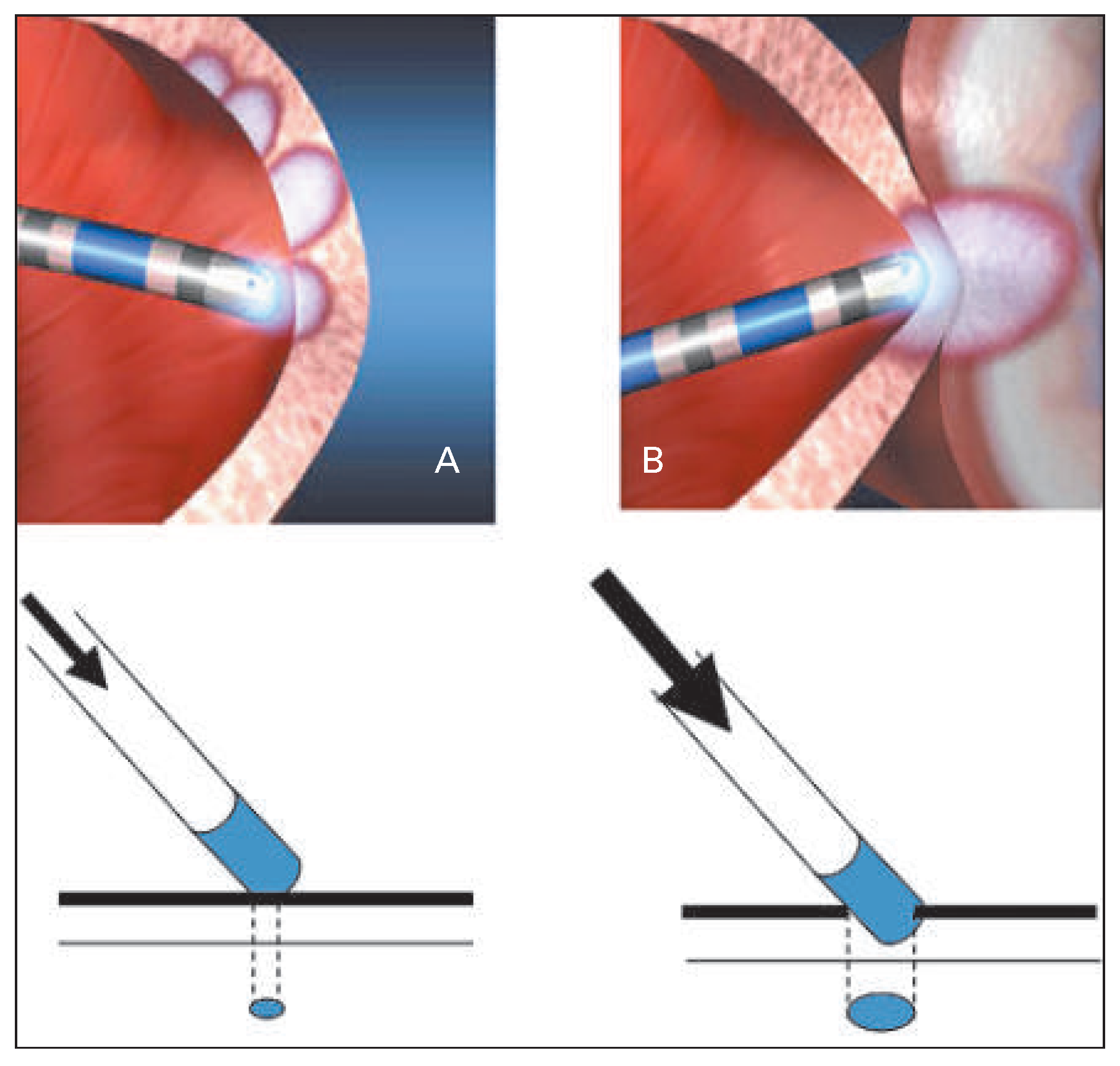
Figure 1. Better contact results in deformation of the tissue surface, allowing the ablation electrode to embed slightly and the contact to improve, thereby resulting in more efficient formation and control of the lesion (Figure 1b as compared with Figure 1a).
Cardiovascmed 18 00155 g001
Figure 2. TactiCath™ Ablation Catheter (from: Yokoyama K, et al. Novel contact force sensor incorporated in irrigated radiofrequency ablation catheter predicts lesion size and incidence of steam pop and thrombus. Circ Arrhythm Electrophysiol. 2008;1(5):354–62, reprinted with permission).
Cardiovascmed 18 00155 g002
Figure 3. ThermoCool® SmartTouch™ Ablation Catheter (with authorisation from Biosense Webster Switzerland).
Cardiovascmed 18 00155 g003