Next Article in Journal
Cardiovascular Risk Management in Patients with Rheumatoid Arthritis
Previous Article in Journal
Gastroenterologische Kontraindikationen der Transösophagealen Echokardiographie
 
 
Cardiovascular Medicine is published by MDPI from Volume 28 Issue 1 (2025). Previous articles were published by another publisher in Open Access under a CC-BY (or CC-BY-NC-ND) licence, and they are hosted by MDPI on mdpi.com as a courtesy and upon agreement with Editores Medicorum Helveticorum (EMH).
Review

Assisted Reproduction: A Novel Cardiovascular Risk Factor

by
Emrush Rexhaj
*,
Stefano F. Rimoldi
,
Elisa Bouillet
,
David Cerny
,
Rodrigo Soria
,
Robert Von Arx
,
Yves Allemann
,
Claudio Sartori
and
Urs Scherrer
Department of Cardiology and Clinical Research, Inselspital, CH-3010 Bern, Switzerland
*
Author to whom correspondence should be addressed.
Cardiovasc. Med. 2015, 18(4), 115; https://doi.org/10.4414/cvm.2015.00311
Submission received: 22 January 2015 / Revised: 22 February 2015 / Accepted: 22 March 2015 / Published: 22 April 2015

Abstract

There is abundant evidence indicating that pathological events during early life predispose to cardiovascular and metabolic diseases later in life. Recent evidence suggests that assisted reproductive technologies (ART) represent a novel important example of this problem. Here we will review recent data in humans and animals demonstrating ART-induced cardiac dysfunction, premature vascular ageing and arterial hypertension that appear to be related, at least of part, to epigenetic mechanisms. Therefore, a better comprehension of mechanisms of ART-induced vascular dysfunction and long-term monitoring of the ART population is of upmost importance in order to prevent or treat the long-term cardiovascular consequences. Given the young age of the ART population, we will have to wait for 20–30 years before we will know in how many premature cardiovascular endpoints these ART-induced alterations will result, ART should be taken into account when counseling putting into place preventive strategies.

Introduction

A report from the annual meeting of the Swiss Society of Cardiology 2014
Developmental origin of adult diseases
Epidemiological studies have shown an association between pathological events occurring during early life and the development of cardiovascular and metabolic disease in adulthood, leading to the “Barker’s hypothesis of fetal programming of adult disease” [1]. In line with this hypothesis, we have shown that transient perinatal hypoxia induces pulmonary vascular dysfunction that predisposes to exaggerated hypoxic pulmonary hypertension in young apparently healthy adults [2], and preeclampsia induces pulmonary and systemic vascular dysfunction in young apparently healthy offspring [3]. Taken together, these data demonstrate that environmental insults occurring during the perinatal period and the last trimester of gestation have important long-term consequences for the regulation of the cardiovascular system in humans. Studies in a mouse model of preeclampsia indicate that the altered vascular responsiveness in the offspring is related to epigenetic changes [4]. Assisted reproductive technologies (ART) involve the manipulation of early embryos during a period when they may be particularly vulnerable. In line with this concept, recent evidence indicates that ART represents a novel important example of this problem [5,6,7].

Studies in humans

Vascular dysfunction in children conceived by means of assisted reproductive technologies
The prevalence of infertility has been estimated to be 9% worldwide [8]. ART has allowed millions of infertile couples to have children, and in developed countries ART children now make up for 2% to 5% of births [9].
In a recent study, we found that ART children display marked generalised vascular dysfunction, as evidenced by a roughly 30% higher hypoxic pulmonary artery pressure, a 25% lower flow-mediated dilation (FMD), a markedly faster pulse-wave velocity (PWV) and increased intima-media thickness (IMT) in ART than in control children (Figure 1) [7]. In order to exclude confounding factors that could contribute to ART-induced vascular dysfunction, we studied vascular function in parents of ART children, children conceived after ovarian hyperstimulation alone and naturally conceived siblings of ART children. Vascular function was normal in all these groups (Figure 1), suggesting that ART per se induces vascular dysfunction. In the pulmonary circulation, this defect predisposes to exaggerated hypoxic pulmonary hypertension, already during childhood. In the systemic circulation, endothelial dysfunction, increased vascular stiffness and IMT represent early steps in the development of atherosclerosis and are independent risk factors for premature cardiovascular morbidity [10].
Recent data suggest that vascular dysfunction in ART children may be reversible [11]. In ART mice, vascular dysfunction is related to decreased vascular endothelial nitric oxide (NO) synthase expression and NO synthesis (see below) [5]. We speculated that a similar mechanism underpins vascular dysfunction in ART children. We found that vascular dysfunction in ART children was associated with increased oxidative stress and decreased plasma NO [11]. Antioxidant administration normalised oxidative stress and plasma NO and improved endothelial function in the systemic and pulmonary circulation. These data show that in young individuals ART-induced vascular dysfunction is subject to redox-regulation and potentially reversible [11].
Blood pressure in children conceived by means of assisted reproductive technologies
Among the potential long-term consequences of ARTmediated vascular dysfunction, arterial hypertension may represent an important problem [12]. In our study, arterial blood pressure was not different between ART and control children (mean age 11.1 years) [7]. Belva et al. also found no difference in arterial blood pressure between 14-year-old ART and naturally conceived adolescents [13]. However, in a larger study (225 ART children and 225 spontaneously conceived control children) including older subjects (age 8–18 years), Ceelen et al. found that office systolic and diastolic blood pressure were higher in ART children than in controls [14]. Taken together these data suggest that the prevalence of HTA in this population may increase with increasing age. Consistent with this concept, adult ART mice display arterial hypertension (see below).
Additional studies using ambulatory blood pressure monitoring (ABPM) are needed in this population in order to follow the evolution of arterial blood pressure and to determine whether ART-induced vascular dysfunction translates into arterial hypertension.
Cardiac dysfunction in children conceived by means of assisted reproductive technologies
A very recent study showed that children conceived by means of ART manifest cardiac and vascular remodelling in utero that persists in early postnatal life [15]. This study compared cardiac function and morphology in 100 foetuses conceived with ART and 100 control pregnancies. In ART foetuses, the myocardial wall was thicker and the longitudinal heart function was decreased, as shown by a decreased tricuspid ring displacement, impaired relaxation and dilated atria. These signs of cardiovascular remodelling persisted at 6 months of age. However, it is not known how they will evolve later in life. To provide such information, we recently assessed cardiac function in older ART children and adolescents (Von Arx R et al.). We found that, whereas left ventricular function was normal and comparable to that of naturally conceived children, right ventricular end-diastolic area was increased (cardiac remodelling) and right ventricular diastolic function was impaired, particularly under stressful conditions (high altitude exposure). These data suggest that ART-induced cardiac remodelling persists into adulthood and may predispose to cardiac dysfunction later in life.
To assess long-term consequence of ART on cardiovascular function and mortality, and examine potential underlying mechanisms, we studied mice generated by ART.

Studies in mice

Vascular dysfunction in mice generated by assisted reproductive technologies
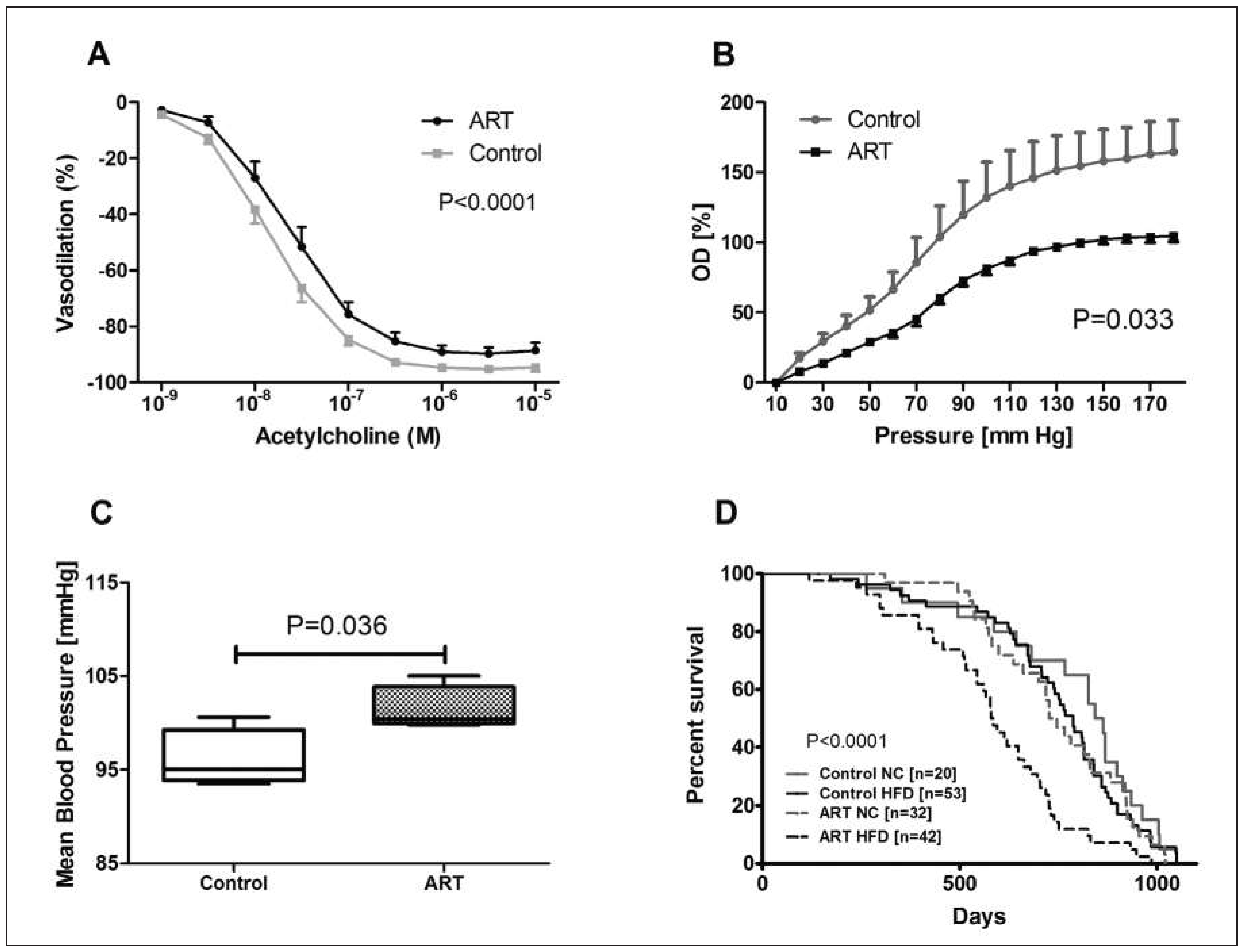
Consistent with data in humans, we found that ART induces systemic vascular dysfunction also in mice, as evidenced by decreased endothelial-dependent mesenteric artery vasodilation (Figure 2 panel A) and increased carotid artery stiffness (Figure 2 panel B) [5]. Notably, this vascular dysfunction in vitro translated into arterial hypertension in vivo in adult ART mice (fig. 2 panel C). To study the long-term consequences of ART-induced vascular dysfunction, we compared the lifespan of ART mice with that of control mice fed a Western-style high-fat diet. The lifespan was roughly 25% shorter in ART than in control mice, demonstrating the long-term consequences of ARTinduced cardiovascular dysfunction (Figure 2 panel D).
Underlying mechanisms
The demonstration of ART-induced cardiovascular dysfunction in normal fertile mice strengthens the concept that ART per se is the main culprit causing this problem.
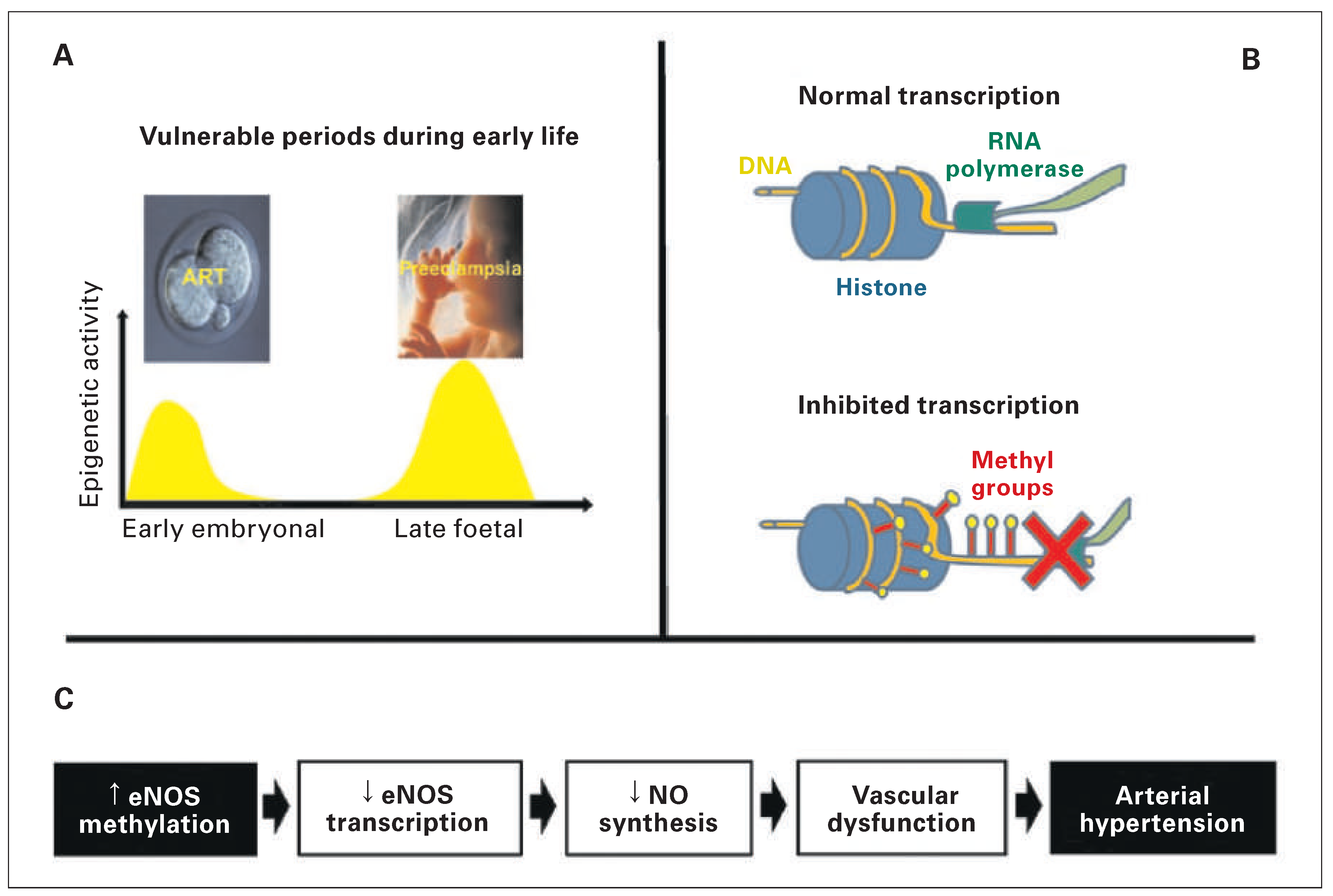
ART implies manipulation of the embryo during a vulnerable period with increased epigenetic activity (Figure 3 panel A) [6]. The term “epigenetics” refers to chromatin-based mechanisms that regulate gene expression without affecting the DNA sequence and can be transmitted to the next generation(s). The best studied epigenetic mechanisms involve DNA methylation (Figure 3 panel B) and histone modifications (acetylation and methylation) [16]. Several observations indicate that epigenetic mechanisms underpin ARTinduced cardiovascular alterations. First, male ART mice transmit vascular dysfunction to the next generation. Second, the methylation of the promoter of the endothelial nitric oxide synthase (eNOS) gene in vascular tissue is increased in ART mice. Third, DNA dysmethylation of the eNOS translated into decreased eNOS expression in the vasculature and decreased vascular NO synthesis in ART compared with control mice. Taken together, these findings indicate that in mice, ART alters the entire chain of events starting from eNOS promoter methylation and ending in premature vascular aging and arterial hypertension (Figure 3 panel C) [6]. Finally, epigenetic alterations can be reversed by drugs such as histone deacethylase inhibitors (i.e., butyrate) [4]. We found that butyrate administration normalised eNOS promoter methylation and vascular function in ART mice, and prevented the transmission of this problem to the next generation [5]. Collectively these data indicate that ART induces premature vascular ageing and hypertension in mice by an epigenetic mechanism.
We then started out to test for specific events during embryo culture that may contribute to ART-induced epigenetic and cardiovascular alterations. The time passed by the embryo on the culture medium before its transfer to the recipient mother may play a role. We therefore compared vascular function in mice born after implantation of two cell embryos (24 h in culture media) and blastocysts (86 h in culture media). Vascular dysfunction was comparable in the two groups, suggesting that shortening the culture time does n0t prevent ART-induced vascular dysfunction. In line with this observation, altered methylation patterns and gene expression are already detectable in two cell stage embryos [17]. Taken together, these data suggest that suboptimal culture conditions contribute to ARTinduced epigenetic alterations and vascular dysfunction. In line with this hypothesis, preliminary data in mice suggest that modification of the culture media may attenuate ART-induced epigenetic alterations and vascular dysfunction in mice [18].

Conclusion

In conclusion, these observations suggest that ART per se induces vascular dysfunction both in humans and animals. In mice, endothelial dysfunction and arterial hypertension are induced during the embryo development in culture media by an epigenetic mechanism. A similar mechanism may also underpin ART-induced vascular dysfunction in humans. Comparison with young populations with similar vascular dysfunction at a young age and known cardiovascular complications later in life (i.e., preeclampsia), suggest that ART may represent an important novel risk factor predisposing to premature cardiovascular morbidity and mortality (Figure 4) [19]. Given the young age of this population (the first ART child was born in 1978), the magnitude of the problem is not yet known, since clinically manifest cardiovascular disease has not had time to develop. Now, however, it is already important to avoid traditional cardiovascular risk factors in this population and to detect early cardiovascular alterations with the aim to prevent or at least optimally treat cardiovascular complications.

Funding / potential competing interests:

This work was supported by the Swiss National Science Foundation, the Swiss Society of Cardiology, the Swiss Society of Hypertension and the Placide Nicod Foundation.

References

  1. Barker, D.J. Maternal and fetal origins of coronary heart disease. J R Coll Physicians Lond. 1994, 28, 544–551. [Google Scholar] [CrossRef] [PubMed]
  2. Sartori, C.; Allemann, Y.; Trueb, L.; Delabays, A.; Nicod, P.; Scherrer, U. Augmented vasoreactivity in adult life associated with perinatal vascular insult. Lancet. 1999, 353, 2205–2207. [Google Scholar] [CrossRef] [PubMed]
  3. Jayet, P.Y.; Rimoldi, S.F.; Stuber, T.; Salmon, C.S.; Hutter, D.; Rexhaj, E.; et al. Pulmonary and systemic vascular dysfunction in young offspring of mothers with preeclampsia. Circulation. 2010, 122, 488–494. [Google Scholar] [CrossRef] [PubMed]
  4. Rexhaj, E.; Bloch, J.; Jayet, P.Y.; Rimoldi, S.F.; Dessen, P.; Mathieu, C.; et al. Fetal programming of pulmonary vascular dysfunction in mice: role of epigenetic mechanisms. Am J Physiol Heart Circ Physiol. 2011, 301, H247–52. [Google Scholar] [CrossRef] [PubMed]
  5. Rexhaj, E.; Paoloni-Giacobino, A.; Rimoldi, S.F.; Fuster, D.G.; Anderegg, M.; Somm, E.; et al. Mice generated by in vitro fertilization exhibit vascular dysfunction and shortened life span. J Clin Invest. 2013, 123, 5052–5060. [Google Scholar] [CrossRef] [PubMed]
  6. Rimoldi, S.F.; Sartori, C.; Rexhaj, E.; Cerny, D.; Von Arx, R.; Soria, R.; et al. Vascular dysfunction in children conceived by assisted reproductive technologies: underlying mechanisms and future implications. Swiss Med Wkly. 2014, 144, w13973. [Google Scholar] [CrossRef] [PubMed]
  7. Scherrer, U.; Rimoldi, S.F.; Rexhaj, E.; Stuber, T.; Duplain, H.; Garcin, S.; et al. Systemic and pulmonary vascular dysfunction in children conceived by assisted reproductive technologies. Circulation. 2012, 125, 1890–1896. [Google Scholar] [PubMed]
  8. Boivin, J.; Bunting, L.; Collins, J.A.; Nygren, K.G. International estimates of infertility prevalence and treatment-seeking: potential need and demand for infertility medical care. Hum Reprod. 2007, 22, 1506–1512. [Google Scholar] [CrossRef] [PubMed]
  9. Sutcliffe, A.G.; Ludwig, M. Outcome of assisted reproduction. Lancet. 2007, 370, 351–359. [Google Scholar] [CrossRef] [PubMed]
  10. Engelen, L.; Ferreira, I.; Stehouwer, C.D.; Boutouyrie, P.; Laurent, S. Reference intervals for common carotid intima-media thickness measured with echotracking: relation with risk factors. Eur Heart J. 2013, 34, 2368–2380. [Google Scholar] [CrossRef] [PubMed]
  11. Rimoldi, S.F.; Sartori, C.; Rexhaj, E.; Bailey, D.M.; Marchi, S.F.; McEneny, J.; et al. Antioxidants improve vascular function in children conceived by assisted reproductive technologies: A randomized double-blind placebo-controlled trial. Eur J Prev Cardiol 2014. [CrossRef] [PubMed]
  12. Hart, R.; Norman, R.J. The longer-term health outcomes for children born as a result of IVF treatment: Part I General health outcomes. Hum Reprod Update. 2013, 19, 232–243. [Google Scholar] [CrossRef] [PubMed]
  13. Belva, F.; Roelants, M.; De Schepper, J.; Roseboom, T.J.; Bonduelle, M.; Devroey, P.; et al. Blood pressure in ICSI-conceived adolescents. Hum Reprod. 2012, 27, 3100–3108. [Google Scholar] [CrossRef] [PubMed]
  14. Ceelen, M.; van Weissenbruch, M.M.; Vermeiden, J.P.; van Leeuwen, F.E.; Delemarre-van de Waal, H.A. Cardiometabolic differences in children born after in vitro fertilization: follow-up study. J Clin Endocrinol Metab. 2008, 93, 1682–1688. [Google Scholar] [CrossRef] [PubMed]
  15. Valenzuela-Alcaraz, B.; Crispi, F.; Bijnens, B.; Cruz-Lemini, M.; Creus, M.; Sitges, M.; et al. Assisted reproductive technologies are associated with cardiovascular remodeling in utero that persists postnatally. Circulation. 2013, 128, 1442–1450. [Google Scholar] [CrossRef] [PubMed]
  16. Cowley, A.W.; Jr Nadeau, J.H.; Baccarelli, A.; Berecek, K.; Fornage, M.; Gibbons, G.H.; et al. Report of the National Heart, Lung, and Blood Institute Working Group on epigenetics and hypertension. Hypertension. 2012, 59, 899–905. [Google Scholar] [CrossRef] [PubMed]
  17. Shi, W.; Haaf, T. Aberrant methylation patterns at the two-cell stage as an indicator of early developmental failure. Mol Reprod Dev.. 2002, 63, 329–334. [Google Scholar] [CrossRef] [PubMed]
  18. Rexhaj, E.R.S.; Giacobino, A.; Sartori, C.; Allemann, Y.; Scherrer, U. Epigenetically-induced systemic vascular dysfunction and hypertension by in vitro fertilization; prevention by addition of melatonin to the culture media. FASEB J. 2012, 26, 874. [Google Scholar]
  19. Rexhaj, E.R.S.; Brenner, R.; Pireva, A.; Cerny, D.; Von Arx, R.; Sartori, C.; et al. Role of foetal programming and epigenetic mechanisms in the pathogenesis of arterial hypertension. Cardiovascular Medicine. 2014, 42–45. [Google Scholar]
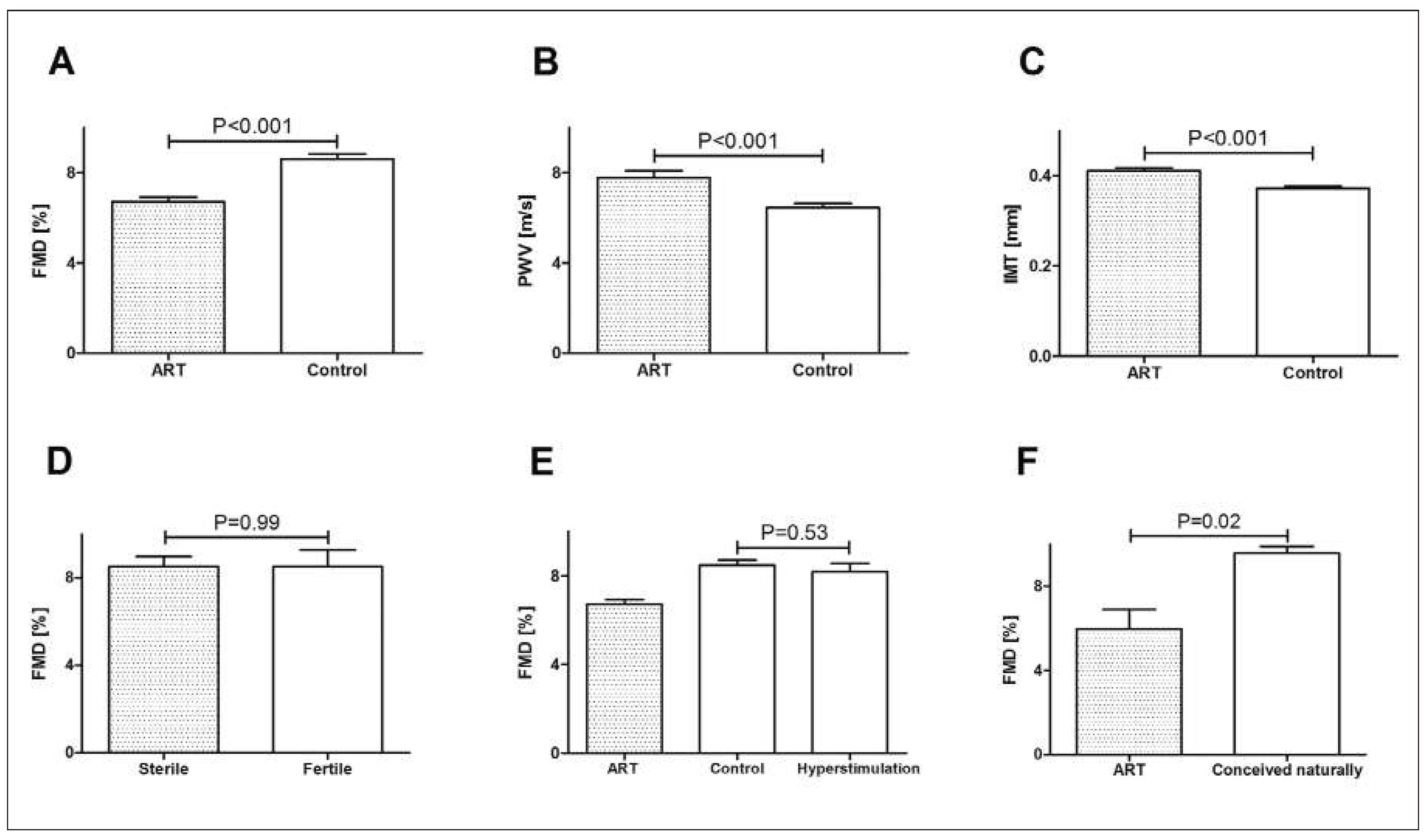
Figure 1. Vascular function in children conceived by means of assisted reproductive technologies (ART), control children and their parents. Flow-mediated dilation (FMD, panel A), pulse wave velocity (PWV, panel B), intima-media thickness (IMT, panel C) in children conceived by ART or conceived naturally (control). FMD in sterile and fertile parents (panel D). FMD in children conceived after ovarian hyperstimulation, ART or control (panel E). FMD in siblings, one conceived naturally and the other by ART (panel F).
Cardiovascmed 18 00115 g001
Figure 2. Endothelial function, arterial stiffness, arterial blood pressure and life span in mice generated with ART and control mice. Acetylcholine-induced mesenteric-artery vasodilation in vitro (panel A), carotid artery pressure diameter relationship in vitro (panel B; OD, outer diameter) and mean arterial blood pressure measured by telemetry in vivo (panel C). Kaplan-Meier survival curves of ART and control mice fed with normal chow (NC) or with a high-fat diet (HFD) (panel D).
Cardiovascmed 18 00115 g002
Figure 3. Role of epigenetic mechanisms in vascular dysfunction in mice generated with assisted reproductive technologies (ART). Vulnerable periods during pregnancy coincide with high epigenetic activity (panel A). DNA hypermethylation, one of the main epigenetic mechanism, inhibits DNA transcription (panel B). In ART mice the entire chain, starting with eNOS gene hypermethylation and ending up with vascular dysfunction and arterial hypertension (panel C). From: Rimoldi SF, Sartori C, Rexhaj E, Cerny D, Von Arx R, et al. Vascular dysfunction in children conceived by assisted reproductive technologies: underlying mechanisms and future implications. Swiss Med Wkly. 2014;144:w13973, with permission.
Cardiovascmed 18 00115 g003
Figure 4. Schematic representation of prenatal, perinatal and childhood factors that may lead to epigenetic alterations that alter cardiovascular function and determine cardiovascular risk later in life.
Cardiovascmed 18 00115 g004