Next Article in Journal
The Early Development of Cardiology in Zurich–A Personal Account Part I
Previous Article in Journal
Supracardiac Total Anomalous Pulmonary Venous Connection with Right Ascending Vertical Vein
 
 
Cardiovascular Medicine is published by MDPI from Volume 28 Issue 1 (2025). Previous articles were published by another publisher in Open Access under a CC-BY (or CC-BY-NC-ND) licence, and they are hosted by MDPI on mdpi.com as a courtesy and upon agreement with Editores Medicorum Helveticorum (EMH).
Review

Pregnancy-Associated Cardiomyopathies

by
Denise Hilfiker-Kleiner
* and
Johann Bauersachs
Department of Cardiology and Angiology, Medical School Hannover, Carl-Neuberg-Str. 1, 30625 Hannover, Germany
*
Author to whom correspondence should be addressed.
Cardiovasc. Med. 2014, 17(9), 245; https://doi.org/10.4414/cvm.2014.00262
Submission received: 24 June 2014 / Revised: 24 July 2014 / Accepted: 24 August 2014 / Published: 24 September 2014

Summary

Cardiovascular disease is a major cause of complications in pregnancy worldwide and the number of patients who develop cardiac problems during pregnancy is increasing. The physiological changes occurring during pregnancy and in the peripartum period provide a challenge to the cardiovascular system of all women. Understanding the morphological and functional changes in normal pregnancy is therefore important for the timely recognition of cardiovascular pathology during this vulnerable period. However, evidence-based clinical data in this field are scarce and there is a deficit in understanding general physiological and pathophysiological processes operating in the maternal heart around pregnancy. This review focuses on novel aspects of physiological and pathophysiological changes of the maternal cardiovascular system with a special focus laid on hypertensive complications in pregnancy, i.e., pre-eclampsia. A second focus is set on peripartum heart failure, especially peripartum cardiomyopathy (PPCM) which is a potentially life-threatening heart disease emerging towards the end of pregnancy or in the first postpartal months in previously healthy women. We present clinical and basic science data on the current state of knowledge of normal pregnancy, pre-eclampsia and PPCM and bring them into context thereby highlighting promising novel diagnostic tools and therapeutic approaches.

Introduction

At present, 0.2 to 4% of all pregnancies in western industrialised countries are complicated by cardiovascular diseases (CVD) and the number of the patients who develop cardiac problems during pregnancy is increasing [1]. Pregnancy, delivery and the peripartum period provide a challenge to the cardiovascular system. Understanding normal pregnancy is therefore important for the timely recognition of cardiac pathology during this vulnerable period. In fact, heart failure is the most common complication during pregnancy in women with pre-existing cardiovascular disease, and occurs typically at the end of the second trimester, or after birth. It is most common in women with pre-existing cardiomyopathies or pulmonary hypertension and is associated with pre-eclampsia and an adverse maternal and perinatal outcome [2].
Apart from pre-existing cardiovascular disease that is challenged by pregnancy stress, pregnancy-associated factors themselves may induce disease of the cardiovascular system. The most prevalent ones are hypertensive complications such as pre-eclampsia and the HELLP syndrome (H = haemolysis, EL = elevated liver enzymes, LP = low platelets counts), previously unrecognised underlying genetic diseases that manifest as heart failure during pregnancy as the “cardiac stress” model, and peripartum cardiomyopathy (PPCM).
Cardiovascular disease around pregnancy provides substantial challenges for the treating physician especially due to a lack of evidence-based clinical data, as well as a deficit in understanding general physiological and pathophysiological processes operating in the maternal heart during pregnancy.
This review briefly summarises normal physiological changes in pregnancy. The major focus will be on new onset cardiovascular disease around pregnancy and less on pregnancy with already pre-existing cardiovascular disease. Finally, it will discuss state-of-theart treatment options, prognosis and novel insights in potential pathophysiologic mechanisms behind pregnancy-mediated heart disease.

Normal physiological changes during pregnancy

Pregnancy poses a physiological “stress test” to the cardiovascular system as haemodynamic changes increase cardiac workload throughout pregnancy associated with increased cardiac output especially close to term and at delivery. The relationship between haemodynamic alterations, hormonal and metabolic changes and cardiac pathologies during pregnancy and the postpartum period are not well studied. Chung et al. [3] recently summarised how the maternal cardiovascular system copes with pregnancy. Marked haemodynamic changes in the maternal circulation in the first trimester of pregnancy cause a profound decline in systemic vascular resistance (SVR) that, in turn, abets a reciprocal increase in cardiac output (CO) of approximately 40% or 2 l/min lasting throughout pregnancy [3]. These circulatory changes are thought to condition the maternal system for the rapid growth phase of the foetus and placenta in the 2nd half of pregnancy, when oxygen and nutrient demands are rising exponentially. These changes need powerful dilatory mechanism(s) to counteract compensatory structural and functional hypertrophy for which the pregnancy hormone relaxin (RLN) may be responsible [4].
The hormonal changes during pregnancy impact not only on the cardiovascular system, but also alter the propensity to blood clotting and haemorrhage [5]. In addition, since glucose is the preferred substrate of the fetus, pregnancy reprogrammes the maternal metabolism to maximise shunting of glucose to the fetus by developing some degree of insulin resistance in the mother shifting her metabolism more towards fatty acids [5].
Thus, pregnancy represents one of the most profound mechanisms of system-wide hormonal, haemodynamic and metabolic reprogramming. To date, our understanding of these pregnancy-induced changes, and especially alterations in the maternal cardiovascular system during pregnancy, remains limited and more research in this field is warranted to benefit mother and fetus health as well as women’s health in general.

Hypertensive complications in pregnancy and impact on the cardiovascular system

Hypertensive complications such as pre-eclampsia are a common maternal complication of mid/late gestation that affects up to 8% of pregnant women worldwide [6,7,8]. Pre-eclampsia has been defined as onset of sustained hypertension (>140 mm Hg systolic or >90 mm Hg diastolic blood pressure) with development of proteinuria of at least 1+ on dipstick or >300 mg per 24 hours after 20 weeks of gestation. Severe disease is defined as blood pressure >160 mm Hg systolic or >110 mm Hg diastolic, proteinuria >5 g per 24 hours, neurological symptoms such as seizures, pulmonary oedema, hepatic or renal dysfunction, thrombocytopenia or fetal growth restriction [4]. Pre-eclampsia is a leading cause for premature delivery with high risk for maternal, fetal and neonatal morbidity and mortality [4]. To date, no specific therapies for pre-eclampsia are available and pharmacological control of blood pressure is limited to only a few compounds as summarised in the guidelines for treatment of cardiovascular disease in pregnancy [1]. In fact, (premature) delivery remains often the only “cure” for (pre-)eclampsia. Although acute symptoms and renal damage resolve relatively quickly after delivery, proteinuria and hypertension may take up to two years to disappear implying that endothelial injury may be long-lasting. Women with pre-eclampsia have 3- to 8-fold increased risk of cardiovascular disease (including ischaemic heart disease, hypertension and stroke), obesity, dyslipidaemia and end-stage renal disease later in life [9,10,11]. Moreover, we observed that almost half of the women with PPCM experienced hypertensive disorders and preeclampsia during pregnancy [12]. As outlined below, pre-eclampsia and PPCM share common pathomechanisms including endothelial damage suggesting that pre-eclampsia may predispose women to PPCM [13,14]. Finally, there are observations that not only the mother’s CVD risk is increased by pre-eclampsia but also the child’s cardiovascular health is affected with higher risks for high blood pressure and stroke (reviewed by Ahmed et al. 2014) [8].

Familial predisposition and genetic forms of pregnancy-associated cardiomyopathies
Pregnancy displays a physiological stress test for the human heart. Thus, it is not surprising that a subset of patients with peripartum heart failure turned out to be carriers of mutations associated with familial forms of dilated cardiomyopathies (DCM), including mutations MYH7, SCN5A, PSEN2, MYH6, TNNT2, cardiac troponin C (TNNC1), and MYBPC3 [15,16]. Previously not detected non-compaction cardiomyopathy is also present in some of these patients [17,18]. A transient condition of borderline non-compaction cardiomyopathy resolving after functional recovery suggests that it may not be so easy to distinguish non-genetic forms like PPCM from genetically caused or at least fostered forms of peripartum heart failure [19]. Additional genetic factors may also contribute to the susceptibility to peripartum heart failure, a feature that is especially interesting in the light of the higher incidence of the disease observed in patients with African ancestry [20,21].
In the German PPCM registry only about 16% of patients have a positive family history for cardiomyopathies [12]. Thus, the vast majority of patients with peripartum heart failure who do not report pre-existing cardiac disease are considered to have PPCM. Since it is not easy to distinguish non-genetic from genetic forms of peripartum heart failure and since “true” nongenetic PPCM patients seem to have a higher chance for recovery compared to the genetic forms, careful familial history taking is important in patients with peripartum heart failure. Novel techniques such as next generation sequencing may be helpful in these patients to identify disease-causing factors and co-factors.
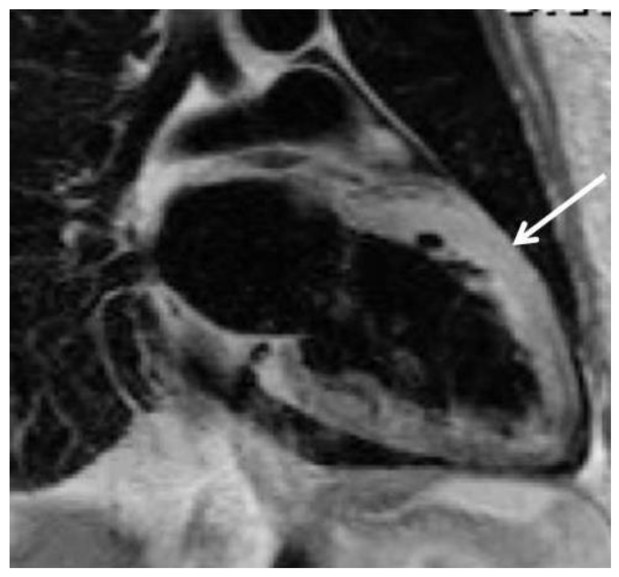
Figure 1. Cardiac magnetic resonance imaging (MRI) of the heart of a patient with acute PPCM. The T2-weighted image demonstrates intramyocardial oedema detectable in some patients at the time of initial diagnosis (arrow).
Cardiovascmed 17 00245 g001

Peripartum cardiomyopathy (PPCM)

In contrast to the above-mentioned genetic forms, PPCM is defined as a non-familial form of peripartum heart failure characterised as “an idiopathic cardiomyopathy presenting with heart failure secondary to left ventricular (LV) systolic dysfunction towards the end of pregnancy or in the months following delivery, where no other cause of heart failure is found” as proposed by the Working Group on PPCM from the Heart Failure Association of the ESC [22]. Clinically, PPCM resembles dilated cardiomyopathy (DCM) but the LV may not always be dilated. The ejection fraction (EF) is nearly always reduced below 45% [22]. PPCM is considered an independent disease, whose diagnosis relies on its relation to pregnancy and the exclusion of other cardiomyopathies [22,23].
PPCM is increasingly recognised as an important condition for women’s health and its incidence in the USA (and also in Europe) is rising (from 1 in 4350 between 1990 and 1993 to 1 in 2229 between 2000 and 2002) [24]. Mielniczuk et al. attributed this mainly to rising maternal age and a substantial increase in multifoetal pregnancies due to reproductive techniques as well as increasing awareness. In fact, the rising interest in pregnancy-related cardiac research [13,25,26], the international registry on PPCM as part of the EURObservational Research Programme (http://www. eorp.org) [27] and other national and international reporting facilities [12,28,29] may contribute to greater awareness and the larger number of PPCM cases diagnosed in recent years.
PPCM can present very dramatically with acute heart failure needing intensive care admission, or it may develop subtly over several weeks. Especially in the slow developing form of PPCM, physicians are often faced with the situation on how to distinguish between peripartum discomfort in healthy women, such as fatigue, mild shortness of breath or mild oedema, from the pathological symptoms of PPCM. Unspecific symptoms of cardiac congestion, such as abdominal discomfort, pleuritic chest pain and palpitations can also occur. Establishing the diagnosis of PPCM therefore relies on a high index of suspicion, as early signs and symptoms of heart failure and peripartum-associated physiological discomfort are often not easy to distinguish which can markedly delay diagnosis [20,22,30]. Experts in the field suggest careful history taking with the exact onset of symptoms in relation to pregnancy and a low threshold for diagnostic confirmation or exclusion of LV systolic dysfunction using echocardiography (and/or MRI) [20,21].

Pathomechanisms of PPCM

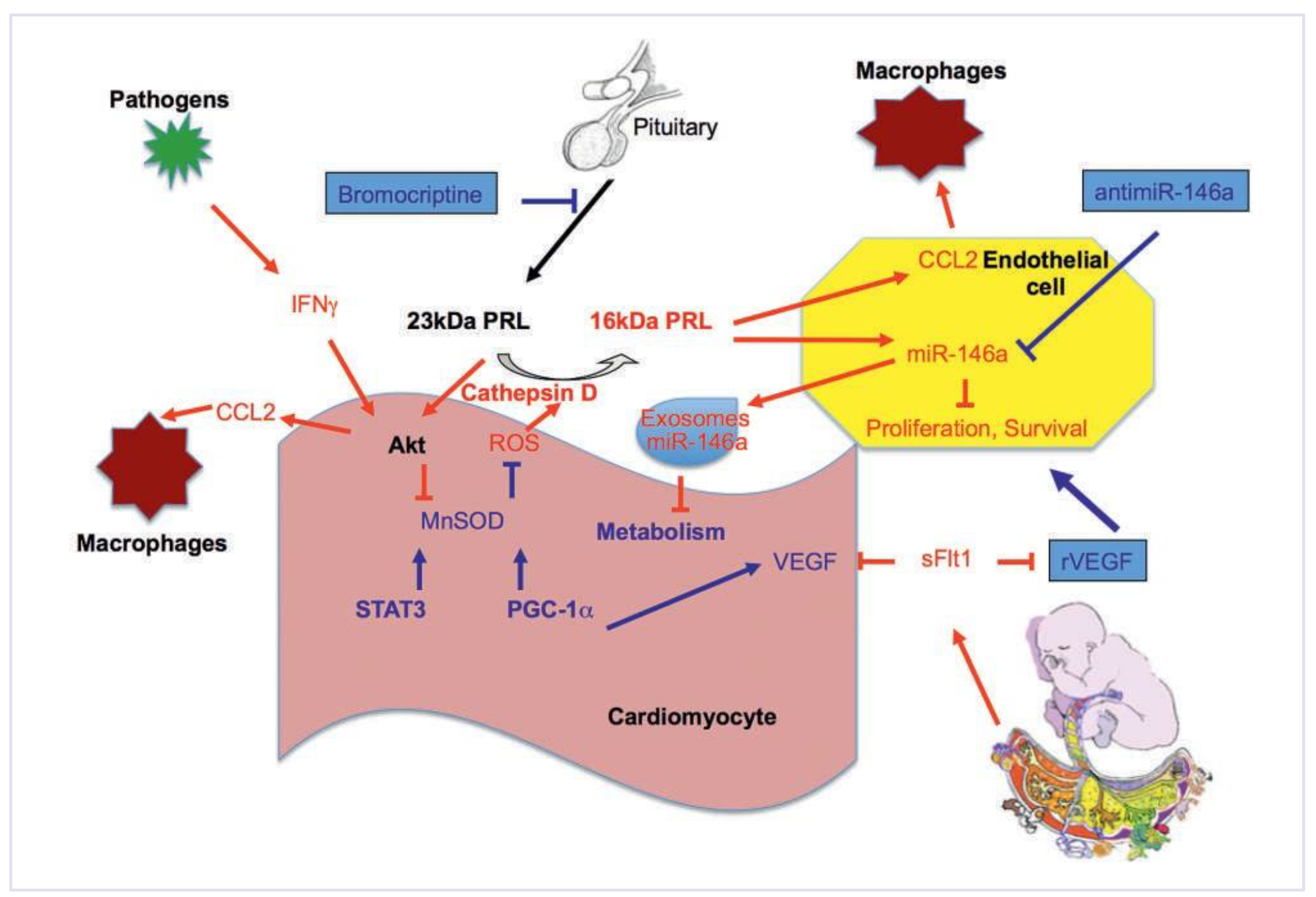
In recent years great advances were made in discovering pathomechanisms that induce and drive PPCM. Among them low selenium levels, various viral infections, stress-activated cytokines, inflammation and autoimmune reaction and a pathologic response to haemodynamic stress are suspected [20,31]. More recent studies suggest that different factors may induce PPCM and finally merge into a common pathway which includes the coincidental presence of unbalanced oxidative stress and high levels of the nursing hormone Prolactin (PRL) leading to the production of an angiostatic and pro-apoptotic 16-kDa PRL [25,26]. The 16kDa PRL mainly impacts on the endothelium and may, together with additional anti-angiogenic factors such as soluble fms-like tyrosine kinase-1 (sFlt1), disturb the angiogenic balance in the peripartum phase, thereby leading primarily to damage of the endothelium. This notion emphasises that PPCM may initially start as a disease of the endothelial layer [13,14]. Subsequently, the impaired vasculature affects also the cardiac muscle in part by lowering survival signals and by inducing metabolic shortage, which finally impact on cardiac function [13,26].
Interestingly, oxidative stress rises during uneventful pregnancies [32] and may be needed to enhance the maternal defence potential against pathogens during pregnancy where the immune system is compromised and the risk of infections is high, especially around term and during delivery. In normal pregnancy a parallel increase in total antioxidant capacity with a peak early postpartum is observed [32]. In addition, experimental models suggest an increase in organ-specific anti-oxidative defence mechanisms that seems specifically important in the maternal heart during the peripartum phase [13,26]. These experimental studies discovered the involvement of signalling pathways including signal transducer and activator of transcription 3 (STAT3) and peroxisome proliferator-activated receptor gamma co-activator 1-alpha (PGC1α) which increase the expression of antioxidative enzymes such as manganese sodium dismutase (MnSOD) [13,26]. Moreover, the maternal heart also seems to up-regulate the expression of pro-angiogenic factors, i.e., vascular endothelial growth factor (VEGF), for additional protection from anti-angiogenic factors including the 16-kDa PRL, its downstream mediator microRNA-146a and sFlt1 [13,26].
During late pregnancy, sFlt1 is released from the placenta and abnormally high levels of this factor have been associated with pre-eclampsia. Clinically, preeclampsia causes cardiac dysfunction independent of blood pressure [6,33]. We observed that sFlt1 is increased in the serum of patients with PPCM even several days or weeks after delivery [13]. Furthermore, in our German PPCM registry, pre-eclampsia during pregnancy was frequently present in patients who later developed PPCM [12]. These data suggest a potential connection between the two diseases which involves increased sFlt1 and an insufficient up-regulation of cardiac expression of VEGF [13]. Supporting this notion, a therapeutic concept using the prolactin blocker bromocriptine and recombinant VEGF could prevent experimental PPCM in mice [13].
Figure 2. Scheme depicting pathophysiological concepts in PPCM. Coincidental presence of Prolactin (23-kD PRL) released from the pituitary gland is under conditions of enhanced oxidative stress (ROS) proteolytically cleaved to a 16-kDa PRL fragment by proteases, such as cathepsin D, which is at least in part released by cardiomyocytes. In PPCM this process is induced by a shift in the redox balance towards increased oxidative stress due to the down-regulation of transcription factors STAT3 and PGC1α and/or by increased Akt activation, which all lead to suppression of anti-oxidative enzymes such as MnSOD. The 16-kDa PRL exerts angiostatic effects in endothelial cells, which are mediated mainly by miR-146a, the microRNA induced by 16-kDa PRL. The 16-kDa PRL induces also the shedding of miR-146a containing exosomes, which are impairing metabolic activity in cardiomyocytes. Furthermore, 16-kDa promotes via activation of NFkappaB endothelial inflammation. In some PPCM patients IFNg (possibly induced by pathogens) and/or 23-kDa PRL may in addition promote cardiac inflammation via up-regulation of the pro-inflammatory chemokine CCL2 in cardiomyocytes, a setting that seems to be specifically detrimental. STAT3 and PGC1α are also needed to protect the cardiac vasculature from sFlt1, an additional anti-angiogenic factor present in the peripartum phase, by increasing the cardiac expression of the pro-angiogenic factor VEGF that is neutralising the adverse effects of sFlt1. Thus, blocking prolactin by bromocriptine, neutralising miR-146a by antagomir strategies and/or VEGF agonist treatment could be novel disease-specific therapeutic concepts for PPCM.
Cardiovascmed 17 00245 g002
Taken together, these data indicate that PPCM may often start as a disease of the endothelium, leading to loss or damage of the vasculature. Moreover, PPCM may be a multifactorial disease caused by the coincidental presence of unbalanced oxidative stress, impaired cardioprotective and pro-angiogenic signalling and high expression of anti-angiogenic factors. Part of these mechanisms may already be initiated during pregnancy for example by pre-eclampsia. The current understanding of pathomechanisms inducing PPCM is explained in more detail in a recent review and summarised in figure 2 [14].

Biomarkers

As mentioned above, the diagnosis of PPCM is often delayed and complicated by the fact that symptoms of heart failure overlap to some degree with normal pregnancy-associated discomfort. In addition, PPCM patients are frequently not seen first by cardiologists. Therefore, biomarkers would help to identify PPCM patients and refer them to expert physicians for further diagnostic assessment. NT-proBNP, a frequently used marker for heart failure is almost always increased in patients with peripartum heart failure but not in peripartum women without heart failure [12,34] and would therefore be an easy marker to be tested in any peripartum woman reporting discomfort. However, NT-proBNP is a rather unspecific marker for heart failure. In turn, the recently discovered miR-146a is a direct downstream effector of the PPCM-causing factor 16-kDa PRL [25]. We recently showed that circulating miR-146a is specifically up-regulated in patients with PPCM, but not in healthy peripartum women or in patients with DCM [12,25]. The fact that 16-kDa PRL induces shedding of miR-146a containing endothelial exosomes fits well with the observation that the endothelial microparticles are up-regulated in patients with PPCM [35].
Thus, currently NT-proBNP is a commercially available marker for screening of peripartum heart failure. Experimental data are promising to develop more disease-specific markers that may help in the future to distinguish PPCM from other types of heart failure allowing optimal early management and risk stratification for these young patients.

Therapeutic concepts and management for PPCM

Currently, peripartum heart failure is treated according to the ESC guidelines for heart failure in pregnancy [1]. In brief, late in pregnancy therapeutic interventions need to consider the health of the mother and the fetus while after delivery standard therapy for heart failure (beta-blockers, ACE-inhibitors/AT1-blockers diuretics, mineralocorticoid receptor antagonists) is recommended. The more recent insights into the pathophysiology of peripartum heart failure and especially PPCM provide novel and more disease-specific therapeutic concepts. The potential role for the angiostatic and pro-apoptotic 16-kDa PRL in the initiation and progression of PPCM leads to the idea that blocking prolactin in PPCM patients might be beneficial. Indeed, the suppression of PRL release by the dopamine D2 receptor agonist bromocriptine could prevent the onset of disease in several animal models of PPCM [13,26]. In addition, several case reports and a small clinical study showed that the addition of bromocriptine to standard heart failure therapy was associated with improved LV function and a composite clinical outcome in women with acute severe PPCM [22,26,36,37]. Currently the efficacy of the bromocriptine therapy concept is investigated in larger controlled randomised multicentre trials in Germany (randomisation of 60 PPCM patients to standard therapy for heart failure with or without bromocriptine, the study is registered at ClinicalTrials.gov, study number: NCT00998556).
It is important to note that PPCM patients are at a relatively high risk for sudden death [34,38]. First observations suggest that PPCM patients with severely depressed cardiac function and/or ventricular arrhythmias may benefit from wearable cardioverter/defibrillator (WCD). In a single centre observational study, we treated all patients with LV ejection fraction <35% with a WCD and observed several appropriate shocks during the following months (unpublished observations). Thus, we suggest considering early WCD therapy in PPCM patients with severely depressed LV function. As many PPCM patients recover from the disease or display at least marked improvement of systolic LV function, operative ICD or CRT-D implantation for primary prophylaxis is not recommended during the first six months after instituting appropriate medical heart failure therapy.
An additional important point concerns future pregnancies in PPCM patients. Due to the risk for relapse of heart failure PCCM patients should be discouraged from becoming pregnant again. Our and other investigators’ observations from PPCM patients with subsequent pregnancies suggest that these patients frequently tolerate the pregnancy state quite well, especially if they enter the subsequent pregnancy with fully recovered cardiac function [22,39]. However, cardiac dysfunction re-emerges often in the periand postpartum phase [22,39]. Therefore, subsequent pregnancies in PPCM patients should always be followed in experienced centres with close collaboration between obstetricians and heart failure cardiologists. This is especially important in PPCM patients who become pregnant without complete recovery of LV function.
PPCM patients should be informed about contraceptive options (we recommend IUD since hormonal contraceptives may interact with heart failure medication, and counsel them about the risk for relaps in subsequent pregnancies).

Conclusion

In recent years the awareness for PPCM has increased for the benefit of these patients. Larger clinical data sets are collected and analysed, thus allowing more insight into the pathophysiology of the disease and providing important information for diagnosis and management of these patients. Large clinical registries on peripartum heart failure (ESC EUROOBS program (www.escardio.org) [40] together with experimental research are needed to further broaden our understanding of PPCM with regard to its aetiology, risk factors, diagnosis and, most importantly, optimised treatment strategies and management.

Funding

Experimental work was supported by the BMBF and the DFG.

Potential Competing Interests

Experimental work was supported by the BMBF and the DFG.

References

  1. Regitz-Zagrosek, V.; Blomstrom Lundqvist, C.; Borghi, C.; Cifkova, R.; Ferreira, R.; Foidart, J.M.; et al. ESC Guidelines on the management of cardiovascular diseases during pregnancy: the Task Force on the Management of Cardiovascular Diseases during Pregnancy of the European Society of Cardiology (ESC). Eur Heart J. 2011, 32, 3147–3197. [Google Scholar] [PubMed][Green Version]
  2. Ruys, T.P.; Roos-Hesselink, J.W.; Hall, R.; Subirana-Domenech, M.T.; GrandoTing, J.; Estensen, M.; et al. Heart failure in pregnant women with cardiac disease: data from the ROPAC. Heart 2014, 100, 231–238. [Google Scholar] [CrossRef] [PubMed]
  3. Chung, E.; Leinwand, L.A. Pregnancy as a cardiac stress model. Cardiovasc Res. 2014, 101, 561–570. [Google Scholar] [CrossRef] [PubMed]
  4. Conrad, K.P.; Davison, J.M. The renal circulation in 1 normal pregnancy and preeclampsia. Is there a place for relaxin? Am. J. Physiol. Ren. Physiol. 2014, 306, F1121–35. [Google Scholar] [CrossRef] [PubMed]
  5. Liu, L.X.; Arany, Z. Maternal cardiac metabolism in pregnancy. Cardiovasc Res. 2014, 101, 545–553. [Google Scholar] [CrossRef] [PubMed]
  6. Bello, N.; Rendon, I.S.; Arany, Z. The relationship between pre-eclampsia and peripartum cardiomyopathy: a systematic review and meta-analysis. J Am Coll Cardiol. 2013, 62, 1715–1723. [Google Scholar] [CrossRef] [PubMed]
  7. Rana, S.; Powe, C.E.; Salahuddin, S.; Verlohren, S.; Perschel, F.H.; Levine, R.J.; et al. Angiogenic factors and the risk of adverse outcomes in women with suspected preeclampsia. Circulation 2012, 125, 911–919. [Google Scholar] [CrossRef] [PubMed]
  8. Ahmed, R.; Dunford, J.; Mehran, R.; Robson, S.; Kunadian, V. Pre-eclampsia and future cardiovascular risk among women: a review. J Am Coll Cardiol. 2014, 63, 1815–1822. [Google Scholar] [CrossRef] [PubMed]
  9. Mannisto, T.; Mendola, P.; Vaarasmaki, M.; Jarvelin, M.R.; Hartikainen, A.L.; Pouta, A.; et al. Elevated blood pressure in pregnancy and subsequent chronic disease risk. Circulation 2013, 127, 681–690. [Google Scholar] [CrossRef] [PubMed]
  10. Vikse, B.E. Pre-eclampsia and the risk of kidney disease. Lancet 2013, 382, 104–106. [Google Scholar] [CrossRef] [PubMed]
  11. Williams, D. Long-term complications of preeclampsia. Semin Nephrol. 2011, 31, 111–122. [Google Scholar] [CrossRef] [PubMed]
  12. Haghikia, A.; Podewski, E.; Libhaber, E.; Labidi, S.; Fischer, D.; Roentgen, P.; et al. Phenotyping and outcome on contemporary management in a German cohort of patients with peripartum cardiomyopathy. Basic Res Cardiol. 2013, 108, 366. [Google Scholar] [CrossRef] [PubMed]
  13. Patten, I.S.; Rana, S.; Shahul, S.; Rowe, G.C.; Jang, C.; Liu, L.; et al. Cardiac angiogenic imbalance leads to peripartum cardiomyopathy. Nature 2012, 485, 333–338. [Google Scholar] [CrossRef] [PubMed]
  14. Hilfiker-Kleiner, D.; Sliwa, K. Pathophysiology and epidemiology of peripartum cardiomyopathy. Nat Rev Cardiol. 2014, 11, 364–370. [Google Scholar] [CrossRef] [PubMed]
  15. Morales, A.; Painter, T.; Li, R.; Siegfried, J.D.; Li, D.; Norton, N.; et al. Rare variant mutations in pregnancy-associated or peripartum cardiomyopathy. Circulation 2010, 121, 2176–2182. [Google Scholar] [CrossRef] [PubMed]
  16. van Spaendonck-Zwarts, K.Y.; van Tintelen, J.P.; van Veldhuisen, D.J.; van der Werf, R.; Jongbloed, J.D.; Paulus, W.J.; et al. Peripartum cardiomyopathy as a part of familial dilated cardiomyopathy. Circulation 2010, 121, 2169–2175. [Google Scholar] [CrossRef] [PubMed]
  17. Bahl, A.; Swamy, A.; Sharma, Y.; Kumar, N. Isolated noncompaction of left ventricle presenting as peripartum cardiomyopathy. Int J Cardiol. 2006, 109, 422–423. [Google Scholar] [CrossRef] [PubMed]
  18. Lea, B.; Bailey, A.L.; Wiisanen, M.E.; Attili, A.; Rajagopalan, N. Left ventricular noncompaction presenting as peripartum cardiomyopathy. Int J Cardiol. 2012, 154, e65–6. [Google Scholar] [CrossRef] [PubMed]
  19. Hilfiker-Kleiner, D.; Struman, I.; Hoch, M.; Podewski, E.; Sliwa, K. 16-kDa prolactin and bromocriptine in postpartum cardiomyopathy. Curr. Heart Fail. Rep. 2012, 9, 174–182. [Google Scholar] [CrossRef] [PubMed]
  20. Sliwa, K.; Fett, J.; Elkayam, U. Peripartum cardiomyopathy. Lancet 2006, 368, 687–693. [Google Scholar] [CrossRef]
  21. Sliwa, K.; Hilfiker-Kleiner, D.; Petrie, M.C.; Mebazaa, A.; Pieske, B.; Buchmann, E.; et al. Current state of knowledge on aetiology, diagnosis, management, and therapy of peripartum cardiomyopathy: a position statement from the Heart Failure Association of the European Society of Cardiology Working Group on peripartum cardiomyopathy. Eur J Heart Fail. 2010, 12, 767–778. [Google Scholar] [CrossRef] [PubMed]
  22. Sliwa, K.; Hilfiker-Kleiner, D.; Petrie, M.; Mebazaa, A.; Pieske, B.; Buchmann, E.; et al. Current state of knowledge on aetiology, diagnosis, management, and therapy of peripartum cardiomyopathy: a position statement from the Heart Failure Association of the European Society of Cardiology Working Group on Peripartum Cardiomyopathy. Eur Heart J. 2010, 12, 767–778. [Google Scholar] [CrossRef] [PubMed]
  23. Pearson, G.D.; Veille, J.C.; Rahimtoola, S.; Hsia, J.; Oakley, C.M.; Hosenpud, J.D.; et al. Peripartum cardiomyopathy: National Heart, Lung, and Blood Institute and Office of Rare Diseases (National Institutes of Health) workshop recommendations and review. JAMA 2000, 283, 1183–1188. [Google Scholar] [CrossRef] [PubMed]
  24. Mielniczuk, L.M.; Williams, K.; Davis, D.R.; Tang, A.S.; Lemery, R.; Green, M.S.; et al. Frequency of peripartum cardiomyopathy. Am J Cardiol. 2006, 97, 1765–1768. [Google Scholar] [CrossRef] [PubMed]
  25. Halkein, J.; Tabruyn, S.P.; Ricke-Hoch, M.; Haghikia, A.; Nguyen, N.Q.; Scherr, M.; et al. MicroRNA-146a is a therapeutic target and biomarker for peripartum cardiomyopathy. J Clin Invest. 2013, 123, 2143–2154. [Google Scholar] [CrossRef] [PubMed]
  26. Hilfiker-Kleiner, D.; Kaminski, K.; Podewski, E.; Bonda, T.; Schaefer, A.; Sliwa, K.; et al. A Cathepsin D-cleaved 16 kDa form of prolactin mediates postpartum cardiomyopathy. Cell 2007, 128, 589–600. [Google Scholar] [CrossRef] [PubMed]
  27. Sliwa, K.; Hilfiker-Kleiner, D.; Mebazaa, A.; Petrie, M.C.; Maggioni, A.P.; Regitz-Zagrosek, V.; et al. EURObservational Research Programme: a worldwide registry on peripartum cardiomyopathy (PPCM) in conjunction with the Heart Failure Association of the European Society of Cardiology Working Group on PPCM. Eur J Heart Fail. 2014, 16, 583–591. [Google Scholar] [CrossRef] [PubMed]
  28. Elkayam, U. Clinical characteristics of peripartum cardiomyopathy in the United States: diagnosis, prognosis, and management. J Am Coll Cardiol. 2011, 58, 659–670. [Google Scholar] [PubMed]
  29. Safirstein, J.G.; Ro, A.S.; Grandhi, S.; Wang, L.; Fett, J.D.; Staniloae, C. Predictors of left ventricular recovery in a cohort of peripartum cardiomyopathy patients recruited via the internet. Int J Cardiol. 2012, 154, 27–31. [Google Scholar] [CrossRef] [PubMed]
  30. Selle, T.; Renger, I.; Labidi, S.; Bultmann, I.; Hilfiker-Kleiner, D. Reviewing peripartum cardiomyopathy: current state of knowledge. Future Cardiol. 2009, 5, 175–189. [Google Scholar] [PubMed]
  31. Sliwa, K.; Forster, O.; Libhaber, E.; Fett, J.D.; Sundstrom, J.B.; Hilfiker-Kleiner, D.; et al. Peripartum cardiomyopathy: inflammatory markers as predictors of outcome in 100 prospectively studied patients. Eur Heart J. 2006, 27, 441–446. [Google Scholar]
  32. Toescu, V.; Nuttall, S.L.; Martin, U.; Kendall, M.J.; Dunne, F. Oxidative stress and normal pregnancy. Clin Endocrinol 2002, 57, 609–613. [Google Scholar] [CrossRef] [PubMed]
  33. Powe, C.E.; Levine, R.J.; Karumanchi, S.A. Preeclampsia, a disease of the maternal endothelium: the role of antiangiogenic factors and implications for later cardiovascular disease. Circulation 2011, 123, 2856–2869. [Google Scholar] [CrossRef] [PubMed]
  34. Forster, O.; Hilfiker-Kleiner, D.; Ansari, A.A.; Sundstrom, J.B.; Libhaber, E.; Tshani, W.; et al. Reversal of IFN-gamma, oxLDL and prolactin serum levels correlate with clinical improvement in patients with peripartum cardiomyopathy. Eur J Heart Fail. 2008, 10, 861–868. [Google Scholar] [CrossRef] [PubMed]
  35. Walenta, K.; Schwarz, V.; Schirmer, S.H.; Kindermann, I.; Friedrich, E.B.; Solomayer, E.F.; et al. Circulating microparticles as indicators of peripartum cardiomyopathy. Eur Heart J. 2012, 33, 1469–1479. [Google Scholar] [CrossRef] [PubMed]
  36. Habedank, D.; Kuhnle, Y.; Elgeti, T.; Dudenhausen, J.W.; Haverkamp, W.; Dietz, R. Recovery from peripartum cardiomyopathy after treatment with bromocriptine. Eur J Heart Fail. 2008, 10, 1149–1151. [Google Scholar] [CrossRef] [PubMed]
  37. Meyer, G.P.; Labidi, S.; Podewski, E.; Sliwa, K.; Drexler, H.; Hilfiker-Kleiner, D. Bromocriptine treatment associated with recovery from peripartum cardiomyopathy in siblings: two case reports. J Med Case Reports. 2010, 4, 80. [Google Scholar] [CrossRef] [PubMed]
  38. Sliwa, K.; Forster, O.; Tibazarwa, K.; Libhaber, E.; Becker, A.; Yip, A.; et al. Long-term outcome of peripartum cardiomyopathy in a population with high seropositivity for human immunodeficiency virus. Int J Cardiol. 2011, 147, 202–208. [Google Scholar] [CrossRef] [PubMed]
  39. Elkayam, U.; Tummala, P.P.; Rao, K.; Akhter, M.W.; Karaalp, I.S.; Wani, O.R.; et al. Maternal and fetal outcomes of subsequent pregnancies in women with peripartum cardiomyopathy. N Engl J Med. 2001, 344, 1567–1571. [Google Scholar] [CrossRef] [PubMed]
  40. Sliwa, K.; Hilfiker-Kleiner, D.; Mebaaza, A.; Petrie, M.C.; Maggioni, A.; RegitzZagrosek, V.; et al. EURObservational Research Programme: a worldwide registry on peripartum cardiomyopathy (PPCM) in conjunction with the Heart Failure Association of the European Society of Cardiology Working Group on PPCM. Eur J Heart Fail. 2014, 16, 583–591. [Google Scholar] [CrossRef]