Anatomie und Pathologie Einführung
Die Mitralklappe (MK) besteht aus dem Anulus, dem anterioren und posterioren Segel, den Chordae tendineae und den Papillarmuskeln (welche zusammen den subvalvulären Apparat bilden) und, im weiteren Sinne, dem linken Ventrikel. Primär valvuläre Erkrankungen der Mitralklappe, auch organische Mitralinsuffizienz (MI) genannt, sind strukturelle Schäden an Anulus, Segeln und Chordae. Funktionelle Erkrankungen beziehen sich auf Veränderungen am linken Ventrikel, welche sekundär (d.h. via Geometriestörung) auf die morphologisch intakte Klappe einwirken [
1]. Zu nennen sind hier die verschiedenen Formen von dilatativen und ischämischen Kardiomyopathien (KMP) [
2]. Als dritte Entität findet sich in der Literatur die gemischte Form, wobei funktionelle und organische Aspekte oft kombiniert mit Verkalkungen unterschiedlicher Ausprägung vorliegen [
3,
4].
Anulus
Der bindegewebige Mitralring stellt die Grenze zwischen dem linken Atrium und dem linken Ventrikel dar. Man unterscheidet einen vorderen und einen hinteren Anteil; die Unterteilung wird durch die Kommissuren definiert. Der anteriore, kürzere Anteil ist zwischen den beiden Trigona (Trigonum dexter und sinister) aufgespannt; der hintere Anteil läuft parallel zum posterioren Segel und stellt etwa zwei Drittel des Gesamtumfangs des Anulus dar. Der Anulus umschliesst die Mitralklappe vollständig und definiert ihre dreidimensionale Geometrie, die einem Reitsattel ähnelt (s. Abbildung 1b) [
5]. Die sekundäre Deformation und Dilatation des Anulus bei der funktionellen und degenerativen [
2] sowie die Verkalkung bei der gemischten Mitralinsuffizienz spielen zentrale Rollen in der Entstehung der Mitralinsuffizenz. Für Angaben zu anulären Dimensionen, gemessen mittels MRI sowie Echokardigraphie, siehe Tabellen 1 und 2.
Abbildung 1.
Mitralklappensegel und Anulus. Anatomie der Mitralklappe im Überblick. Abb. 1a: Schematische Ansicht der Mitralklappe inklusive der einzelnen Segmente, adaptiert nach Biaggi et al. [
36], Abdruck mit freundlicher Genehmigung von JACC: Cariovascular Imaging (Elsevier). Abb. 1b: 3D-Rekonstruktion der Mitralklappe. Rot markiert sind die wichtigsten Distanzen. Abkürzungen: A1, A2, A3 = respektives Segment des anterioren Mitralsegels, AV = Aortenklappe, Ca = anterolaterale Kommissur, Cp = posteromediale Kommissur, AP = anteroposteriorer Abstand; COM = Interkommissuraler Abstand, Cs = Koronarsinus, H = Sattelhöhe; LCC, RCC und NCC = linkskoronare, rechtskoronare respektive nicht-koronare Tasche der Aortenklappe.
Tabelle 1.
Anulusund Papillarmuskel-Geometrie bei Patienten mit ischämischer Mitralinsuffizienz im Vergleich zu gesunden Probanden (Messungen aus CMR, adaptiert nach [
31]).
Tabelle 1.
Anulusund Papillarmuskel-Geometrie bei Patienten mit ischämischer Mitralinsuffizienz im Vergleich zu gesunden Probanden (Messungen aus CMR, adaptiert nach [
31]).
Tabelle 2.
Echokardiographisch ermittelte Mitralklappenund Anulus Werte bei Gesunden sowie verschiedenen Krankheitsbildern. Tabelle nach Kovalova et al. [
50].
Tabelle 2.
Echokardiographisch ermittelte Mitralklappenund Anulus Werte bei Gesunden sowie verschiedenen Krankheitsbildern. Tabelle nach Kovalova et al. [
50].
Segel
Das vordere und das hintere Segel bilden die Klappe als solche. Die Abgrenzung erfolgt wie auch beim Anulus durch die anterolaterale und die posteromediale Kommissur. Das anteriore Segel ist rundlich und mit dem vorderen Anteil des Anulus verwachsen. Das posteriore Segel ist im Vergleich länglich und umfasst das vordere Segel halbmondförmig. Die Segel sind in je drei Segmente unterteilt: das posteriore in das laterale P1-, zentrale P2-und mediale P3-Segment, die gegenüberliegenden Segmente des anterioren Segels analog in A1, A2 und A3 (s. Abbildung 1a). Beim Gesunden beträgt die Länge des anterioren respektive des posterioren MKSegels 24 ± 4 bzw. 10 ± 3 mm [
6], die Öffnungsfläche misst 4–7 cm2 [
7]. Die wichtigsten echokardiographischen Schnittebenen der Mitralklappe und die darauf abgebildeten Segmente sind in Abbildung 2 dargestellt.
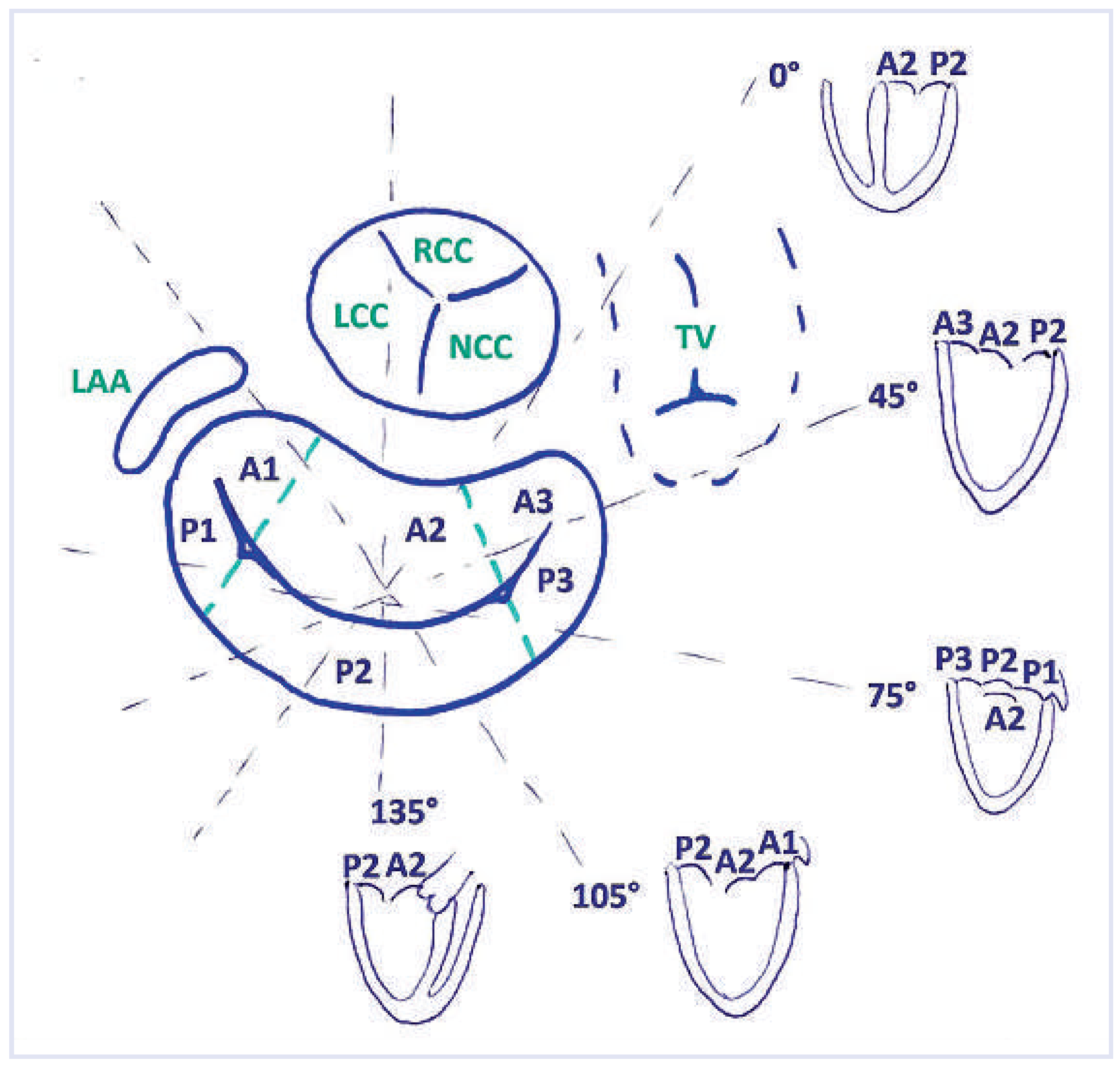
Abbildung 2.
Schematische Darstellung der Mitralklappensegmente im TEE. Die TEE-Untersuchung wird meist in der 0°-Einstellung begonnen. Auf midesophagealer Ebene (Vierkammerschnitt) kommen dabei die Segmente A2 und P2 zur Darstellung. Wird die Schnittebene (auf dieser Darstellung im Uhrzeigersinn) auf ca. 45° rotiert, so sind meist die Segmente A3, A2 sowie P2 abgebildet. Beim commissuralen Schnitt (meist um 75+/−15°) wird durch P3, A2 und P1 geschnitten. In den meisten Fällen kommen in der Langachsenansicht bei ca. 135° die Segmente A2 und P2. zur Darstellung. Die effektiv dargestellten Segmente können aber von den theoretischen abweichen, insbesondere in Abhängigkeit der Lage der TEE-Sonde im Ösophagus, bei Rotation des Herzens und bei den nicht seltenen anatomischen Variationen. Die angegebenen Grad-Zahlen dürfen daher nur als approximativ verstanden werden. Wichtiger ist die Orientierung an der effektiven Anatomie. Abkürzungen: LAA = Left Atrial Appendage, LCC, RCC und NCC: linkskoronare, rechtkoronare respektive nicht-koronare Tasche der Aortenklappe, TV = Tricuspid Valve.
![Cardiovascmed 17 00101 g002 Cardiovascmed 17 00101 g002]()
Bei den Segelpathologien unterscheiden wir organische, funktionelle und gemischte Ursachen. Bei der organischen Mitralinsuffizienz kommt es zu einer mehr oder minder ausgeprägten Form von Prolaps (MKP), verursacht durch eine Bindegewebedegeneration. Auf der einen Seite des Spektrums steht dabei die «fibroelastic deficiency» (FED) mit Ausdünnung der Segel und mildem, meist singulärem Prolaps und schliesslich Sehnenfadenabriss. Das andere Ende des Spektrums stellt der Morbus Barlow dar, verursacht durch myxomatöse Degeneration. Hierbei reisst das Elastin, es wird vermehrt Kollagen gebildet und Glykoprotein eingelagert [
8]. Dadurch verdicken und verlängern sich die Segel und ein Grossteil der Sehnenfäden, es kommt zum Prolaps praktisch aller Segmente (siehe Tabelle 3). Zwischen diesen beiden Extremen gibt es eine Unzahl von Formen mit sehr unterschiedlicher, teilweise auch asymmetrischer Ausprägung beider Aspekte (sogenannte «forme fruste») [
9].
Bei der funktionellen Mitralinsuffizienz ist die Klappe per se nicht wesentlich degeneriert. Vielmehr ist der fehlende Klappenschluss (ungenügenden Koaptation) eine Folge der veränderten Ventrikeloder Papillarmuskelgeometrie. Dies kann bei allen Formen der dilatativen KMP auftreten [
2].
Bei der gemischten Mitralinsuffizienz findet sich am häufigsten eine Kombination von Prolaps und Anulusverkalkung, wobei die Verkalkung teilweise auch auf die Segel übergreift. Eine zweite häufige Form der gemischten Mitralinsuffizienz findet sich bei der rheumatisch veränderten Mitralklappe. Klassischerweise dominiert hierbei der stenotische Aspekt. Je nach Kombination von Verkalkungen, Restriktion und Elongation der Segel kann aber auch die Mitralinsuffizienz führend und schwer sein [
10].
Subvalvuläre Strukturen
Die unelastischen Chordae tendineae und die Papillarmuskeln werden als subvalvulärer Apparat zusammengefasst. Die Einmündung der Papillarmuskeln in den Ventrikel stellt sich geflechtoder wurzelartig dar oder imponiert in Extremfällen als Netzwerk. Aus beiden Papillarmuskeln geht eine Vielzahl von Chordae hervor, die von beiden Muskeln aus zu beiden Segeln ziehen. Wir unterscheiden primäre (Insertion am freien Segelrand), sekundäre (Insertion in den Bauch des Segels) sowie tertiäre Chordae (nicht vom Papillarmuskel, sondern vom freien Myokard ausgehend). Die Geometrie der Klappensegel und damit die Funktion der Mitralklappe hängt vom subvalvulären Apparat ab, während dieser wiederum durch die Form des linken Ventrikels beeinflusst wird. Als Mass für eine funktionelle Pathologie der Papillarmuskeln kann der interpapilläre Abstand gemessen werden (Distanz zwischen den Muskelköpfen) [
1]. Bei der dilatativen KMP nimmt diese Distanz prinzipiell zu (kombiniert mit veränderten Winkeln zwischen Papillarmuskeln und Segeln), und diese ungünstigen Veränderungen der Papillarmuskel-Segel-Geometrie kann die Entstehung einer Mitralinsuffizienz begünstigen [
2]. Die Messung des interpapillären Abstandes hat sich im Alltag allerdings nicht bewährt, da die dreidimensionale Anordnung reproduzierbare Quantifizierungen erschwert und die grossen interindividuellen Variationen das Festlegen von brauchbaren Normwerten kaum zulassen.
Tabelle 3.
Vergleich zwischen ‚fibroelastic deficiency syndrom’ (FED) und Morbus Barlow. Tabelle adaptiert nach Chandra et al. [
51].
Tabelle 3.
Vergleich zwischen ‚fibroelastic deficiency syndrom’ (FED) und Morbus Barlow. Tabelle adaptiert nach Chandra et al. [
51].
Linker Ventrikel
Der linke Ventrikel spielt eine zentrale Rolle in der Geometrie der Mitralklappe. Es ist daher gerechtfertigt, ihn im Zusammenhang mit funktioneller Mitralinsuffizienz als Teil der Mitralklappe zu betrachten. Die veränderte Ventrikelgeometrie bei ischämischer KMP (beispielsweise infolge Akinesie oder gar Aneurysmabildungen) sowie bei idiopathischer dilatativen KMP kann zu unvollständigem Klappenschluss und damit zur Mitralinsuffizienz führen. Die ischämische KMP kann im Extremfall zu einem ischämisch bedingten Papillarmuskel-Abriss führen [
11].
Geometrische Veränderungen sind auch bei der hypertrophen obstruktiven Kardiomyopathie (HOCM) mitbeteiligt an der assoziierten Mitralinsuffizienz. Die nach anterior und apikal verschobenen Papillarmuskeln, die basal-septale Hypertrophie sowie die Verdickung und Elongation des anterioren Mitralsegels führen in Kombination zur systolischen Obstruktion des Ausflusstrakts durch das anteriore MK Segel (sogenannte «systolic anterior motion» (SAM) des vorderen Mitralsegels). Die SAM wird verursacht durch den Venturi-Effekt [
12,
13] und führt typischerweise zu einer posterior gerichteten Mitralinsuffizienz.
Quantifizierung der Mitralinsuffizienz Überblick
Der Quantifizierung der Mitralinsuffizienz kommt eine spezielle Bedeutung zu, weil sie eine der Entscheidungsgrundlagen für eine Operation oder perkutane Intervention liefert. Eine schwere symptomatische Mitralinsuffizienz mit erhaltener LV-Funktion stellt per se eine Operationsindikation der Klasse 1A dar, während eine mittelschwere sekundäre Mitralinsuffizienz ausser bei ohnehin stattfindender Herzoperation in der Regel nicht operiert wird (OP-Indikation IIb, C) [
4].
Die Quantifizierung der Mitralinsuffizienz ist anspruchsvoll. Dies hängt zum einen mit den im Folgenden erläuterten technischen Herausforderungen zusammen, zum anderen ist der Schweregrad abhängig von hämodynamischen Parametern wie Blutdruck, Herzfrequenz und Füllungszustand des Patienten. Gewisse Formen der schweren Mitralinsuffizienz treten zudem erst unter Belastung auf. Der wichtigste Grundsatz bei der Beurteilung des Schweregrades der Mitralinsuffizienz lautet daher, dass eine integrative Betrachtungsweise nötig ist. Dabei stimmen im Idealfall Anamnese, Status und Verlaufsgeschichte des Patienten überein mit den gemessenen Parametern aus physikalischer Belastung, Rhythmusüberprüfung und Labor sowie der Bildgebung. Auf letztere soll hier besonders eingegangen werden.
Grundprinzip
Das Regurgitationsvolumen der Mitralinsuffizienz entspricht (in Abwesenheit einer relevanten Aorteninsuffizienz) der Differenz zwischen dem Blutvolumen, welches in der Diastole via Mitralklappe in den linken Ventrikel strömt (MVin), und dem Volumen, welches in der Systole das Herz via Aortenklappe verlässt (LVOT out). Wenn während der Systole mehr als 50% des vorher eingeströmten Bluts wieder in den linken Vorhof zurückfliesst, so gilt die Mitralinsuffizienz als schwer [
14,
15]. Diese Regurgitationsfraktion berechnet sich wie folgt:
Echokardiographie
Traditionell ist die Echokardiographie die Methode der Wahl für die Quantifizierung der Mitralinsuffizienz. Die wichtigsten Methoden können hier nur punktuell wiedergegeben werden, wir verweisen auf die entsprechende Literatur [
16]. So einfach die oben aufgestellte Gleichung scheint, ist die echokardiographische Messung dieser Parameter dennoch eine Herausforderung. Das Hauptproblem aller zweidimensionalen Quantifizierungsmethoden liegt darin, dass zur Berechnung des Regurgitationsvolumens auf geometrische Modelle zurückgegriffen werden muss, welche die eingangs beschriebene komplexe Anatomie des Mitralanulus, der Segel, der Flussrichtungen und der Flussgeschwindigkeiten sowie der Regurgitationsöffnungsfläche (Regurgitant Orifice Area, ROA) nur ungenügend wiedergeben und nur in Spezialsituationen hinreichend genau sind. Die von der europäischen Gesellschaft für Echokardiographie empfohlenen Parameter sind in der Tabelle 4 wiedergegeben. Da ein einzelner Parameter nur selten diagnostisch ausreichend ist, muss im Alltag darauf geachtet werden, dass die Klappenmorphologie die vorliegenden Jets und den abgeleiteten Schweregrad hinlänglich erklären, und dass auch Grösse und Funktion des linken Ventrikels und Vorhofes mit der Quantifizierung der Mitralinsuffizienz übereinstimmen [
17].
Tabelle 4.
Echokardiographische Parameter zur Bestimmung des Schweregrades der Mitralinsuffizienz. Tabelle nach Grayburn et al. [
17], adaptiert nach Zoghbi [
18], Lang [
52], Bonow [
14] und Lancellotti [
16].
Tabelle 4.
Echokardiographische Parameter zur Bestimmung des Schweregrades der Mitralinsuffizienz. Tabelle nach Grayburn et al. [
17], adaptiert nach Zoghbi [
18], Lang [
52], Bonow [
14] und Lancellotti [
16].
Farb-Doppler Echokardiographie
Die Farb-Doppler Echokardiographie stellt das wichtigste Instrument zur Quantifizierung der Mitralinsuffizienz dar (Abbildung 3). Es ist darauf zu achten, dass die Aliasing-Geschwindigkeit möglichst hoch ist (60– 70cm/s), um die Fläche des Insuffizienzjets und damit den Schweregrad der Insuffizienz nicht zu überschätzen. Der Schweregrad einer Mitralinsuffizienz wird bei sehr exzentrischen Jets, stark dilatierten Vorhöfen sowie stark eingeschränkter Funktion des linken Ventrikels tendenziell unterschätzt.
Abbildung 3.
Echokardiographische Zeichen der schweren Mitralklappeninsuffizienz. Abb. 3a: 2D-TTE, Parasternale Längsachse, Vena contracta (roter Pfeil). Abb. 3b und 3c: 4–Kammerund 2–Kammerblick, Mitralinsuffizienzfläche (rot) >40% der Fläche des linken Vorhofs (weiss). Der rote Pfeil in 3b demonstriert die Flusskonvergenzzone. Abb. 3d: Entsprechende continous-wave-Doppler Darstellung des Regurgitationsjets: V-förmig und sehr hohe Signaldichte.
Vena Contracta
Als Vena Contracta (VC) wird der Bereich eines Jets bezeichnet, der die geringste Fläche und die höchste Geschwindigkeit aufweist (Abbildung 4). Typischerweise liegt sie direkt hinter der Regurgitationsöffnungsfläche. In der 2D-Echokardiographie wird die VC mittels Farb-Doppler Untersuchung in der parasternalen Längsachse (und nicht im Vieroder Zweikammerschnitt!) gemessen [
18]. Falls die effektive Regurgitationsöffnungsfläche (EROA) kreisförmig ist, kann sie mittels VC annähernd ermittelt werden (π* [VC/2]2). Meist ist die EROA allerdings ellipsoid bzw. halbmondförmig (bei funktioneller Mitralinsuffizienz) oder von irregulärer Form (bei multiplem Prolaps).
Flow Convergence/Proximal Isovelocity Surface Area (PISA)
Die PISA-Methode beruht auf dem Prinzip der Flussbeschleunigung in der sogenannten Konvergenzzone vor dem Durchtritt durch eine kreisförmige Öffnung. Dabei entstehen halbkugelförmige (in der 2D Bildgebung halbkreisförmige) Ebenen, innerhalb derer dieselbe Flussgeschwindigkeit vorherrscht. Der Radius einer solchen Halbkugel (PISA-Radius) kann mittels Farb-Doppler Echokardiographie gemessen werden. Zusammen mit den Continuous-wave-(CW-)Doppler-Messungen über der Mitralklappe ergeben sich die EROA sowie das Regurgitationsvolumen. Diese Quantifizierungen haben bei ausgewählten Populationen prognostische Bedeutung [
13]; im Praxisalltag sind die zugrunde liegenden geometrischen Annahmen jedoch selten erfüllt [
17], weswegen die klinische Bedeutung der PISA-Methode in der täglichen Routine gering ist.
Abbildung 4.
VC-Area und anatomische Regurgitationsfläche (ROA) bei funktioneller Mitralinsuffizienz. Darstellung der Vena contracta area (VCA) mittels 3D Datensatz (hier 3D TEE). Die grüne (Abb. 4a) und rote Ebene (Abb. 4b) stehen senkrecht zueinander und werden so in den 3D-Datensatz gelegt, dass sie die maximale Regurgitation zeigen. In beiden Abbildungen wird die blaue Ebene nun auf Höhe der Vena contracta gelegt (blaue Linien). In Abb. 4c kann nun die VCA gemessen werden. Abb. 4d zeigt zum Vergleich die anatomische Regurgitationsfläche (AROA) in der 3D-Echokardiographie.
Quantifizierung mittels 3D-Echokardiographie
In den letzten Jahren sind verschiedene dreidimensionale transthorakale und transesophageale Methoden zur Quantifizierung der Mitralinsuffizienz entwickelt worden: Die 3D-VC-Area (Abbildung 4d) [
19], das 3D-Regurgitationsvolumen [
20], die 3D-Planimetrierung der anatomischen ROA (AROA, Abbildung 5) [
21] sowie die 3D-Color-DopplerEchokardiographie mit Flussvolu-metrie (s. Abbildung 6) oder mit 3D PISA-Methode [
22]. Alle Methoden überzeugen gegenüber den 2D-Berechnungen, weil sie nicht mehr auf geometrischen Annahmen basieren und damit einen gewichtigen Fehler ausschalten. Prinzipiell ermöglichen sie auch die Quantifizierung von komplexen und multiplen Regurgitationsjets. Die reduzierte zeitliche und räumliche Auflösung sowie die teilweise sehr zeitaufwändigen Techniken limitieren jedoch deren breite Anwendung im Alltag. Speziell die 3D-PISA-Methode mittels der neu entwickelten single-beat real-time 3D-Color-Doppler-Technik ist eine vielversprechende Methode bei Trikuspidal- [
23] und Mitralinsuffizienz [
24]. Dies insbesondere daher, weil erstmals die verschiedenen Beschleunigungsareale, welche über die Zeit entstehen, integriert werden und damit der Zeitpunkt der Messung nicht mehr ins Gewicht fällt. Bisher sind die Arbeitsschritte dieser Methode aber noch nicht vollautomatisiert. Es müssen immer noch betrachterabhängige Korrekturen gemacht werden, welche die Reproduzierbarkeit auch dieser Methode limitieren.
Abbildung 5.
Anatomische Regurgitationsfläche (AROA) bei Mitralklappenprolaps 3D-Rekonstruktion der Mitralklappe mittels zusätzlicher Software (3D Quantification Software 8.1, Philips Ultrasounds). Abb. 5a: 2D-Darstellung der AROA (gelb markiert). Roter und blauer Pfeil: max. Höhe und Breite. Abb. 5b: Längsachse: Diese Ebene steht senkrecht zur blauen Achse in 5a. Roter Pfeil: Max. Höhe des Prolaps. Abb. 5c: 3D-Darstellung der Klappe. Die AROA ist deutlich abgrenzbar (weisse Markierung), ebenso die Durchmesserlinien. Abb. 5d: 3D-Darstellung des Prolaps analog zu 5b.
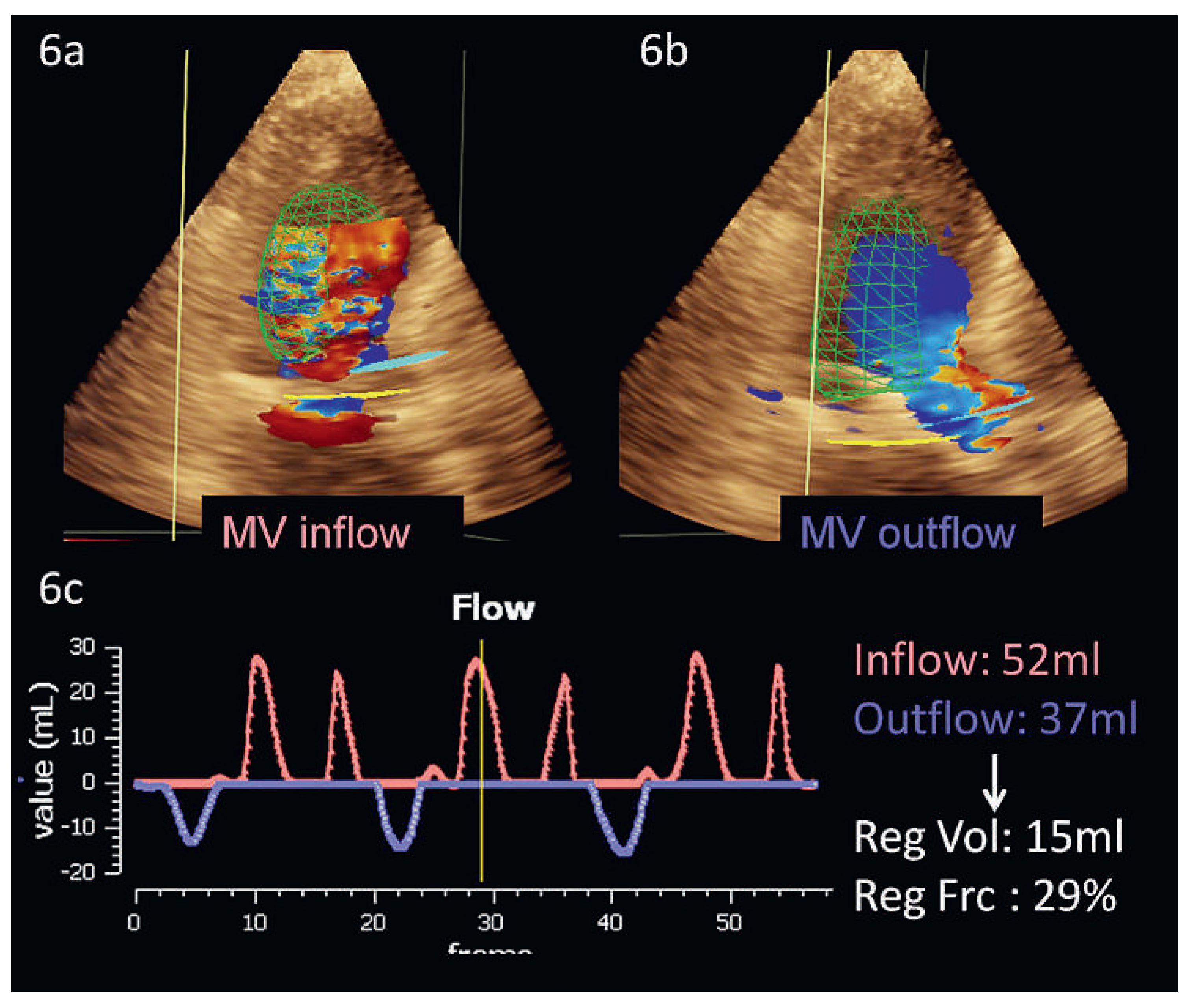
Abbildung 6.
Überschrift: 3D-Color-Flow-Doppler: Quantifizierung der Mitralinsuffizienz. Abb. 6a und 6b: Messung des Mitralklappen Einstromvolumens (6a) und LVOT Ausflussvolumens (6b) mittels 3D-Color-Flow-Doppler Quantifizierung. In Abb. 6c sind Inflow und Outflow über die Zeit aufgetragen, aus der Differenz rechnet sich das Regurgitationsvolumen sowie die Regurgitationsfraktion. Abkürzungen: MV = Mitralklappe, RegVol = Regurgitationsvolumen, RegFrc = Regurgitationsfraktion.
Computertomographie
Eine Mitralklappenuntersuchung mittels Computertomographie (CT) wird aufgrund der limitierten zeitlichen Auflösung und der fehlenden Möglichkeit der Flussdarstellung sowie der damit verbundenen Strahlenbelastung nur in Ausnahmefällen empfohlen. Prinzipiell ist aber eine Quantifizierungen der Mitralinsuffizienz mittels Messung der Regurgitationsfläche [
25] sowie des Regurgitationsvolumens [
26] möglich. Die CT kann insbesondere von Vorteil sein bei Patienten mit starken anulären oder valvulären Verkalkungen und bei schwerem Übergewicht [
27]. Limitierend ist die Strahlenbelastung für die Patienten, v.a. bei Protokollen, die eine Aufnahme des gesamten Herzzyklus und somit eine umfassende Evaluation der Mitralklappenbewegung erlauben. Neue strahlenarme Aufnahmeprotokolle erlauben nur statische Bilder, die meist in der Diastole, dem Zeitpunkt der geringsten Koronarbewegung erfolgen und somit die Mitralklappe nur in geöffnetem Funktionszustand zeigen [
28,
29].
Magnetresonanztomographie
Die kardiale Magnetresonanztomographie (CMR) erlaubt eine exzellente Darstellung der Herzanatomie in allen Ebenen, was eine Visualisierung der Herzklappen mit deren Einund Ausflussgebiet ermöglicht. Zudem stellt sie den Goldstandard in der Quantifizierung der ventrikulären Volumina und Masse dar, welche für die Beurteilung von Klappenerkrankungen bei funktioneller Mitralinsuffizienz essentiell ist. Die Quantifizierung der Mitraklappenregurgitation mittels CMR kann sowohl indirekt als auch direkt mittels Bestimmung der ROA erfolgen. Die CMR stellt mittlerweile die bevorzugte Alternative zur Echokardiographie dar; die Anwendung im klinischen Alltag ist jedoch vor allem durch deren Verfügbarkeit, die relativ hohen Kosten und die für die Beurteilung notwendige Erfahrung limitiert [
30]. Vorteile der CMR sind v.a. die hohe örtliche Auflösung und die Möglichkeit der direkten Flussquantifizierung; Nachteile sind die gegenüber der 2D Echokardiographie geringere zeitliche Auflösung und die nach wie vor notwendigen Atempausen. Letztere verlangen eine gute Kooperation vom Patienten und können bei sprachlichen Barrieren oder bei Dyspnoe ein Hindernis darstellen [
31].
Indirekte Quantifizierung
Die indirekte Bestimmung des Regurgitationsvolumens erfolgt mittels Subtraktion des Vorwärtsschlagvolumens, welches über der Aorta ascendens mittels «Phase Contrast Velocity Encoding» (VENC) bestimmt wird, vom aus den anatomischen Kurzachsenschnitten berechneten totalen Schlagvolumen. Analog der Echokardiographie ist diese Methode nur in Abwesenheit einer relevanten Aorteninsuffizienz oder eines Ventrikelseptumdefektes korrekt. Nachteilig wirkt sich aus, dass durch den Einsatz von zwei verschiedenen CMR-Techniken die Fehlerwahrscheinlichkeit steigt – eine fehlerhafte Messung bei einer der beiden Techniken verändert bereits das Resultat [
32]. Hinzu kommt, dass diese Methode (theoretisch) die Mitralinsuffizienz gegenüber der Echokardiographie überschätzt, da das Vorwärtsschlagvolumen in der Aorta ascendens um den Koronarfluss kleiner ausfällt als das im LVOT gemessene Schlagvolumen der Echokardiographie. Im praktischen Alltag ist dieser Unterschied aber vernachlässigbar klein.
Direkte Quantifizierung – Phasenkontrast-Flussmessung
Die direkte Messung des Regurgitationsvolumens erfolgt mittels VENC direkt oberhalb der Mitraklappenebene. Aufgrund der Verschiebung der Klappenebene während des Herzzyklus und der oft exzentrischen Jets ist eine akkurate Bestimmung des Flusses oft schwierig. Zudem können durch die hohen Flussgeschwindigkeiten von bis zu 6m/s Artefakte verursacht werden, welche eine genaue Quantifizierung weiter erschweren. Bei regelmässigem Herzrhythmus stimmen die Messwerte der direkten Methode mit denjenigen der indirekten Methode mit einer guten Genauigkeit überein; irreguläre Rhythmen wirken sich speziell auf Phasenkontrastmessungen aus [
33].
Planimetrie der ROA
Die Ausmessung der ROA mittels CMR ist möglich, wird jedoch selten routinemässig eingesetzt [
34]. Die mit CMR ausgemessene ROA zeigt gute Korrelation mit der Regurgitationsfraktion respektive dem Regurgitationsvolumen [
35].
Differentielle Bildgebung der Mitralinsuffizienz Allgemein
Die Wahl der bildgebenden Methode für die Darstellung der Mitralinsuffizienz richtet sich nach dem präferenziell darzustellenden Aspekt: dem anatomischen oder eher funktionellen, dem diagnostischen oder dem therapeutischen. Alle Methoden haben ihre Vorund Nachteile und müssen daher gezielt eingesetzt werden. Insgesamt wird die überwiegende Mehrzahl der Patienten mit Mitralinsuffizienz primär mittels Echokardiographie abgeklärt. CT- oder CMR-Untersuchungen werden nur bei speziellen Fragestellungen herbeigezogen, jedoch selten als primäre Abklärungsmodalität eingesetzt. Bei Unsicherheit empfiehlt sich die Rücksprache mit einem Spezialisten für kardiale Bildgebung, um unnötige Untersuchungen zu vermeiden.
Zu berücksichtigen gilt es auch die lokalen personellen und finanziellen Ressourcen. Fortgeschrittene 3D-Echokardiographie-, kardiale CTund MR-Bildgebung verlangen vertiefte Weiterbildung, um genügend Expertise zu erlangen. Und während ein Ultraschallgerät mobil in jedes Patientenzimmer gefahren warden kann, sind die Standorte mit CMR limitiert. Schlussendlich müssen auch finanzielle Überlegungen einfliessen: eine Standard-Echokardiographie kostet rund 360 Sfr., eine CMR 800 Sfr. (mit Kontrastmittel zirka 1000 Sfr.) und ein CT bis zu 1000 Sfr.
Abbildung 7.
Mitralklappen-Prolaps (Segment P2) im Vergleich zwischen Echokardiographie und kardialer Magnetresonanztomographie. Abb. 7a: P2–Prolaps (roter Pfeil) mit Flail bei schwerer Mitralinsuffizienz im TEE. Abb. 7b: Schwere Regurgitation mittels Farb-Doppler. Abb. 7c: Im CMR erkennbarer Prolaps des P2–Segments (roter Pfeil) bei leichter Mitralinsuffizienz (Patient nicht identisch). Abkürzungen: CMR = kardiale Magnetresonanztomographie, MI = Mitralinsuffizienz, TEE = Transesophageales Echo.
Organische Mitralinsuffizienz
Aufgrund der hohen zeitlichen
und räumlichen Auflösung ist die 2D-Echokardiographie das primäre diagnostische Instrument für den Mitralklappenprolaps (Abbildung 7a). Die 2D-Echokardiographie erlaubt es, auch abgerissene Sehnenfäden (Millimeterbereich) zu verfolgen, die während eines Herzschlages mehrfach hin und her schlagen, oder die Details der verschiedenen prolabierenden Segmente zu erkennen [36, 37]. Keine andere Untersuchungstechnik erlaubt solche Präzision am schlagenden Herzen. Gleichzeitig kann mittels der Farb-Doppler-Echokardiographie auch die Funktion beurteilt werden (Abbildung 7b). Insbesondere qualitative Aspekte werden dabei abgedeckt: Anzahl, Richtung und zeitliches Auftreten der verschiedenen Insuffizienzjets. Hingegen kann die Quantifizierung der Mitralinsuffizienz aufgrund der oben beschriebenen technischen Limitation (insbesondere durch unzutreffende geometrische Annahmen) anspruchsvoll oder unmöglich sein. Die Quantifizierung der Mitralinsuffizienz wird in Zukunft wohl in die Domäne der 3D-Echokardiographie fallen. Ein zusätzlicher Vorteil der 3D transesophagealen Echokardiographie (3D-TEE) liegt darin, dass die komplexe anatomische Struktur der Mitralklappe gut verständlich demonstriert werden kann. Die Übereinstimmung mit dem chirurgischen Befund ist dabei selbst bei komplexen Klappenveränderungen verblüffend [
37]. Besonders bei der Quantifizierung der mit Morbus Barlow assoziierten multiplen Jets ist die 3D- der 2D-Technik überlegen [
38]. Die 3D-TEE geschieht in Echtzeit (‚real-time’) und kann bei Geräten der neusten Generation auch mit dem Farb-Doppler-Modus ergänzt werden, was wiederum für die Identifikation von sehr exzentrischen Jets ein Vorteil sein kann. 3D-TEE zeigt gegenüber 2D eine höhere Sensitivität der einzelnen Segmente beim MKP sowie bei Chordaeruptur [
37]. Die grosse Datenmenge, welche gleichzeitig vom Ultraschallgerät verarbeitet werden muss, bringt allerdings gerade bei der 3D transthorakalen Echokardiographie (3D-TTE) eine deutliche Einbusse der räumlichen Auflösung mit sich. Dies führt zu einer schlechteren Sensitivität für die Erkennung von prolabierenden Segmenten und Chordarupturen gegenüber der 3D-TEE [
39], so dass die 3D-TTE im Alltag oft enttäuschend wenig anatomische Zusatzinformation bringt.
Einer der grössten Vorteile der TEE liegt aber zweifelsohne darin, dass sie nicht nur zur Basisdiagnostik, sondern auch perioperativ zur Beurteilung des Resultates einer Mitralklappenoperation oder-intervention eingesetzt werden kann. Im Falle der perkutanen Mitralklappenrekonstruktion mittels MitraClip ist sie sogar unabdingbare Voraussetzung für eine erfolgreiche Behandlung (Abbildung 8).
Auch die CMR kann für diagnostische Zwecke bei organischer Mitralinsuffizienz genutzt werden. Mittels direkter Visualisierung der ROA lässt sich prinzipiell eine funktionelle von einer organischen Ursache abgrenzen [
35]. Die CMR hat eine mit Echokardiographie vergleichbare diagnostische Aussagekraft bei der Identifikation der verschiedenen Segmente eines Prolaps (Abbildung 7c). Bei der Darstellung von Sehnenfädenabrissen ist sie aufgrund schlechterer zeitlicher Auflösung der Echokardiographie unterlegen; überdies fehlt im Gegensatz zur Echokardiographie die Möglichkeit, die Schnittebene während der Untersuchung zu wechseln. Indiziert ist die CMR vor allem bei schlechter Ultraschallqualität [
30].
Abbildung 8.
MitraClip: Erfolgskontrolle mittels 3D-TEE. Abb. 8a und d: 3D-Darstellung der Mitralklappe, von anterior gesehen. 8b und 8c entsprechen den Schnittebenen in 8a. 8e und 8f entsprechen den Schnittebenen in 8d. Die posteriore Ablösung des medialen MitraClips ist gut erkennbar (weisser Stern, 8b und c), wohingegen der laterale Clip gut an beiden Segeln verankert ist (8f, weisse Pfeile).
Die CT spielt in der Diagnostik und Therapie des MKP eine untergeordnete Rolle. In geübten Händen weist die CT zur Diagnose eines MKP eine mit der Echokardiographie vergleichbare Sensitivität und Spezifität (93% bzw. 96%) auf. Die Sensitivität zur Identifikation einzelner prolabierender Segmente (Scallops) ist durch die tiefere zeitliche Auflösung aber deutlich niedriger (ca. 69%, Spez.: 95%). Ein Vorteil der CT ist allerdings die Darstellung von verkalkten Strukturen [
40].
Funktionelle Mitralinsuffizienz
Nicht-ischämische Kardiomyopathie
Das bevorzugte bildgebende Verfahren zur Diagnose einer durch eine KMP verursachte Mitralinsuffizienz ist die Echokardiographie [
41]. Die Grösse des linken Ventrikels sowie die exakte Störung seiner Geometrie können dargestellt werden (Abbildung 9a). Ebenfalls erkennbar ist das Ausmass der Anulusdilatation und des Tetherings der Sehnenfäden. Zudem können die «tenting height» und «tenting area» gemessen werden, welche für die chirurgische Sanierung eine prognostische Aussagekraft besitzen [
42]. Die Quantifizierung der Mitralinsuffizienz birgt die eingangs erwähnten Schwierigkeiten. Gewisse Formen der KMP ergeben ein typisches, in der Echokardiographie gut zu erkennendes Myokardmuster ab (z.B. Amyloidose).
Für echokardiographisch unerklärte Formen kann in einigen Fällen die CMR weiterhelfen, welche eine zusätzliche Gewebecharakterisierung erlaubt [
43]. Die CMR bietet insbesondere die Möglichkeit, die myokardiale Ursache der Mitralinsuffizienz weiter zu evaluieren: Myokardiale Ödeme, kleinere und grössere Fibroseherde sowie das Ausmass von Narben. In der Regel ist zudem die räumliche Auflösung exzellent und er-möglicht insbesondere bei schlechter Schallqualität die Quantifizierung der Ventrikeldimensionen (Abbildung 9b), der LV-Funktion sowie des Schweregrades [
44], der‚ tenting area’ und der Anulusfläche [
35].
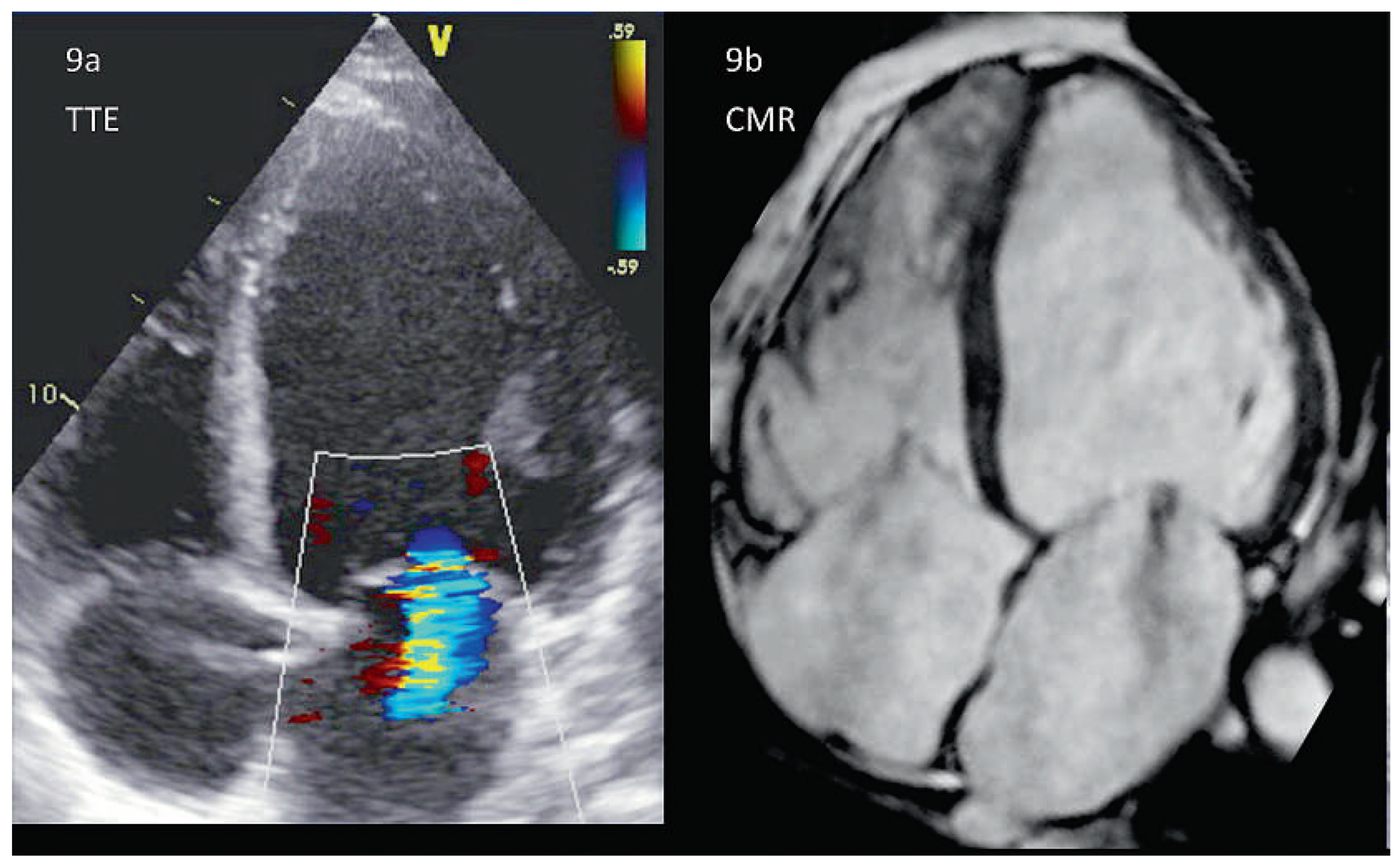
Abbildung 9.
Leichte funktionelle Mitralinsuffizienz, Vergleich CMR und TTE am selben Patienten. Die 4–Chamber-View erlaubt die Beurteilung der Ventrikelgeometrie, der Myokardtextur sowie des Schweregrades der Mitralinsuffizienz (hier leicht). Echokardiographisch (9a) ist insbesondere die apikale und laterale Endokardgrenze schwer zu erkennen, was bei der CMR (9b) optimal zur Darstellung kommt. In der CMR kann auch bei einem dilatierten Herz gleichzeitig der rechte Ventrikel (und die hier leichte Trikuspidalinsuffizienz) gezeigt werden. Bei der Echokardiographie muss dies meist in mehreren Schritten geschehen, um eine genügende zeitliche Auflösung zu bewahren.
Die Computertomografie ist auch bei der funktionellen Mitralinsuffizienz keine Primärmodalität, erlaubt jedoch die Beurteilung der Koronararterien zum Ausschluss einer Koronarerkrankung als Ursache von kardialen Beschwerden und linksventrikulärer Dysfunktion [
43].
Hypertrophe obstruktive Kardiomyopathie (HOCM)
Die 2D TTE stellt die Methode der Wahl dar zur Diagnose oder zum Ausschluss einer HOCM. Nicht nur können die Wanddicken meist sehr adäquat gemessen werden, vielmehr ermöglicht die M-Mode-Echokardiographie die Darstellung des SAM aufgrund seiner ungeschlagenen zeitlichen Auflösung. Zur Diagnose einer relevanten Obstruktion bei HOCM eignet sich der CW-Doppler zur Bestimmung des Druckgradienten über dem linksventrikulären Ausflusstrakt (LVOT) oder innerhalb des Ventrikels. Besonders interessant und diagnostisch wichtig sind dabei die hämodynamischen Veränderungen (s. Abbildung 10a und b) während gewissen Manövern wie Valsalva oder dem sogenannten‚ leg-rise’-Test. Die Echokardiographie ist auch bei der Therapie der HOCM essenziell: Bei der septalen Alkoholablation kann sie durch Injektion von Kontrastmittel in den entsprechenden Koronarast das zu abladierende Gebiet darstellen und unmittelbar nach Ablation den Therapie-Erfolg messen (Rückgang der LVOT-Obstruktion und damit Rückgang der Mitralinsuffizienz). Bei der chirurgischen Resektion erlaubt die perioperative TEE die exakte septale Ausmessung vor der Myektomie.
Die CMR gehört zur Routinediagnostik bei HOCM. Dies allerdings weniger, um die Mitralinsuffizienz zu quantifizieren, als vielmehr das exakte Ausmass und die Lokalisation der Hypertrophie aufzuzeigen. Zudem kann der Fibroseanteil am Myokardgewebe mittels Late-Gadolinium-Enhancement (LGE) quantifiziert werden. [
45]. Die Cine-CHR erlaubt eine hervorragende Beurteilung der SAM (s. Abbildung 10c) [
12,
44]. Zusätzlich kann die Phasenkontrast-Flussmessung den Druckgradienten über dem LVOT und damit den Schweregrad der Obstruktion beurteilen; diese ist jedoch aufgrund der zeitlichen Auflösung der Echokardiographie unterlegen.
Mit der CT lässt sich ein SAM zwar erkennen, da die CT aber nicht in der Lage ist, den Druckgradienten zu messen, lassen sich daraus nur bedingt Schlüsse über den Schweregrad und folglich den Therapiebedarf ziehen [
12]. Eine alternative Anwendung ergibt sich für das CT als intraprozedurale Kontrolle bei der septalen Alkoholablation, falls die Echokardiographie aus anatomischen Gründen nicht optimal verwendet werden kann [
46].
Ischämische Kardiomyopathie
Analog zu den nicht ischämischen KMP ist die Echokardiographie bei einer ischämischen Mitralinsuffizienz die primäre diagnostische Methode der Wahl. Dabei dient sie der Messung der Ventrikelgrösse und-funktion und der Diagnose von regionalen Wandmoti-litätsstörungen sowie von Herzwand-Aneurysmen. Die Erkennung dieser Veränderungen ist äusserst wichtig, weil sie via Geometriestörung den eigentlichen Mechanismus für die Mitralinsuffizienz darstellen. Die Diagnose einer fortwährenden Ischämie, z.B. mittels StressEchokardiographie, ist Voraussetzung für die gezielte Therapie (in diesem Falle primär die Revaskularisation). Die zweiund dreidimensionale Echokardiographie erkennt auch die Ischämie-bedingten Komplikationen wie eine ischämische Septumruptur oder den Papillarmuskelabriss, welcher plötzlich zum akuten Lungenödem führen kann. Differentialdiagnostisch erlaubt die 2D- oder 3D-TEE die Darstellung der Papillarmuskeln, welche bei einer ischämischen KMP asymmetrisch verschoben sind und häufig einen exzentrischen Jet zur Folge haben [
15].
Abbildung 10.
Überschrift: Hypertrophe obstruktive Kardiomyopathie, Vergleich TEE und CMR. Abb. 10a: Im TEE dargestellte systolic anterior motion des anterioren Mitralklappensegels (SAM). Mittels Farb-Doppler kann die Flussbeschleunigung im LVOT infolge SAM sowie der typische, nach posterior gerichtete Jet visualisiert werden (Abb. 10b). Abb. 10c: Neben dem SAM ist insbesondere das Ausmass der Hypertrophie erkennbar (Patient nicht identisch). Abkürzungen: Ao = Aorta, AML = anteriores Mitralsegel, PML = posteriores Mitralsegel, SAM = Systolic Anterior Motion.
Mit LGE-CMR lässt sich das Ausmass der Vernarbung im LV sowie die LV Volumina quantifizieren [
30,
47], welche indirekte Rückschlüsse auf eine funktionelle Mitralinsuffizienz zulassen.
Die CT bleibt im Alltag bei Mitralinsuffizienz im Rahmen einer ischämischen KMP die Modalität dritter Wahl. Meist kommt die CT zusammen mit der Abklärung der Koronaranatomie zum Einsatz [
48].
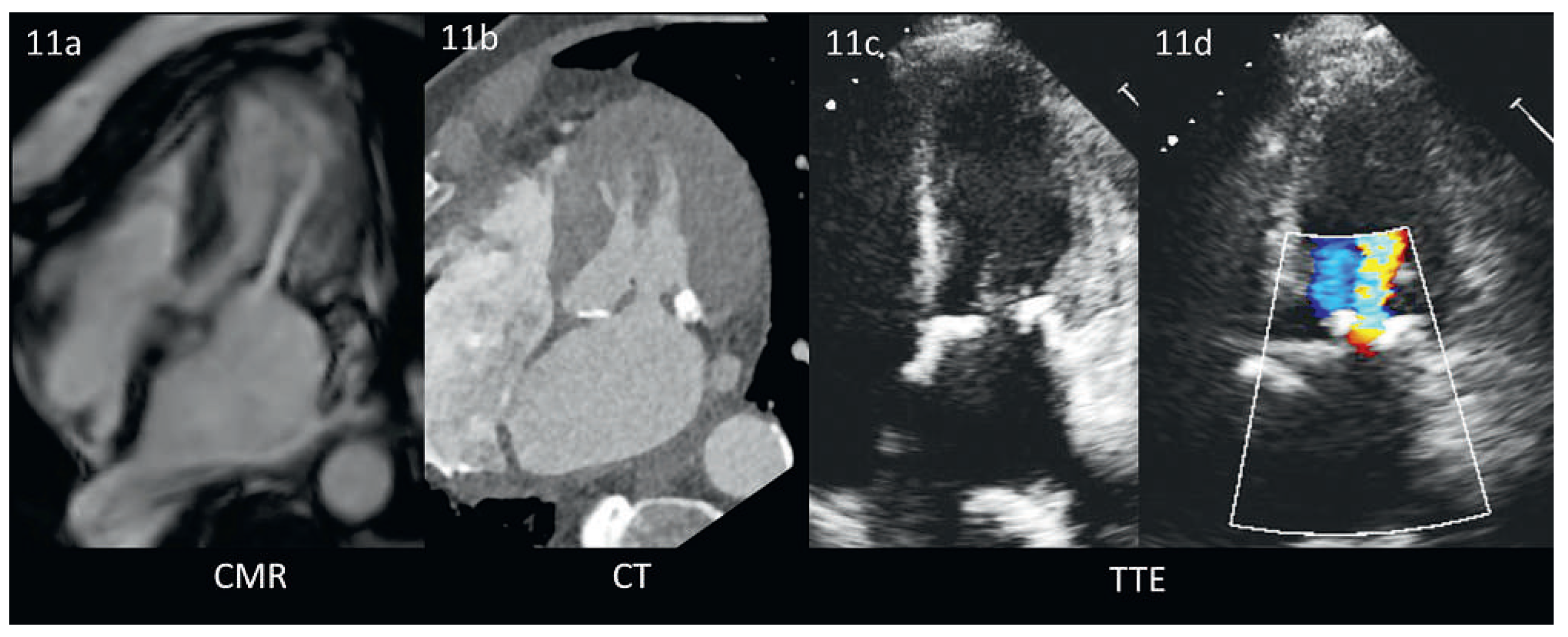
Abbildung 11.
Mitralklappenstenose bei verkalkten Segeln im CMR, CT und TTE (identischer Patient). 11a: Die CMR ist zur Darstellung von Verkalkungen wenig geeignet, zeigt aber ähnlich wie die Farb-Doppler Echokardiographie (11d) die Flussbeschleunigung über der stenotischen Klappe gut. Abb. 11b: Im CT lassen sich die verkalkten Strukturen präzise vom umliegenden Gewebe abgrenzen. Abb. 11c/d: Diese Verkalkungen sind auch in der Echokardiographie gut darstellbar. Die gleiche Darstellung ist aber selbst beim identischen Patienten eine Herausforderung.
Abbildung 12.
Papilläres Fibroelastom, Vergleich CT, 2D-TEE, 3D-TEE (gleiche Patientin). 12a: das Fibrolastom lässt sich im CT wegen ungenügender zeitlicher Auflösung nicht darstellen. 12b und c: kleines Fibroelastom (histologisch gesichert) am posterioren Mitralklappensegel (roter Stern). Abkürzungen: PML, posteriores Mitralklappensegel; Ao, Aortenwurzel.
Gemischte Mitralinsuffizienz
Wenn ausgeprägte Verkalkungen das Bild der gemischten Mitralinsuffizienz beherrschen, so kann die Qualität der transthorakalen Echokardiographie massgebend beeinträchtigt sein. Das kann bis hin zur völligen Verkennung einer Mitralinsuffizienz im Schallschatten des verkalkten Anulus reichen. Hier ist bei klinischer Symptomatik eine transesophageale Echokardiographie, ggf. auch eine CM- Roder CT-Untersuchung zu erwägen (Abbildung 11). Speziell die CT ist eine hervorragende Modalität zur Darstellung der Verkalkungen, die exakte Lokalisation der Verkalkung hilft bei der operativen Planung (Resektion bzw. Rekonstruktion des Anulus) [
49]. Bei weniger ausgeprägten Veränderungen hingegen stellt die TTE oder TEE das subtile Zusammenspiel zwischen Kalk, Fibrose, Segelverdickung und -beweglichkeit oft sehr genau dar – eine notwendige Voraussetzung, um die exakte Regurgitationsursache zu definieren und eine geeignete Therapie abzuleiten. Die geringe zeitliche Auflösung limitiert hier insbesondere den Einsatz der CT. Die vor allem diastolisch aufgenommenen Bilder können dazu führen, dass ein Mitralklappenprolaps übersehen wird. Infolge der geringen zeitlichen Auflösung können zudem Verdickungen oder Tumore als Ursache einer Mitralinsuffizienz in der CT verpasst werden (Abbildung 12).


