Next Article in Journal
Hypertensive Herzkrankheit – zur Diagnoseist der Nachweis Einer Linksventrikulären Hypertrophie Nicht Nötig
Previous Article in Journal
Arterial Hypertension in Elderly Patients
 
 
Cardiovascular Medicine is published by MDPI from Volume 28 Issue 1 (2025). Previous articles were published by another publisher in Open Access under a CC-BY (or CC-BY-NC-ND) licence, and they are hosted by MDPI on mdpi.com as a courtesy and upon agreement with Editores Medicorum Helveticorum (EMH).
Interesting Images

Lost in Space - Left Lateral Cardiac Displacement Due to an Unusual Cause
by
Niels Holm
1,*,
Esther Bächli
2 and
Tobias Hoefflinghaus
3
1
Universitätsspital Zürich, Zürich, Switzerland
2
Spital Uster, Zürich, Switzerland
3
Stadtspital Waid, Zürich, Switzerland
*
Author to whom correspondence should be addressed.
Cardiovasc. Med. 2014, 17(2), 55; https://doi.org/10.4414/cvm.2014.00223
Submission received: 19 November 2013 / Revised: 19 December 2013 / Accepted: 19 January 2014 / Published: 19 February 2014
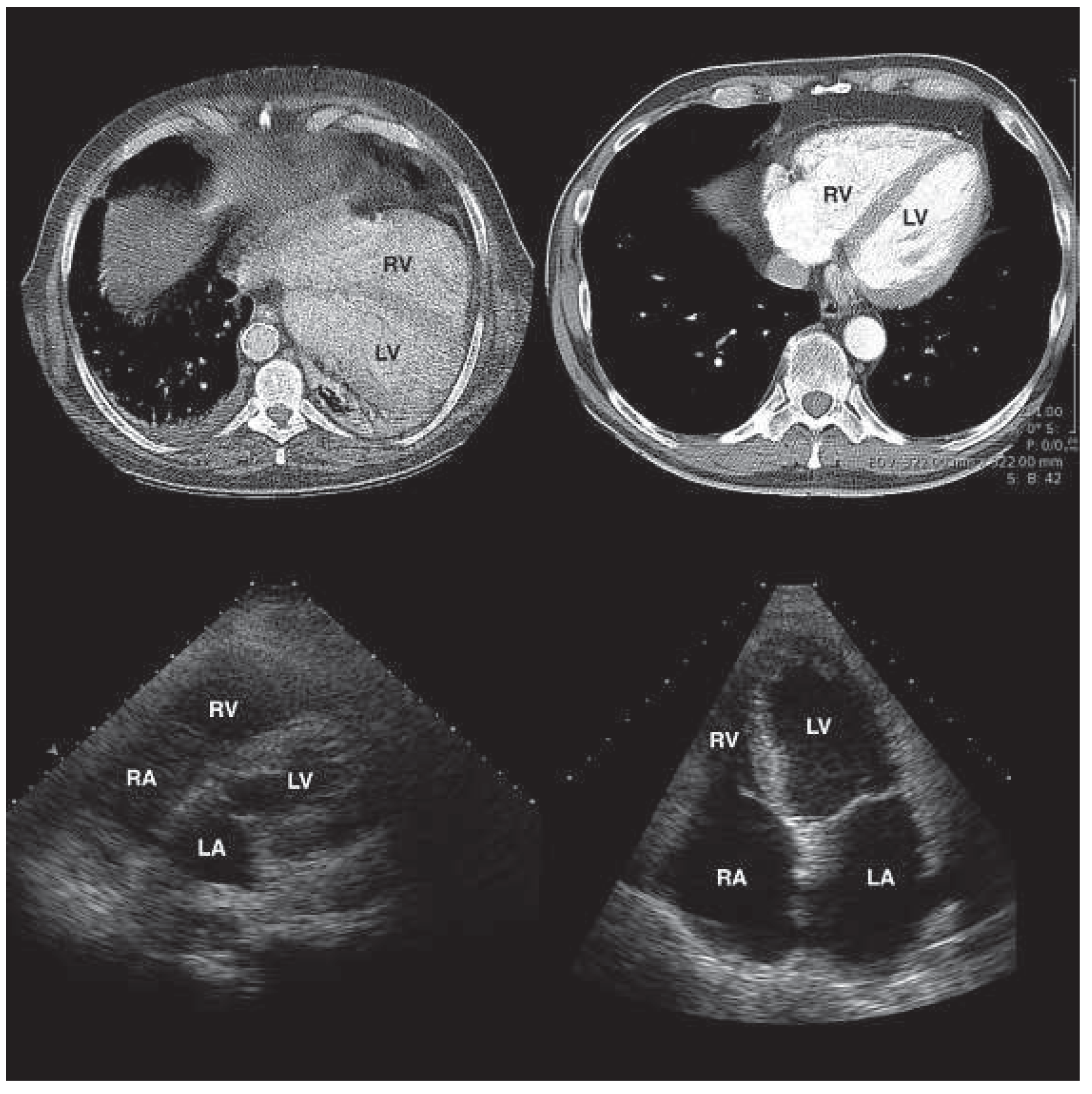
We present a case of an incidental finding of congenital absent pericardium (CAP). A 69–year-old patient was admitted to the intensive care unit because of septic shock of unknown cause. The patient’s history was remarkable for alcoholic liver disease with cirrhosis and lung emphysema. The chest X-ray (CXR) revealed an enlarged and displaced cardiac silhouette of atypical shape without signs of congestive heart failure. Electrocardiogram (ECG) showed right axis deviation, leftward displaced transition zone in the precordial leads and significant changes of QRS axis and R wave amplitude between admission and follow-up. Echocardiographic images were hard to obtain and atypical lateral views were required. Computed tomography of the chest revealed cardiac displacement into the left thoracic cavity. Congenital absent pericardium (CAP) was assumed. Shortly after, the patient died of lung failure. Autopsy confirmed pneumonia as cause of death and incidentally revealed a complete isolated absence of the pericardium and a displaced heart to the left.
CAP was first described by Realdus Colombus (1559) [1]. The aetiology of this anomaly is an incomplete development of the transversal septum and the pleuropericardial folding in the early embryonic stage [2]. The prevalence is unknown; various authors have described isolated forms in surgical or pathological series of 0.0001% to 0.044%. One third of all cases were associated with other cardiac lesions, like atrial septal defect, bicuspid aortic valve and tetralogy of Fallot. Patients can present asymptomatic or with atypical chest pain, largely nonexertional and with postural dependency. Partial defects can lead to cardiac herniation with shock or sudden death. Clinical examination may show lateral displacement of the heart. ECG often displays a partial right bundle block pattern and a deviated transition zone to the left. CXR can show a levoshift with loss of the right heart border. Often lung tissue is interposed between main pulmonary artery and aorta. Although echocardiographic examination requires unusual views and is difficult to interpret because of cardiac hypermobility leading to abnormal swinging and ventricular septal motion, the change of the position of the heart from left side to supine position is a key finding for this diagnosis by echocardiography. CXR combined with magnetic resonance imaging are key to establish the diagnosis. In patients with debilitating symptoms pericardial reconstruction is a curative option [3].
Figure 1. Cardiac displacement to the left (above left) compared with normal cardiac position (above right). Unusual echocardiographic findings (below left) with the transducer held in usual left lateral position for apical four-chamber view compared with a normal echocardiographic orientation (below right) [4]. LA left atrium, LV left ventricle, RA right atrium, RV right ventricle.
Cardiovascmed 17 00055 g001

Funding / potential competing interests

No financial support and no other potential conflict of interest relevant to this article were reported.

References

  1. Columbus, M.R. De re anatomica; Beurlaque, N., Ed.; Venice, 1559; Vol 15, pp. 265–269. [Google Scholar]
  2. Garnier, F.; Eicher, J.C.; Philip, J.L.; Lalande, A.; Bieber, H.; Voute, M.F.; et al. Congenital complete absence of the left pericardium: a rare cause of chest pain or pseudo-right heart overload. Clin Cardiol. 2010, 33, E52–E57. [Google Scholar] [CrossRef] [PubMed]
  3. Gatzoulis, M.A.; Munk, M.D.; Merchant, N.; Van Arsdell, G.S.; McCrindle, B.W.; Webb, G.D. Isolated congenital absence of the percardium: clinical presentation, diagnosis, and management. Ann Thorac Surg. 2000, 69, 1209–1215. [Google Scholar] [CrossRef] [PubMed]
  4. Connolly, H.M.; Click, R.L.; Schattenberg, T.T.; Seward, J.B.; Tajik, A.J. Congenital absence of the pericardium: echocardiography as a diagnostic tool. J Am Soc Echocardiogr. 1995, 8, 87–92. [Google Scholar] [CrossRef]