Next Article in Journal
Almanac 2013: Cardiac Arrhythmias and Pacing—An Editorial Overview of Selected Research That Has Driven Recent Advances in Clinical Cardiology
Previous Article in Journal
Wann Ist Bei Hypertonie-Patienten Eine Echokardiographie Nötig?
 
 
Cardiovascular Medicine is published by MDPI from Volume 28 Issue 1 (2025). Previous articles were published by another publisher in Open Access under a CC-BY (or CC-BY-NC-ND) licence, and they are hosted by MDPI on mdpi.com as a courtesy and upon agreement with Editores Medicorum Helveticorum (EMH).
Case Report

Mechanical Thrombectomy After Embolic Internal Carotid Artery Occlusion in Acute Stroke

by
Christophe A. Wyss
1,
Anahita Dastoor
2 and
Roberto Corti
1,*
1
Herzklinik Hirslanden, Witellikerstrasse 40, CH-8032 Zürich, Switzerland
2
Spital Glarus, Glarus, Switzerland
*
Author to whom correspondence should be addressed.
Cardiovasc. Med. 2014, 17(2), 51; https://doi.org/10.4414/cvm.2014.00204
Submission received: 19 November 2013 / Revised: 19 December 2013 / Accepted: 19 January 2014 / Published: 19 February 2014

Case presentation

A 34-year old woman presented to the emergency department with a wake-up stroke, after onset of a sensomotoric paresis of the right arm and motoric aphasia. Her National Institutes of Health Stroke Scale was 12. She was haemodynamically stable with a blood pressure of 100/60 mm Hg and a heart rate of 90/min.
The initial CT scan showed an occlusion of the left internal carotid artery (ICA) with a delayed perfusion (elevated time-to-peak) in the left-side, middle cerebral artery (MCA) and anterior cerebral artery (ACA) areas. The MR scan showed an extensive diffusion-weighted MR (DWI) hyperintense lesion in one-third to one-half of the MCA territory (Figure 1). T2-weighted images could not document any flow void in distal cervical and intracranial segments of the left ICA.
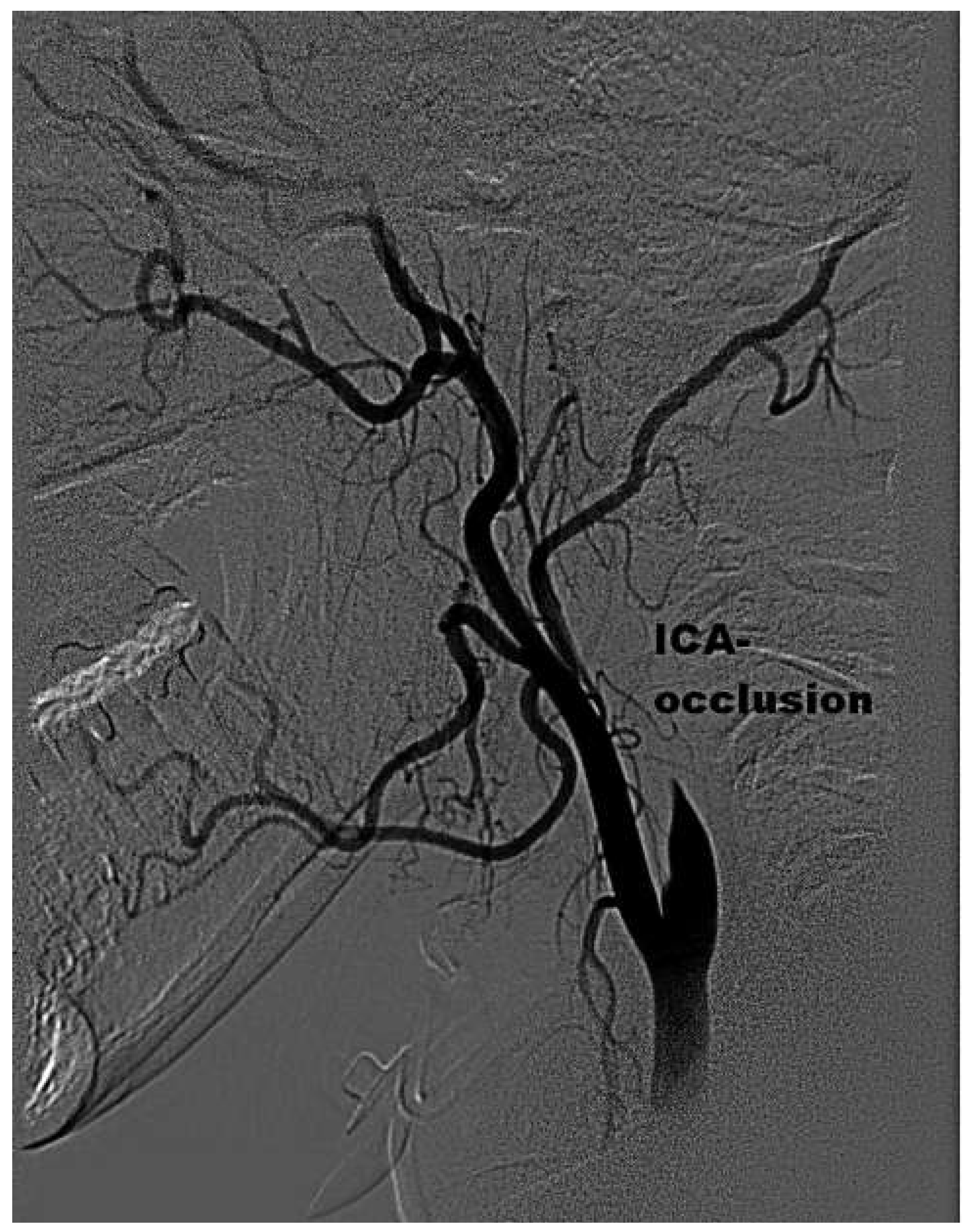
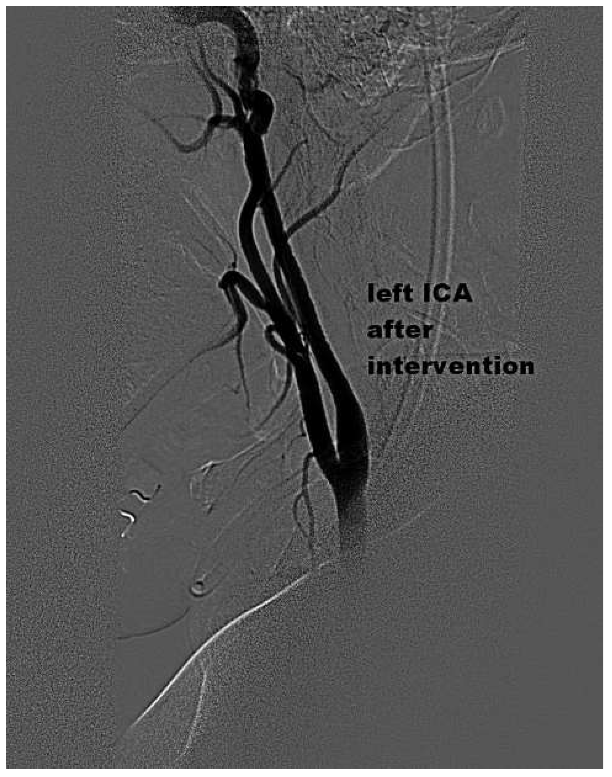
The time of onset of the stroke was uncertain, with an estimated delay of seven hours. As the patient was not eligible for fibrinolytic therapy anymore, she was given intravenous acetylsalicylic acid (ASA) and unfractioned heparin (UFH) and brought to the catheterisation laboratory. Angiograms confirmed a thrombotic occlusion of the left ICA just after the bifurcation (Figure 2). There was only moderate collateral filling in contralateral injections. Because of concern about haemorrhagic transformation of the extensive area at risk, neither rtPA nor glycoprotein IIb/IIIa-antagonist were administered. Through a femoral 5F-access, a coronary Judkins R4 catheter (Cordis Corporation, Bridgewater, NJ, USA) was placed in the left external carotid artery over a MagicTorque 0.035-guidewire (Boston Scientific, Miami, FL, USA). Using standard technique, an 8F shuttle sheath (Cook Medical Inc., Bloomington, IN, USA) was placed in the common carotid artery and the occlusion of the ICA was passed with a hydrophilic coronary wire (Whisper MS coronary 0.014 guidewire, Abbott, IL, USA). We tried to place a distal embolic protection system (Emboshield, Abbott, IL, USA), but the Emboshield Wire failed to cross the lesion. After sev-eral passages of manual thrombus aspiration using a 7F Export-catheter (Medtronic, Santa Rosa, CA, USA), a noticeable amount of thrombus was aspirated. Finally, complete flow restoration could be documented in the ICA and MCA without angiographic signs of distal embolisation (Figure 3, Figure 4 and Figure 5). Neither a stenosis nor a dissection of the ICA could be documented at the point of former occlusion. The patient showed an impressively rapid recovery and started to talk a few minutes after flow restoration. Therapeutic anticoagulation with intravenous UFH was initiated. Prolonged cardiac rhythm monitoring documented no arrhythmias, we found no coagulopathies, the patient had no traditional cardiovascular risk factors. Transoesophageal echocardiography showed a tunnel-shaped patent foramen ovale (PFO) with a spontaneous right-left-shunt and an interatrial septum aneurysm. The aetiology of the stroke was considered as paradoxical embolism trough a PFO and the patient underwent successful percutaneous PFO-closure with an Amplatzer 25-mm PFO Occluder (AGA Medical, Minnesota, USA) 4 days after the stroke.
The patient recovered clinically within one week with complete restoration of the motoric function of the right arm (with slight sensoric deficits) and with moderate residual neuropsychological deficits (verbal flow, cognitive flexibility).

Discussion

The natural history of cerebral infarctions of the MCA territory with acute occlusion of the ICA is of poor outcome, with estimates of 16% to 55% mortality and 40% to 69% significant morbidity [1]. Acute carotid occlusions usually contain large clot volume [2], therefore an advantage of clot removal therapy is the potential ability to retrieve some large or partially organised firm clots that can rarely be disrupted by pharmacologic thrombolysis alone. Furthermore, lysis of a large clot burden entails prolonged rtPA infusion time and – depending on the quality of contralateral flow – delays the rtPA action of possible concommittant arterio-arterial embolic intracerebral occlusions. Therefore, immediate restoration of flow in the ICA by a mechanical intervention seems promising. In this case, an impressively rapid recovery of the motoric aphasia was achieved. Thus, we proposed the thrombotic occlusion of the ICA responsible for the hyperintensive lesion in the MRI. More common and reasonable is an embolisation in a peripheral artery (middle cerebral artery or even more peripheral) with the occluded internal carotid artery as the source of the embolus. The neurological recovery could also be due to a spontaneous lysis of the peripheral embolisation in combination with heparin and aspirin. Considering this hypothesis, the intervention at the internal carotid artery would be as a secondary prevention and not therapeutic.
In analogy to the treatment of acute coronary syndromes, early reperfusion therapy in the setting of acute thromboembolic stroke should consider mechanical thrombus removal strategies. These approaches which require well-trained personel are likely to be more effective than thrombolysis on selected patients and deserve further evaluation in clinical trial settings [3].
Several devices like aspiration through balloon guiding catheters and snares have been described for clot removal in the ICA in small series [4]. Mechanical rheolytic thrombectomy using dedicated devices has been reported [5]. There are promising results of treatment with newer devices with or without previous adjunctive therapy (intra-arterial tissue plasminogen activator) [6]. Wake-up stroke still remains a challenge. Mechanical intervention to remove large clots in large vessel-occlusions in carefully selected patients with wake-up stroke seems promising, but still needs further evaluation in clinical trial settings [7,8].
We report on successful manual thrombus aspiration in an embolic occlusion of the ICA using the Export-catheter, which is well established in intracoronary thrombectomy in acute myocardial infarction. Although risks and benefits of clot removal therapy in the treatment of acute embolic stroke are still in evaluation [9], clot removal therapy by manual aspiration in selected patients seems feasible and effective in the treatment of patients with acute embolic occlusion of the ICA.
Despite occlusions of the ICA which may often occur in the proximal part of the ICA, only in a minority of cases is this type of procedure limited to the extracranial carotid and so the specialist carrying out the intervention should be ready to perform an intracranial procedure.
The most common causes of arterial occlusion involving the major cerebral arteries are emboli, most commonly arising from atherosclerotic arterial narrowing at the bifurcation of the common carotid artery, from cardiac sources, or from atheroma in the aortic arch and a combination of atherosclerotic stenosis and superimposed thrombosis [10]. Thus, in this young patient, there was no evidence for any of this type of atherosclerosis. Pronged cardiac rhythm monitoring documented no arrhythmias, we found no coagulopathies.
The patient had no traditional cardiovascular risk factors. All these points and the sudden manifestation give a strong suggestion to a cryptogenic stroke due to a paradoxical embolism. That is why further investigations were made.
Transoesophageal echocardiography showed a tunnel-shaped patent foramen ovale (PFO) with a spontaneous right-left-shunt and an interatrial septum aneurysm. According to the current management data at the time of the event, there was good evidence to perform a percutaneous PFO-closure. (The incident occured before Closure I [11], RESPECT [12] and PC [13] Trials were published).

Funding / potential competing interests

No financial support and no other potential conflict of interest relevant to this article were reported.

References

  1. Meyer, F.B.; Sundt, T.M., Jr.; Piepgras, D.G.; Sandok, B.A.; Forbes, G. Emergency carotid endarterectomy for patients with acute carotid occlusion and profound neurological deficits. Ann Surg. 1986, 203, 82–89. [Google Scholar] [CrossRef]
  2. Endo, S.; Kuwayama, N.; Hirashima, Y.; Akai, T.; Nishijima, M.; Takaku, A. Results of urgent thrombolysis in patients with major stroke and atherothrombotic occlusion of the cervical internal carotid artery. AJNR Am J Neuroradiol. 1998, 19, 1169–1175. [Google Scholar]
  3. Rebecca, M. Sugg, Marc D. Malkoff, Elizabeth A. Noser, Hashem M. Shaltoni, Raymond Weir et al. Endovascular Recanalization of Internal Carotid Artery Occlusion in Acute Ischemic Stroke. AJNR Am J Neuroradiol. 2005, 26, 2591–2594. [Google Scholar]
  4. Imai, K.; Mori, T.; Izumoto, H.; Takabatake, N.; Kunieda, T.; et al. Clot removal therapy by aspiration and extraction for acute embolic carotid occlusion. AJNR Am J Neuroradiol. 2006, 27, 1521–1527. [Google Scholar] [PubMed]
  5. Bellon, R.J.; Putman, C.M.; Budzik, R.F.; Pergolizzi, R.S.; Reinking, G.F.; Norbash, A.M. Rheolytic thrombectomy of the occluded internal carotid artery in the setting of acute ischemic stroke. AJNR Am J Neuroradiol. 2001, 22, 526–530. [Google Scholar] [PubMed]
  6. Wade S. Smith, Gene Sung, Jeffrey Saver, Ronald Budzik, Gary Duckwiler, David S. Liebeskind, et al. Mechanical Thrombectomy for Acute Ischemic Stroke. Final Results of the Multi MERCI Trial. Stroke. 2008, 39, 1205–1212.
  7. Natarajan, S.K.; Snyder, K.V.; Siddiqui, A.H.; Ionita, C.C.; Hopkins, L.N.; Levy, E.I. Safety and effectiveness of endovascular therapy after 8 hours of acute ischemic stroke onset and wake-up strokes. Stroke. 2009, 40, 3269–3274. [Google Scholar] [CrossRef] [PubMed]
  8. Kuruvilla, A.; Norris, G.M.; Xavier, A.R. Acute endovascular recanalization therapy in wake-up stroke. J Neurol Sci. 2011, 300, 148–150. [Google Scholar] [CrossRef] [PubMed]
  9. Ciccone, A.; Valvassori, L.; Nichelatti, M.; Sgoifo, A.; Ponzio, M.; et al. Endovascular treatment for acute ischemic stroke. N Engl J Med. 2013, 368, 904–913. [Google Scholar] [CrossRef] [PubMed]
  10. Draga Jichici. Alison Elizabeth Baird, Anterior Circulation Stroke; Medscape 21.6.2013.
  11. Furlan, A.J.; Reisman, M.; Massaro, J.; Mauri, L.; Adams, H.; Albers GWet, a.l. Closure or medical therapy for cryptogenic stroke with patent foramen ovale. N Engl J Med. 2012, 366, 991–999. [Google Scholar] [CrossRef] [PubMed]
  12. Carroll, J.D.; Saver, J.L.; Thaler, D.E.; Smalling, R.W.; Berry, S.; MacDonald, L.A.; et al. Closure of Patent Foramen Ovale versus Medical Therapy after Cryptogenic Stroke. N Engl J Med. 2013, 368, 1092–1100. [Google Scholar] [CrossRef] [PubMed]
  13. Meier, B.; Kalesan, B.; Mattle, H.P.; Khattab, A.A.; Hildick-Smith, D.; et al. Percutaneous closure of patent foramen ovale in cryptogenic embolism. N Engl J Med. 2013, 368, 1083–1091. [Google Scholar] [CrossRef] [PubMed]
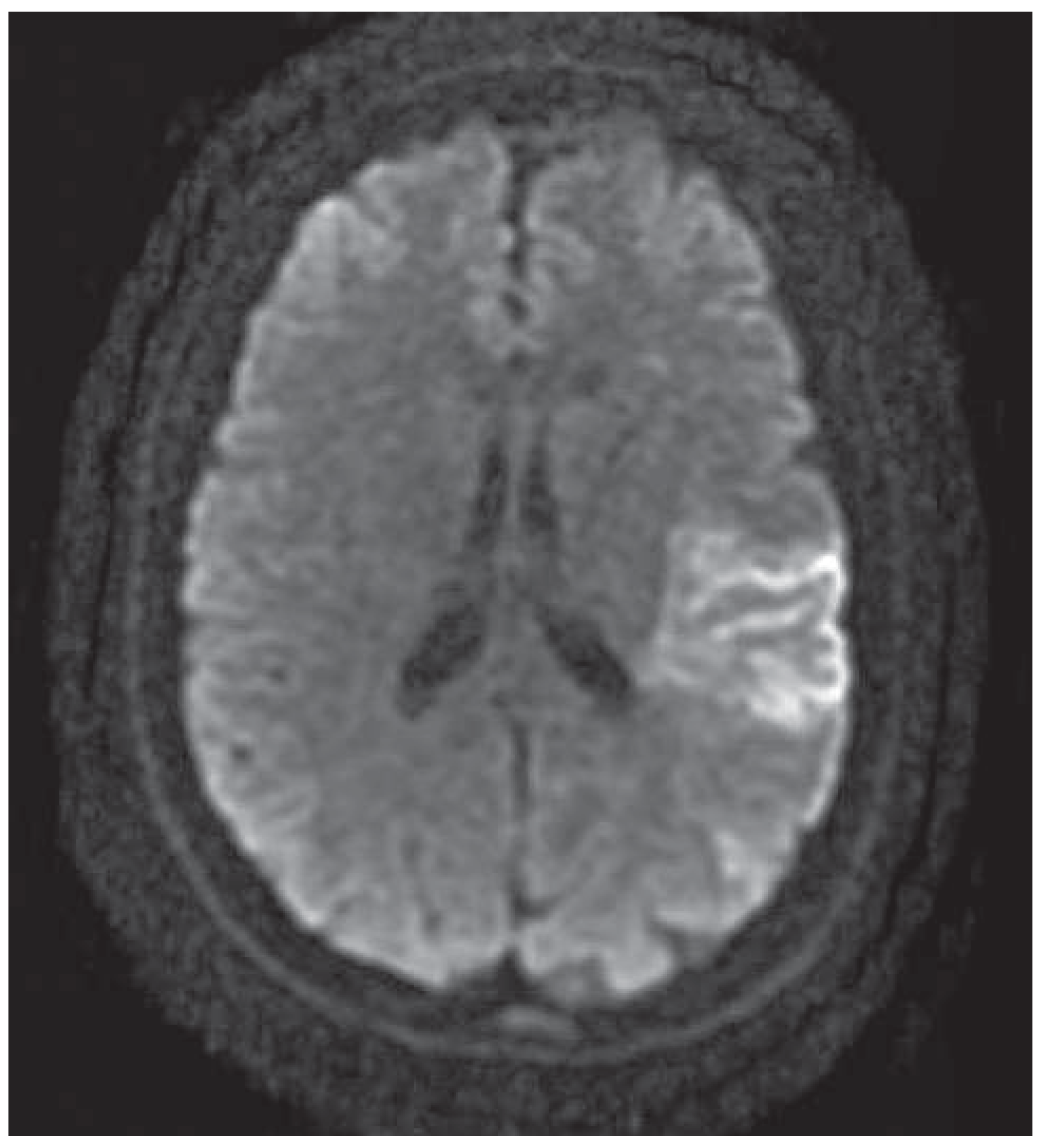
Figure 1. MR scan showing an extensive diffusion-weighted MR (DWI) hyperintense lesion in one-third to one-half of the MCA territory.
Cardiovascmed 17 00051 g001
Figure 2. Thrombotic occlusion of the left ICA just after the bifurcation.
Cardiovascmed 17 00051 g002
Figure 3. Left ICA after intervention.
Cardiovascmed 17 00051 g003
Figure 4. Injection into the right carotid artery, showing both anterior cerebral arteries. The left anterior cerebral artery is perfused over the anterior communicating artery of the circle of Willis.
Cardiovascmed 17 00051 g004
Figure 5. Injection into the left carotid artery showing the patent middle cerebral artery.
Cardiovascmed 17 00051 g005