Next Article in Journal
Unusual Complication of Transradial Coronary Angiography
Previous Article in Journal
Swiss Society of Cardiology Cardiovascular Biology Prize
 
 
Cardiovascular Medicine is published by MDPI from Volume 28 Issue 1 (2025). Previous articles were published by another publisher in Open Access under a CC-BY (or CC-BY-NC-ND) licence, and they are hosted by MDPI on mdpi.com as a courtesy and upon agreement with Editores Medicorum Helveticorum (EMH).
Case Report

Quantification of Differential ECMO Return Flow Through an Axillary Artery Anastomosis Graft with Spectral Doppler Echocardiographyphy

by
David G. Platts
1,2,*,
Kiran Shekar
2,3,
Bruce Thomson
2,4 and
John F. Fraser
2,3
1
Department of Echocardiography, Cardiac Investigations Unit, The Prince Charles Hospital, Rode Rd., Chermside Brisbane, QLD, 4032, Australia
2
Critical Care Research Group, The University of Queensland, Brisbane, Australia
3
Adult Intensive Care Service, The Prince Charles Hospital, Brisbane, Australia
4
Department of Cardiothoracic Surgery, The Prince Charles Hospital, Brisbane, Australia
*
Author to whom correspondence should be addressed.
Cardiovasc. Med. 2014, 17(2), 48; https://doi.org/10.4414/cvm.2014.00217
Submission received: 19 November 2013 / Revised: 19 December 2013 / Accepted: 19 January 2014 / Published: 19 February 2014

Summary

Venoarterial extracorporeal membrane oxygenation (VA-ECMO) is a specialised form of advanced cardiac life support that can be utilised in critically ill patients who require short term cardiac support. Cardiac imaging, especially with echocardiography, is fundamental to optimal management of patients supported with ECMO. There are numerous cannulation options available for initiating ECMO. This case report describes the use of a transthoracic echocardiographic transducer and scanner to assess upper limb hyperperfusion in a patient with an axillary artery site for VA-ECMO return flow. The incidence, diagnosis and treatment of this vascular complication of ECMO are discussed.

Case report

A 44-year-old female was transferred to our institution after a late-presentation anterior acute myocardial infarction complicated by severe cardiogenic shock. Transthoracic echocardiography revealed severe segmental left ventricular systolic dysfunction, with an ejection fraction of less than 10%, a left ventricular apical thrombus, and a severely dilated and severely impaired right ventricle. The patient remained in acute pulmonary oedema and refractory shock despite inotropic therapy and an intra-aortic balloon pump. Hence, venoarterial extracorporeal membrane oxygenation (VA-ECMO) was initiated. Because of the risk of upper body hypoxaemia with femoral-femoral access ECMO, the ECMO return flow (which is continuous rather than pulsatile) was delivered with a 22 French Sarns straight aortic cannula inserted into an 8 mm Gortex graft fashioned as an end-to-side anastomosis onto the right axillary artery. The ECMO access cannula was a 25 French multistage cannula inserted percutaneously into the right femoral vein.
The following day, owing to a difference in arterial blood pressure between the left and right upper limbs, there was a concern that right upper limb arterial hyperperfusion was present. The anastomosis site had been surgically explored with no evidence of a compartment syndrome. In order to quantify noninvasively differential flow from the ECMO circuit into the arterial circulation, the flow was evaluated echocardiographically after routine transthoracic echocardiography. A Philips iE33 scanner and S5-1 transducer were used to image the site of the anastomosis of the Gortex graft and axillary artery (Figure 1A). Colour Doppler imaging of this region showed continuous flow through the graft with expected aliasing phenomenon at the anastomosis site where flow became turbulent (Figure 1B). Anatomical and functional data were obtained in order to calculate flows in the axillary artery both before and after the anastomosis site. Real-time imaging using colour flow Doppler demonstrated multiphasic flow down the axillary artery. The vessel diameter was measured as 0.56 centimetres and, by use of spectral Doppler imaging, the velocity time integral (VTI) was measured as 16.2 centimetres (Figure 1C). By use of conventional echocardiographic principles to calculate flow [1], the flow down the axillary artery was measured as 0.32 litres/ minute.
A more proximal scan detected a different Doppler flow profile. There was continuous flow with only minimal pulsatility seen on both colour imaging and spectral Doppler imaging (Figure 1D). On the basis of a measured diameter of 0.56 centimetres and VTI of 166 centimetres (determined by measuring the spectral Doppler signal within one R-R interval on the electrocardiogram), the retrograde flow directed proximally/ centrally was calculated to be 3.28 litres/minute. To minimise error caused by nonparallel alignment of flow and the pulsed Doppler interrogation beam, the transducer position was modified slightly to ensure optimal alignment for assessment of the spectral signal. An alternative method, which is not often used in transthoracic echocardiography and was not utilised in this case, is to modify the interrogation of flow assessment by using an angle correction mechanism. Consequently, differential flow out of the axillary artery anastomosis could be quantified, revealing that 90% of the ECMO return flow was appropriately directed centrally whilst normal flow to the right upper limb was maintained. Hence, the patient was managed conservatively with conventional ongoing observation for neurovascular compromise in the affected limb. VA-ECMO enabled the patient to be successfully bridged to biventricular ventricular assist devices as a bridge to cardiac transplantation.

Discussion

Determining the optimal ECMO cannulation options in patients with cardiogenic shock and hypoxaemia from pulmonary oedema can be difficult. In critically unwell patients, immediate cardiorespiratory support is required. However, the conventional femoral arteryfemoral vein percutaneous approach, although offering rapid support, has the potential disadvantage of upper body hypoxaemia. To overcome this limitation, an axillary artery return ECMO cannulation option can be utilised [2,3]. However, axillary artery side-graft cannulation also has inherent limitations, with recently published work showing that 24.7% of cases are complicated by ipsilateral upper limb hyperperfusion and 17.3% by bleeding from the arterial graft [4]. Upper limb hyperperfusion is present if the arm receiving the ECMO return flow is hyperaemic, warmer and oedematous compared with the contralateral arm. This may then result in an upper limb compartment syndrome. Upper limb hyperperfusion may be have an arterial cause (where there is preferential flow down the arm owing to a proximal arterial narrowing) or a venous cause (where venous drainage of the arm is impeded by haematoma or venous thrombosis).
Management of upper limb hyperperfusion with an arterial cause involves simple measures such as elevation of the upper limb and optimisation of systemic pressures, and the more significant interventions of reducing ECMO flow or even fashioning a new return cannulation site [4]. In a critically unwell patient requiring ECMO support, the decision to reduce the level of support or to fashion a new return site cannot be taken lightly, but may be required to preserve the upper limb. Consequently, as demonstrated in the above case, it is important to make the correct diagnosis. Additionally, management of upper limb hyperperfusion is dependent upon identifying the underlying cause (arterial vs venous aetiology). In this regard, echocardiography may not only have a novel role in assisting with the diagnosis of upper limb hyperperfusion, when integrated with clinical parameters, but may also help in identifying an arterial cause.
Echocardiography plays a fundamental role in the management of patients supported with ECMO [5]. It is particularly useful for the detection of cardiac complications that may arise during ECMO. A conventional echocardiography scanner and transducer also have the potential ability to perform limited vascular assessment, as in this case. By applying standard haemodynamic principles that are employed during every transthoracic echocardiogram, it is possible to calculate flow within a large peripheral vessel. In this case, merely obtaining the VTI on the proximal and dis-tal aspects of the graft anastomosis was sufficient to quantify blood flow relative to the anastomosis site. The VTI represents the total backscatter signal (or Doppler spectral trace) caused by blood flow, with parameters of both magnitude and direction. However, when utilising this technique, it is important that the incident angle between blood flow and the ultrasound beam is considered, as measurement of blood flow velocity is related to this Doppler angle. An incident angle of ≤20° results in an acceptable percentage of error of ≤7% [1]. Hence, Doppler interrogation of the proximal and distal aspects of the axillary artery should involve as close as possible parallel alignment between blood flow and the ultrasound beam, both within an incident angle of 20°.
The diagnosis of upper limb hyperperfusion relies heavily on clinical features, with imaging utilised more to determine aetiology. The authors suggest that transthoracic echocardiography may add incremental value in the diagnosis of arterial upper limb hyperperfusion during ECMO support with an axillary artery anastomosis, and that it can be easily and quickly performed at the bedside. Further research is required to help correlate the quantified differential blood flow with the diagnosis and predictive risk for upper limb hyperperfusion using echocardiography.

Funding / potential competing interests

No financial support and no other potential conflict of interest relevant to this article was reported.

References

  1. Anderson, B. Echocardiography: The Normal Examination and Echocardiographic Measurements, 1st ed.; Wiley-Blackwell; 2002.
  2. Navia, J.L.; Atik, F.A.; Beyer, E.A.; Ruda Vega, P. Extracorporeal membrane oxygenation with right axillary artery perfusion. Ann Thorac Surg. 2005, 79, 2163–2165. [Google Scholar] [CrossRef] [PubMed]
  3. Moazami, N.; Moon, M.R.; Lawton, J.S.; Bailey, M.; Damiano, R., Jr. Axillary artery cannulation for extracorporeal membrane oxygenator support in adults: an approach to minimize complications. J Thorac Cardiovasc Surg. 2003, 126, 2097–2098. [Google Scholar] [CrossRef] [PubMed]
  4. Chamogeorgakis, T.; Lima, B.; Shafii, A.E.; Nagpal, D.; Pokersnik, J.A.; Navia, J.L.; et al. Outcomes of axillary artery side graft cannulation for extracorporeal membrane oxygenation. J Thorac Cardiovasc Surg. 2013, 145, 1088–1092. [Google Scholar] [CrossRef] [PubMed]
  5. Platts, D.G.; Sedgwick, J.F.; Burstow, D.J.; Mullany, D.V.; Fraser, J.F. The role of echocardiography in the management of patients supported by extracorporeal membrane oxygenation. J Am Soc Echocardiogr. 2012, 25, 131–141. [Google Scholar] [CrossRef] [PubMed]
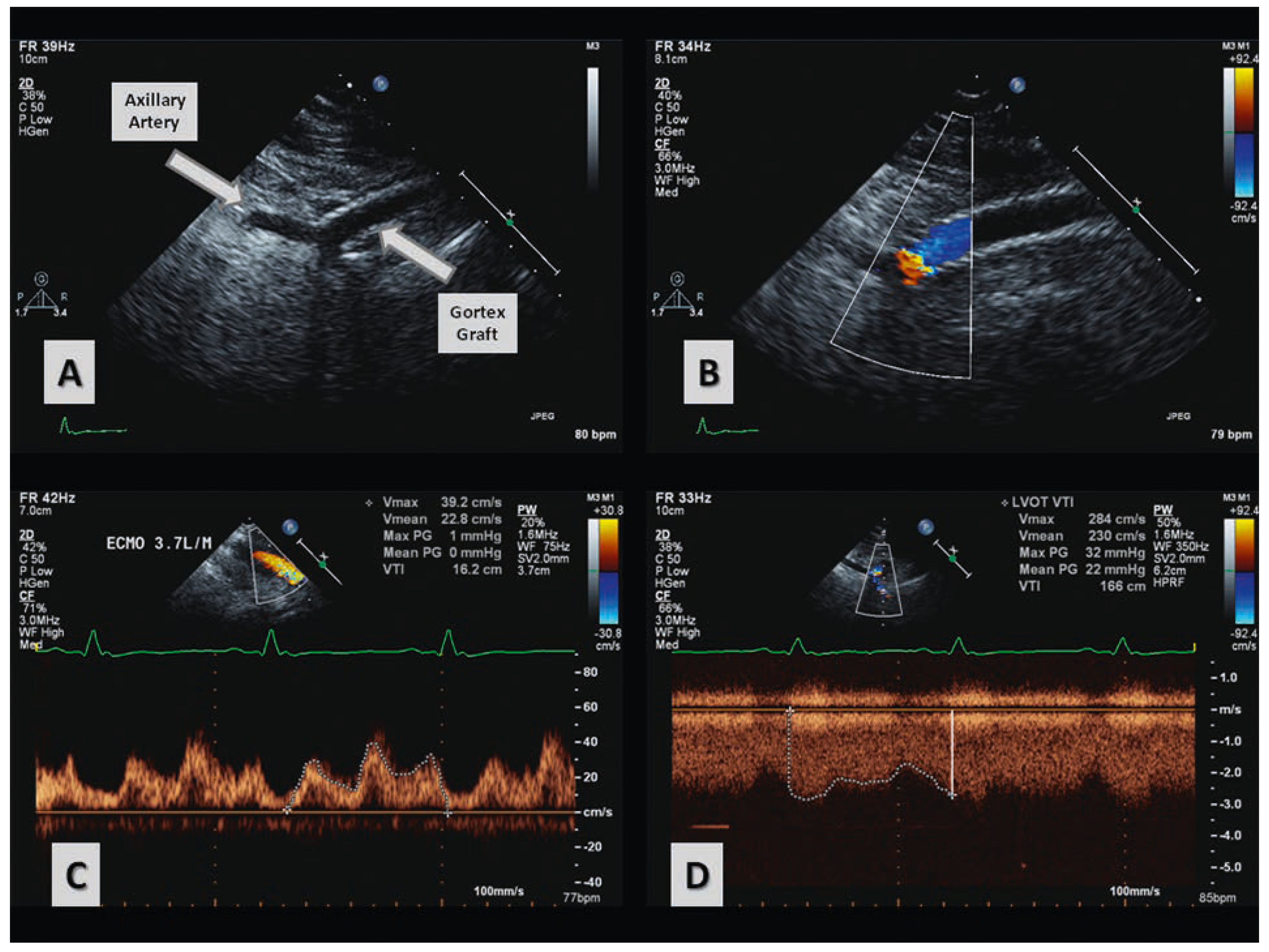
Figure 1. Panel A: Two dimensional echocardiographic imaging of the gortex graft anastomosis site to the axillary artery. Panel B: Colour Doppler imaging of the axillary artery anastomosis site. Panel C: Velocity time integral measurement for antegrade flow in the axillary artery down the arm. Panel D: Velocity time integral measurement for retrograde (centrally directed) flow in the axillary artery.
Cardiovascmed 17 00048 g001