Next Article in Journal
The Early Development of Cardiology in Zurich—A Personal Account Part II
Previous Article in Journal
The L Wave—A Reminder
 
 
Cardiovascular Medicine is published by MDPI from Volume 28 Issue 1 (2025). Previous articles were published by another publisher in Open Access under a CC-BY (or CC-BY-NC-ND) licence, and they are hosted by MDPI on mdpi.com as a courtesy and upon agreement with Editores Medicorum Helveticorum (EMH).
Interesting Images

Atrial Myxoma Resulting in Severe Mitral Stenosis

by
Santosh Desai
1,
Mohmmad Q Najib
2,
Muhammad T. Gill
3,
Umaima Fatima
2 and
Akil Loli
2,*
1
Internal Medicine Residency Program, Banner Goodsamaritan Medical Center/Carl T. Hayden VA Medical Center, Phoenix, AZ 85006, USA
2
Cardiology Fellowship Program, Department of Cardiology, Banner Goodsamaritan Medical Center/Carl T. Hayden VA Medical Center, Phoenix, AZ 85006, USA
3
Mercy Heart Institute, Cinncinnati, OH 45211, USA
*
Author to whom correspondence should be addressed.
Cardiovasc. Med. 2014, 17(10), 269; https://doi.org/10.4414/cvm.2014.00269
Submission received: 22 July 2014 / Revised: 22 August 2014 / Accepted: 22 September 2014 / Published: 22 October 2014
Primary tumours of the heart are mostly benign, consisting mainly of myxomas, with an incidence of less than 0.5% [1,2,3,4]. L eft atrial myxomas are more common and originate at the interatrial septum near the fossa ovalis [1]. Myxomas are often polypoid and pedunculated [1,2,3,4], and in rare instances can cause a mass effect, resulting in the obstruction of blood flow across the mitral valve. We present a case of atrial myxoma, e xtending into the mitral valve orifice and mimicking mitral stenosis physiology secondary to mass effect.
A 59-year-old woman, with a medical history significant for dyslipidemia, was referred from an urgent care centre for further evaluation for shortness of breath and fatigue, which she had developed three weeks prior to presentation. Initial work up included a transthoracic echocardiogram, which demonstrated normal left ventricular size with normal ejection fraction. The mitral valve, however, appeared thickened. A subsequent transesophageal echocardiogram revealed a large prolapsing atrial mass consistent with a myxoma in the left atrium (Figure 1A). Due to the size and positioning of the mass, the myxoma caused obstruction at the mitral valve mimicking severe mitral stenosis (Figure 1B). The mass arose from the fossa ovalis (Figure 1C) and extended into the mitral valve orifice leading to severe mitral stenosis physiology (Figure 1D). The patient was also found to have moderate mitral regurgitation and severe pulmonary hypertension with a right ventricular systolic pressure of 70 mm Hg.
The patient was referred for surgical excision. Intra-operatively a large friable atrial myxoma approximately 2 cm × 4 cm was observed and was excised. The fossa ovalis was also excised and the septal defect was closed with a corMatrix patch (Roswell, GA). After observing a dilated mitral annulus, a decision was made to repair the mitral valve ring. The valve was repaired with a 26 mm memo ring (Sorin; Milan, Italy). A postoperative transesophageal echocardiogram did not show any mitral regurgitation. The patient’s postoperative course was uneventful and she was discharged on the fourth postoperative day.
Common complications arising from the presence of atrial myxomas are embolism and intracardiac obstruction [1], whereas, structural compromise leading to severe mitral valve stenosis is rare [1,2]. Atrial myxomas are typically benign lesions, however, in rare instances, the clinical sequelae related to myxomas can be life-threatening. A sudden alteration in blood flow can cause circulatory collapse, while a chronic alteration in blood flow can lead to the development of pulmonary hypertension [3]. Early detection using imaging modalities such as echocardiography is essential to prevent the harmful sequelea of myxomas. Our case highlights that a myxoma can cause mass effect by extending into the mitral valve orifice resulting in severe mitral valve stenosis physiology.

Funding/potential competing interests:

No financial support and no other potential conflict of interest relevant to this article were reported.

References

  1. Reynen Klaus. Cardiac myxomas. N Engl J Med. 1995, 333, 1610–1617. [Google Scholar] [CrossRef] [PubMed]
  2. Avakian, Solange Desiree, Julio Yoshio Takada, Antonio de Padua Mansur. Giant obstructive left atrial myxoma resembling mitral valve stenosis. Clinics. 2012, 67, 853–854. [Google Scholar] [CrossRef] [PubMed]
  3. Mouine, Najat N, et al. Giant left atrial myxoma mimicking severe mitral valve stenosis and severe pulmonary hypertension. Int Arch Med. 2013, 6, 13. [Google Scholar] [CrossRef] [PubMed]
  4. Ojji, Dike B, et al. Left Atrial Myxoma Mimicking Mitral stenosis. Clinical medicine insights. Case Rep. 2012, 5, 111. [Google Scholar]
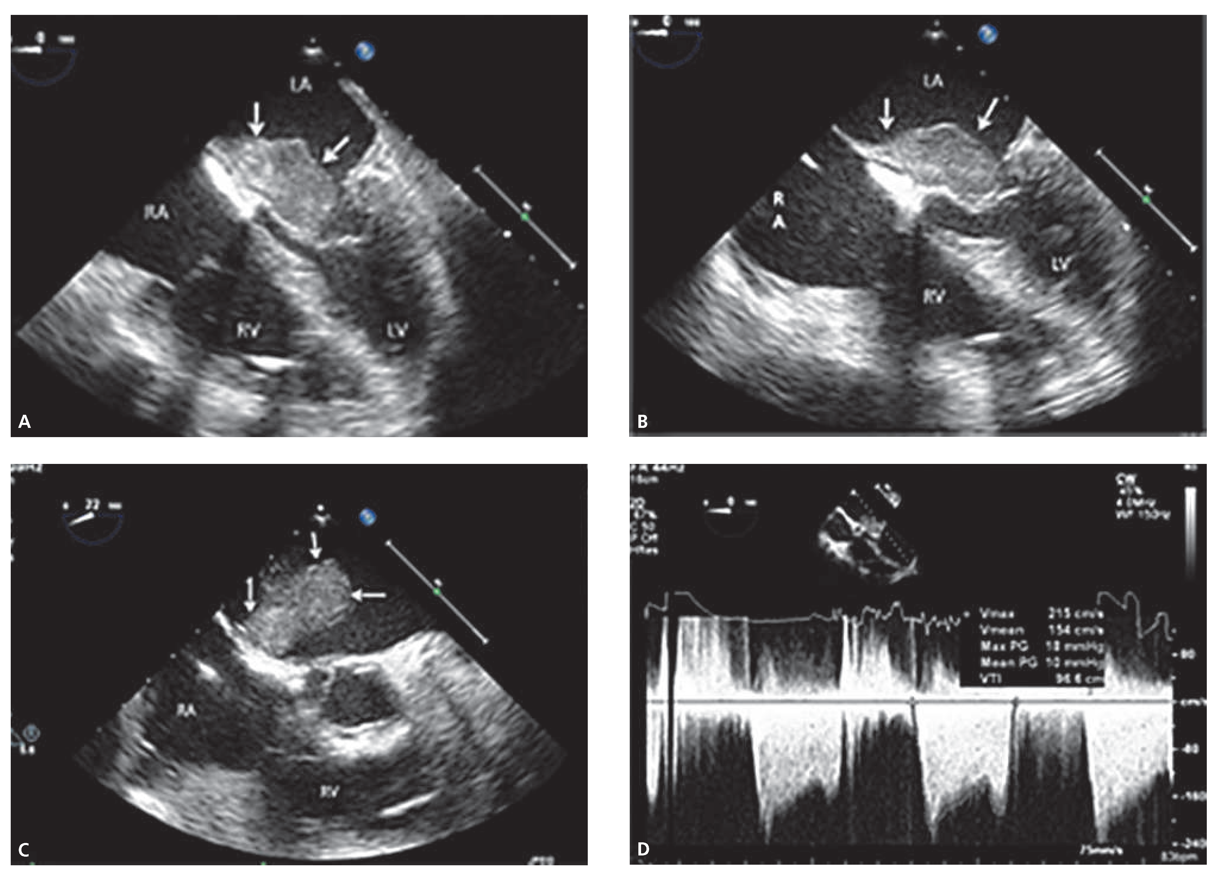
Figure 1. A: Transesophageal echocardiogram four chamber view demonstrating a mass (arrows) in the left atrium producing prolapsing in the mitral valve. B: Transesophageal echocardiogram four chamber view showing a mass (arrows) in the left atrium causing near complete mitral valve obstruction. C: Transesophageal echocardiogram four chamber view, illustrating a mass (arrows) in the left atrium arising from interatrial septum. D: Continuous wave Doppler echocardiogram demonstrating severe mitral stenosis physiology.
Cardiovascmed 17 00269 g001