Next Article in Journal
Do Not Forget Kawasaki Disease
Previous Article in Journal
Klinische Effekte der Katheterbasierten Nierennervenablation bei Therapieresistenter Hypertonie
 
 
Cardiovascular Medicine is published by MDPI from Volume 28 Issue 1 (2025). Previous articles were published by another publisher in Open Access under a CC-BY (or CC-BY-NC-ND) licence, and they are hosted by MDPI on mdpi.com as a courtesy and upon agreement with Editores Medicorum Helveticorum (EMH).
Interesting Images

Recurrent Unexplained Syncope in an Adolescent Girl

by
Stefano Di Bernado
1,*,
Tatiana Boulos-Ksontini
1,
Yvan Mivelaz
1,
Niccole Sekarski
1 and
Etienne Pruvot
2
1
Paediatric Cardiology Unit, Department of Pediatrics, University Hospital, CH-1011 Lausanne, Switzerland
2
Arrhythmia Unit, Service of Cardiology, University Hospital, Lausanne, Switzerland
*
Author to whom correspondence should be addressed.
Cardiovasc. Med. 2014, 17(1), 27; https://doi.org/10.4414/cvm.2014.00200
Submission received: 22 October 2013 / Revised: 22 November 2013 / Accepted: 22 December 2013 / Published: 22 January 2014

Case presentation

This fourteen year-old girl was referred to our outpatient clinic because of recurrent episodes of unexplained syncope. The episodes were of similar presentation. During endurance evaluation at school (the socalled “12-minute running test”), she repetitively developed dizziness, blurred vision and weakness after 8–10 minutes, before passing out for an unusual duration of about 6–8 minutes. After spontaneous recovery, she felt tired and weak for hours and reported neardeath experience suggestive of severe brain hypoperfusion. Apart from these symptoms, no other signs e.g., palpitations or chest pain were noted. Witnesses described the patient as pale and sweaty during the episodes.
Figure 1 shows her ECG at rest. A normal sinus rhythm is observed at 67/min with normal PR interval (138 msec), QRS duration (85 msec), QRS axis (90), and QTc interval (420 msec), and no sign suggestive of ventricular hypertrophy or repolarisation disorder. The echocardiography demonstrated a normal heart as well with left ventricle and right ventricle dimensions and functions within limit. A cardiac MRI with late enhancement allowed us to exclude a congenital coronary anormaly, myocardial scars and signs of arrhythmogenic right ventricular dysplasia.
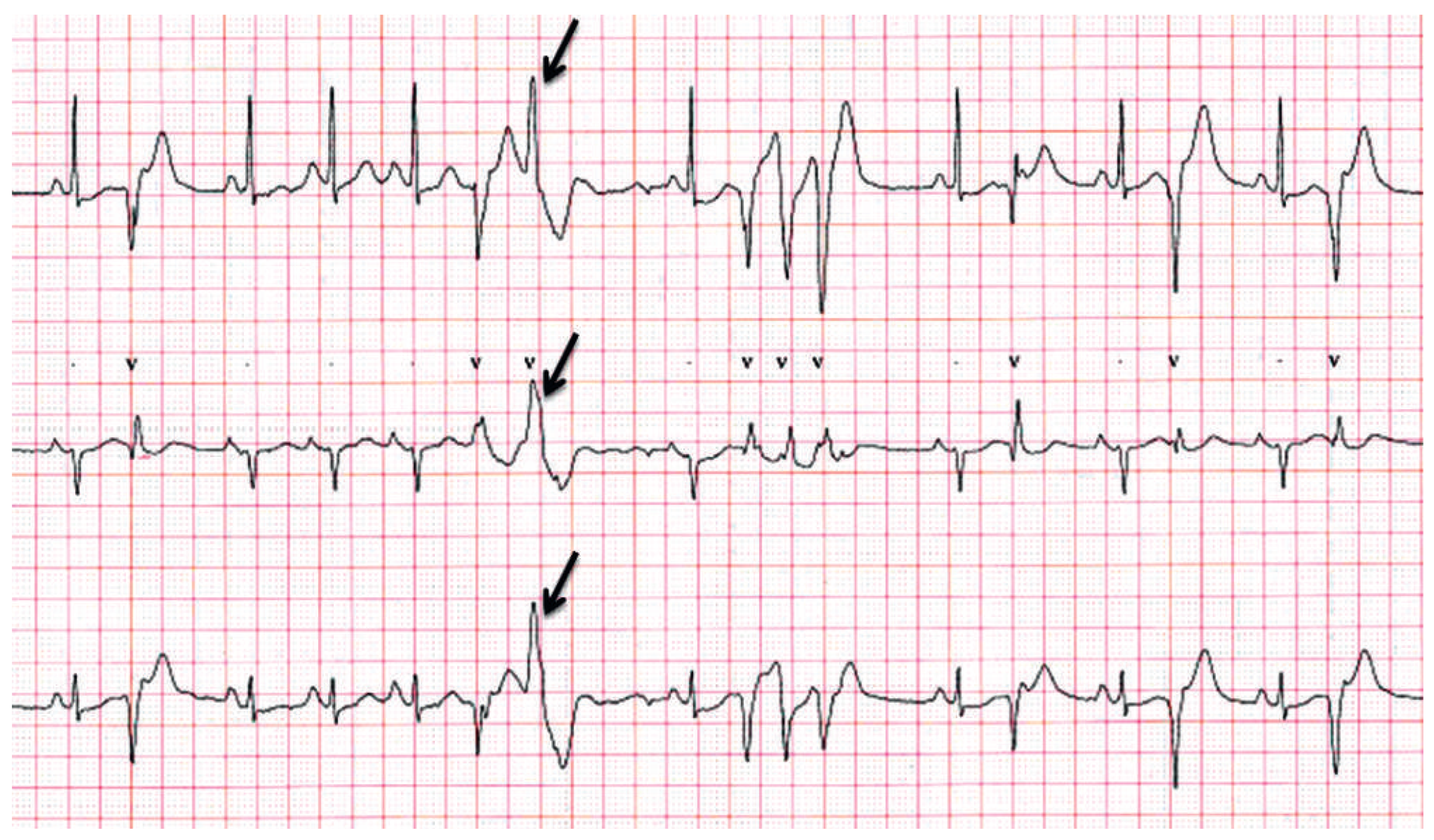
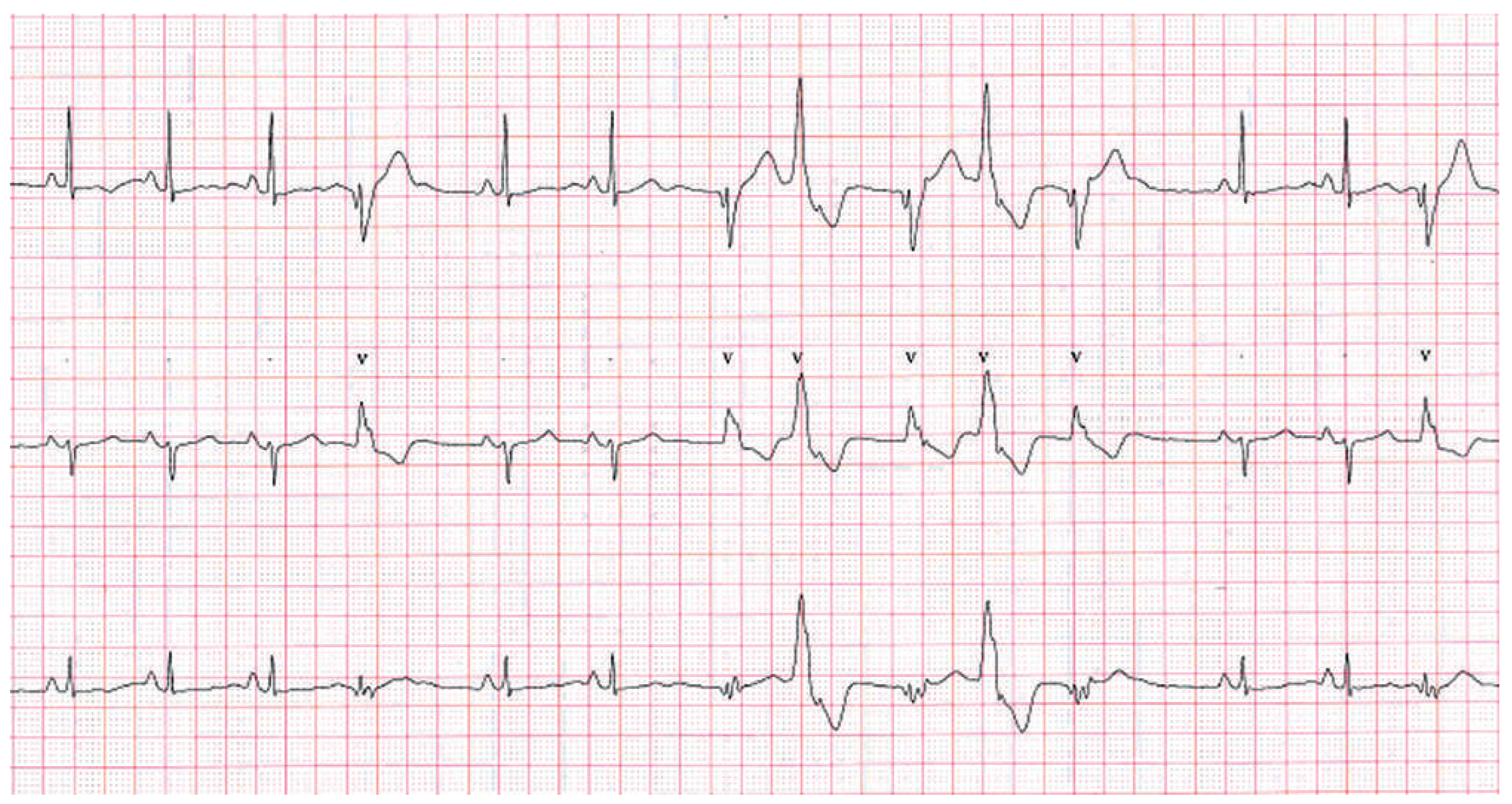
The 24-hour Holter recording revealed several episodes of nonsustained arrhythmia without any symptoms (Figure 2 and Figure 3). Figure 2 shows a tree-derivation Holter recording with short-coupled single ventricular premature beats, doublets and triplets interspaced with sinus beats during daily activity. Figure 3 and Figure 4also shows a ventricular premature beat of a different morphology (arrow) that occasionally occurred during sinus tachycardia. Importantly, these episodes occurred only when the adolescent was involved in some physical activity or during stressful tests at school, while her rhythm was normal at night or at rest.

Question

What are the most likely diagnosis and optimal therapy?

Comments

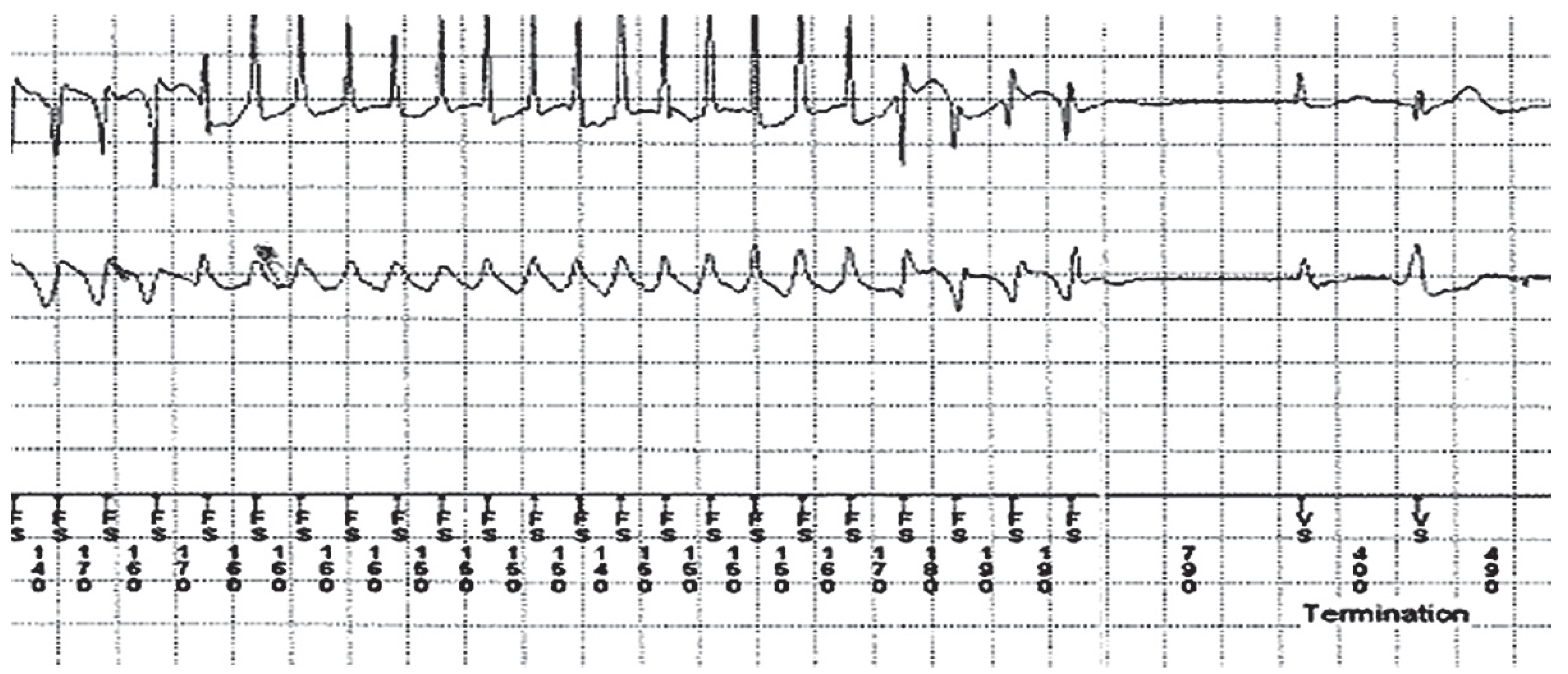
Based on the observation of runs of nonsustained VT and premature beats of two morphologies, the diagnosis of cathecholaminergic polymorphic ventricular tachycardia (CPVT) or LQTS with normal QT interval [1] was highly suspected. Sport was prohibited and beta blocker therapy was initiated first with tapering of propranolol, followed by bisoprolol therapy because of fainting, dizziness and Rayneaud phenomenom. Based on the severity of the recurrent syncopal events and as well as persistence of runs of nonsustained VT despite prescription of beta blockers, an implantable cardioverter-defibrillator (ICD) was implanted. The patient remained asymptomatic for months under treatment. Thirteen months after the ICD implantation, a first presyncopal episode occurred during a stressful mathematical test at school. The first nonsustained polymorphic VT episode lasting 24 beats (coupling interval as short as 150 ms) was recorded by the ICD, that was suggestive of an under dosage of the beta blockers. Medication was changed for metoprolol 25 mg bid. Thirty-three months after the implantation, the patient forgot her medication one night and the next morning, while standing quietly, she presented a near syncope that spontaneously resolved within seconds. Nonetheless, she was transferred to the hospital. Figure 5 shows the interrogation of the ICD at time of presyncope. A nonsustained polymorphic VT of 29 beats compatible with Torsades-de-Pointes is visible. Genetic analyses did not find the typical mutations associated with LQTS1-3 or CPVT.
CPVT occurs in patients without any evidence of heart disease. This disorder typically begins in childhood or adolescence. Affected patients present with life-threatening VT or ventricular fibrillation occurring during emotional or physical stress. The VT morphology may vary continuously, from beat to beat, or may appear as a bidirectional VT [2]. Two mutations have been identified in patients with CPVT: the cardiac ryanodine receptor gene (autosomal dominant form) and the calsequestrin 2 gene (autosomal recessive inheritance) [3]. Both proteins play a major role in the regulation of cardiomyocyte’s intrasarcoplasmic Ca2+. Treatments associate an ICD to terminate sustained arrhythmias and to prevent syncope and/or sudden cardiac death, and antiarrhythmic medication in order to prevent arrhythmias and minimise ICD shocks [4]. Beta blockers form the cornerstone of treatment, in patients with ongoing arrhythmias despite therapy with a beta blocker, the addition of verapamil or flecainide may be effective. In refractory cases, left sympathetic denervation can be an alternative therapeutic option [4,5].
Syncopal episodes are usually benign in nature, in children. The evaluation of syncopal children or adolescents relies on a thorough, detailed history and physical examination. Syncopal episodes that are associated with exercise or sport have to be thoroughly evaluated for their potential danger [6,7].

Funding/potential competing interests:

No financial support and no other potential conflict of interest relevant to this article were reported.

References

  1. Hekkala, A.M.; Viitasalo, M.; Vaananen, H.; Swan, H.; Toivonen, L. Abnormal repolarization dynamics revealed in exercise test in long QT syndrome mutation carriers with normal resting QT interval. Europace: European pacing, arrhythmias, and cardiac electrophysiology: journal of the working groups on cardiac pacing, arrhythmias, and cardiac cellular electrophysiology of the European Society of Cardiology. 2010, 12, 1296–1301. [Google Scholar] [CrossRef] [PubMed]
  2. Richter, S.; Gebauer, R.; Hindricks, G.; Brugada, P. A classic electrocardiographic manifestation of catecholaminergic polymorphic ventricular tachycardia. J Cardiovasc Electrophysiol. 2012, 23, 560. [Google Scholar] [CrossRef] [PubMed]
  3. Nam, G.B.; Burashnikov, A.; Antzelevitch, C. Cellular mechanisms underlying the development of catecholaminergic ventricular tachycardia. Circulation. 2005, 111, 2727–2733. [Google Scholar] [CrossRef] [PubMed]
  4. Van der Werf, C.; Zwinderman, A.H.; Wilde, A.A. Therapeutic approach for patients with catecholaminergic polymorphic ventricular tachycardia: state of the art and future developments. Europace: European pacing, arrhythmias, and cardiac electrophysiology: journal of the working groups on cardiac pacing, arrhythmias, and cardiac cellular electrophysiology of the European Society of Cardiology. 2012, 14, 175–183. [Google Scholar] [CrossRef]
  5. Swan, H.; Laitinen, P.; Kontula, K.; Toivonen, L. Calcium channel antagonism reduces exercise-induced ventricular arrhythmias in catecholaminergic polymorphic ventricular tachycardia patients with RyR2 mutations. J Cardiovas Electrophysiol. 2005, 16, 162–166. [Google Scholar] [CrossRef] [PubMed]
  6. Massin, M.M.; Bourguignont, A.; Coremans, C.; Comte, L.; Lepage, P.; Gerard, P. Syncope in pediatric patients presenting to an emergency department. J Pediatr. 2004, 145, 223–228. [Google Scholar] [CrossRef] [PubMed]
  7. Lewis, D.A.; Dhala, A. Syncope in the pediatric patient. The cardiologist’s perspective. Pediatr Clin North Am. 1999, 46, 205–219. [Google Scholar] [CrossRef] [PubMed]
Figure 1. Surface 12-lead ECG. Normal sinus rhythm, no signs of ventricular hypertrophy, normal intervals and QRS duration.
Cardiovascmed 17 00027 g001
Figure 2. Three-derivation Holter recording. Short-coupled single ventricular premature beats, doublets and triplets interspaced with sinus beats during daily activity. V: ventricular beats.
Cardiovascmed 17 00027 g002
Figure 3. Three-derivation Holter recording. Short-coupled single ventricular premature beats, doublets and triplets interspaced with sinus beats during daily activity, with premature ventricular morphology varying from beat to beat (arrow). V: ventricular beats.
Cardiovascmed 17 00027 g003
Figure 4. Three-derivation Holter recording. Nonsustained bidirectional ventricular tachycardia with QRS axis changing from beat to beat. V: ventricular.
Cardiovascmed 17 00027 g004
Figure 5. ICD interrogation. Nonsustained polymorphic ventricular tachycardia of 29 beats compatible with Torsades-de-Pointes with coupling intervals as short as 150 ms.
Cardiovascmed 17 00027 g005