Next Article in Journal
Cardiology Update 2013 Feiert das 20. Jubiläum. Davos, 10.–15. Februar 2013
Previous Article in Journal
Curing by Burning? What to Expect from Renal Denervation in the Treatment of Arterial Hypertension
 
 
Cardiovascular Medicine is published by MDPI from Volume 28 Issue 1 (2025). Previous articles were published by another publisher in Open Access under a CC-BY (or CC-BY-NC-ND) licence, and they are hosted by MDPI on mdpi.com as a courtesy and upon agreement with Editores Medicorum Helveticorum (EMH).
Case Report

Waiting for Evidence Seems to Make Sense—Except for the One Providing the Evidence

by
Thomas Pilgrim
1,
Rolf Vogel
1 and
Bernhard Meier
1,*
1
Department of Cardiology, Bern University Hospital, CH-3010 Bern, Switzerland
2
ARTORG Cardiovascular Engineering, University of Bern, CH-3010 Bern, Switzerland
*
Author to whom correspondence should be addressed.
Cardiovasc. Med. 2012, 15(9), 258; https://doi.org/10.4414/cvm.2012.01697
Submission received: 26 June 2012 / Revised: 26 July 2012 / Accepted: 26 August 2012 / Published: 26 September 2012

Abstract

A 58-year-old male presented with a history of two prior transient ischaemic attacks and was found to have a patent foramen ovale (PFO) in the absence of atrial fibrillation or relevant carotid atheromatosis. PFO closure was deferred at this stage due to the lack of clinical evidence. Three years later the patient was re-admitted after a major stroke with residual symptoms and finally underwent PFO closure in a minimally invasive procedure using an Amplatzer PFO Occluder.

Case report

A 58-year-old mechanic actively involved in motocross racing was thrown off track four years prior when he experienced a transient left oro-facial palsy that spontaneously resolved within hours. A few months later these symptoms recurred accompanied by temporary motor aphasia and prompted hospitalisation. He admitted to casual smoking and had no significant past medical history except for a mildly elevated lipid profile. Transoesophageal echocardiography identified >25 microbubbles (agitated saline) crossing the atrial septum and appearing in the left atrium within three heart cycles after a Valsalva manoeuver, consistent with a PFO grade III [1]. No atherosclerosis of the aortic arch was documented. Atrial fibrillation was ruled out by in-hospital monitoring and an ambulatory Holter ECG, and Duplex ultrasound and transcranial Doppler studies revealed only minimal atheromatosis of the left carotid bifurcation. Screening for a hypercoagulable state was foregone because there was no personal or family history of venous thromboembolism. Again, the symptoms completely resolved within hours.
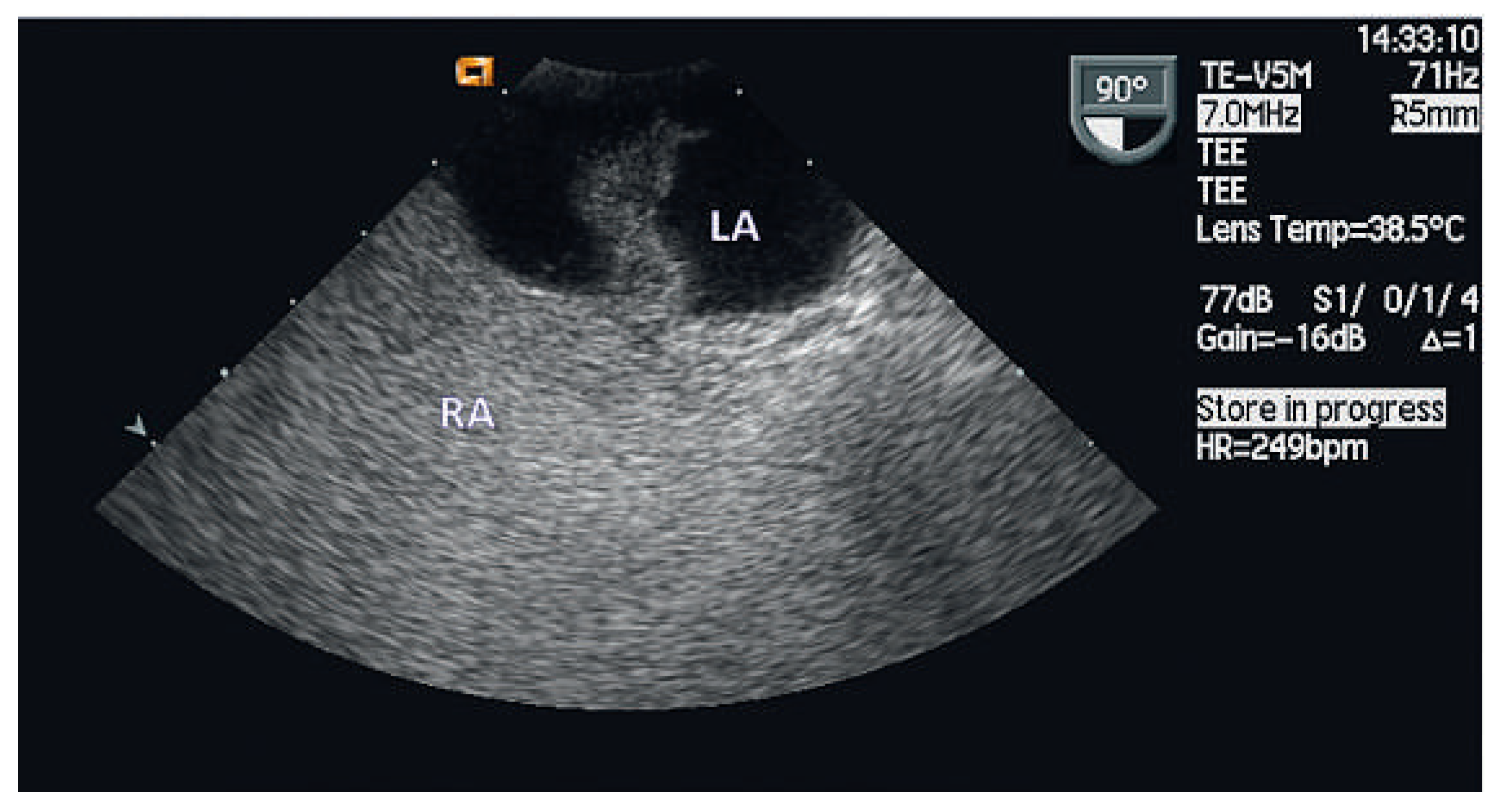
Upon discharge smoking cessation was encouraged, antiplatelet therapy with acetylsalicylic acid was initiated as a secondary prophylaxis and a statin was implemented for lipid control in accordance with current guidelines. The treating physician was reassuring the patient concerning the PFO and refrained from referral for closure. Three years later the patient returned to the emergency room with motoric aphasia that rapidly progressed to a hemisyndrome primarily with paralysis of the left upper extremity corresponding to a NIHSS score of 7/42. After intracranial haemorrhage was ruled out by computed tomography intravenous thrombolysis with alteplase was started two hours after symptom onset. Magnetic resonance angiography identified a small cortical lesion in the right paracentral area as well as a delayed perfusion in the area of the middle cerebral artery and revealed a complete occlusion of the right internal carotid artery suggesting an embolic event (Figure 1). While speech was recovering steadily, residual fine motor skill disabilities of the left hand persisted. A 24-hour-ECG was repeated and again demonstrated regular sinus rhythm with no episodes of atrial fibrillation and a repeated transoesophageal echocardiography was consistent with the previous study showing a PFO grade III suggesting paradoxical embolism as the only obvious mechanism (Figure 2). There was no atrial septal aneurysm. According to the guidelines, the treating physician/neurologist switched the antiplatelet regimen from acetylsalicylic acid to clopidogrel as a secondary prevention and a follow-up carotid Duplex ultrasound with Doppler study was scheduled which was again consistent with minimal atherosclerosis. With respect to the PFO, conflicting recommendations were made by the treating physician and the cardiologist. While the first related the lack of a randomised clinical trial that would dictate PFO closure in this setting, the latter convinced the patient with sound reasoning.
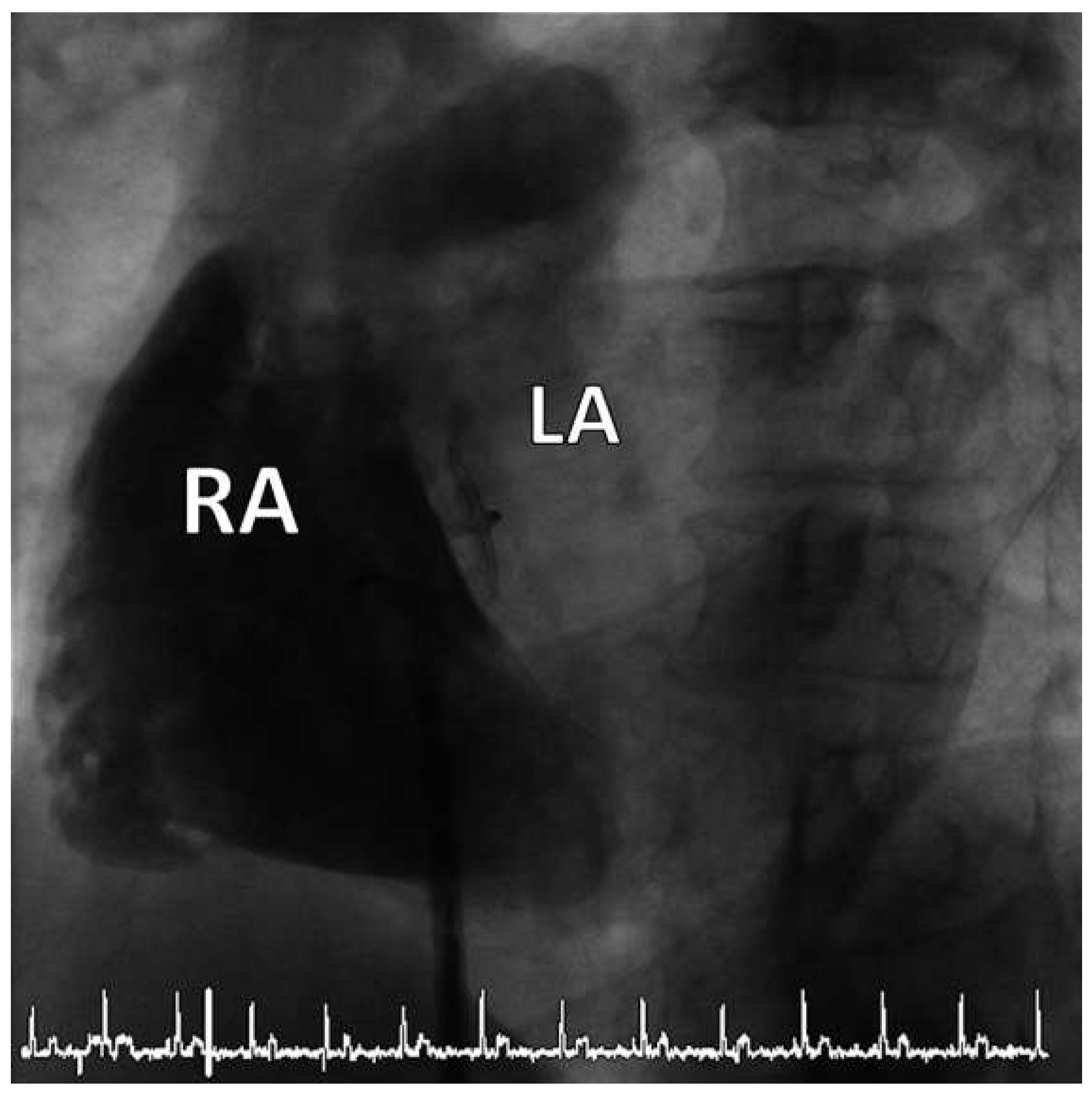
In a minimally invasive outpatient procedure under local anaesthesia the patent foramen ovale was crossed via the right femoral vein under fluoroscopic guidance and occluded using an Amplatzer PFO Occluder with a diameter of 25 mm (Figure 3). Adequate position of the device was confirmed prior to discharge by transthoracic echocardiography and a relevant residual shunt was ruled out.

Discussion

The foramen ovale, an interatrial connection, is a prerequisite for the intrauterine circulation to bypass the immature lungs. It functionally closes after birth due to the subsequent decrease of right atrial pressure that sucks the septum premium against the septum secundum. The prevalence of PFO declines with advancing age and is found in around one quarter of the population during the fourth to eight decade of life. While asymptomatic in most people, the PFO harbours a risk of paradoxical embolism and has also been associated with migraine with aura, decompression illness in divers, platypnoea-orthodeoxia syndrome, obstructive sleep apnoea, and high altitude oedema.
In up to 40% of cases the aetiology of an ischaemic stroke cannot be determined despite extensive work-up [2,3]. Whereas case-control studies revealed a strong correlation between the presence of PFO and cryptogenic stroke in adults aged <55 years, this relationship is still controversial in older age groups [4,5,6,7,8,9]. Even with medical treatment, patients with cryptogenic stroke with PFO are at increased risk for recurrence [10,11,12,13]. Whereas non-randomised data suggest an advantage of percutaneous PFO closure over medical treatment [14,15,16,17], a recent randomised trial comparing PFO closure or medical therapy for cryptogenic stroke did not show a significant benefit with regard to a composite of stroke or TIA at two years of follow-up [18]. Longer follow-up is of essence in this realm.
Transfemoral closure of a PFO can be performed under local anaesthesia as a safe procedure with favourable long-term outcome [14,16,17]. The considerable risk of recurrent cerebrovascular accidents in patients with symptomatic PFO outweighted the infrequent occurrence of periinterventional complications mostly confined to the access site. Transcatheter closure of patent foramen ovale with significant right to left shunt can hence be recommended to patients suffering from cryptogenic stroke based on common sense while awaiting randomised clinical trials in parallel to thorough evaluation of possible other sources of stroke.

Funding/potential competing interests

B. Meier is recipient of research grants and speaker fees of St. Jude Medical.

References

  1. Schneider, B.; Zeinkiewicz, T.; Jansen, V.; Hofmann, T.; Noltenius, H.; Meinertz, T. Diagnosis of patent foramen ovale by transesophageal echocardiography and correlation with autopsy findings. Am J Cardiol 1996, 77, 1202–1209. [Google Scholar] [CrossRef] [PubMed]
  2. Steiner, M.M.; Di Tullio, M.R.; Rundek, T.; Gan, R.; Chen, X.; Liguori, C.; et al. Patent foramen ovale size and embolic brain imaging findings among patinets with ischemic stroke. Stroke 1998, 29, 944–948. [Google Scholar] [CrossRef] [PubMed]
  3. Sacco, R.L.; Ellenberg, J.H.; Mohr, J.P.; Tatemichi, T.K.; Hier, D.B.; Price, T.R.; et al. Infarcts of undetermined cause: the NINCDS Stroke Data Bank. Ann Neurol 1989, 25, 382–390. [Google Scholar] [CrossRef] [PubMed]
  4. Lechat, P.; Mas, J.L.; Lascault, G.; Loron, P.; Theard, M.; Klimczac, M.; et al. Prevalence of patent formane ovale in young stroke patients. N Engl J Med 1988, 2, 11–12. [Google Scholar]
  5. Webster, M.W.; Chancellor, A.M.; Smith, H.J.; Swift, D.L.; Sharpe, D.N.; Bass, N.M.; et al. Patent foramen ovale in young stroke patients. Lancet 1988, 2, 11–12. [Google Scholar] [CrossRef] [PubMed]
  6. De Belder, M.A.; Tourikis, L.; Leech, F.; Camm, A.J. Risk of patent foramen ovale for thomboembolic events in all age groups. Am J Cardiol 1992, 69, 1316–1320. [Google Scholar] [CrossRef] [PubMed]
  7. Di Tullio, M.; Sacco, R.L.; Gopal, A.; Mohr, J.P.; Homma, S. Patent formamen ovale as a risk factor for cryptogenic stroke. Ann Intern Med 1992, 117, 461–465. [Google Scholar] [CrossRef] [PubMed]
  8. Hausmann, D.; Mugge, A.; Becht, I.; Daniel, W.G. Diagnosis of patent formaen ovale by transesophageal echocardiography and association with cerebral and peripheral embolic events. Am J Cardiol 1992, 70, 668–672. [Google Scholar] [CrossRef] [PubMed]
  9. Cabanes, L.; Mas, J.L.; Cohen, A.; Amarenco, P.; Cabanes, P.A.; Oubary, P. Atrial septal aneurysm and patent foramen ovale as a risk factors for cryptogenic stroke in patients less than 55 years of age. A study using transesophageal echocardiography. Stroke 1993, 24, 1865–1873. [Google Scholar] [CrossRef] [PubMed]
  10. Homma, S.; Sacco, R.L.; Di Tullio, M.R.; Sciacca, R.R.; Mohr, J.P. Effect of medical treatment in stroke patients with patent foramen oval. Patent Foramen Ovale in Cryptogenic Stroke Study. Circulation 2002, 105, 2625–2631. [Google Scholar] [PubMed]
  11. Bogousslavsky, J.; Garazi, S.; Jeanrenaud, X.; Aebischer, N.; Van Melle, G. Stroke recurrence in patients with patent foramen ovale: the Lausanne Study. Lausanne Stroke with Paradoxal Embolism Study Group. Neurology 1996, 46, 1301–1305. [Google Scholar] [CrossRef] [PubMed]
  12. Mas, J.L.; Zuber, M. Recurrent cerebrovascular events in patients with patent foramen ovale, atrial septal aneurysm, or both and cryptogenic stroke or transient ischemic attack. French Study Group on Patent Foramen Ovale and Atrial Septal Aneurysm. Am Heart J 1995, 130, 1083–1088. [Google Scholar] [CrossRef] [PubMed]
  13. Nedeltchev, K.; Arnold, M.; Wahl, A.; Sturzenegger, M.; Vella, E.E.; Windecker, S.; et al. Outcome of patients with cryptogenic stroke and patent formane ovale. J Neurol Neurosurg Psychiatry 2002, 72, 347–350. [Google Scholar] [CrossRef] [PubMed]
  14. Wahl, A.; Jüni, P.; Mono, M.; Kalesan, B.; Praz, F.; Geister, L.; et al. Long-term propensity score-matched comparison of percutaneous closure of patent foramen ovale with medical treatment after paradoxical embolism. Circulation 2012, 125, 803–812. [Google Scholar] [CrossRef] [PubMed]
  15. Khairy, P.; O’Donnell, C.P.; Lanzberg, M.J. Transcatheter closure versus medical therapy of patent foramen ovale and presumed paradoxical thromboemboli: a systematic review. Ann Intern Med 2003, 139, 753–760. [Google Scholar] [CrossRef] [PubMed]
  16. Wahl, A.; Praz, F.; Stirnimann, J.; Windecker, S.; Seiler, C.; Nedeltchev, K.; et al. Safety and feasibility of percutaneous closure of patent foramen ovale without intra-procedural echocardiography in 825 patients. Swiss Med Wkly 2008, 138, 567–572. [Google Scholar] [PubMed]
  17. Agarwal, S.; Bajaj, N.S.; Kumbhani, D.J.; Tuzcu Em Kapadia, S.R. Meta- analysis of transcatheter closure versus medical therapy for patent foramen ovale in prevention of recurrent neurological events after presumed paradoxical embolism. JACC Interv 2012, 5, 777–789. [Google Scholar] [CrossRef] [PubMed]
  18. Furlan, A.J.; Reisman, M.; Massaro, J.; Mauri, L.; Adams, H.; Albers, G.W.; et al. Closure or medical therapy for dryptogenic stroke with patent foramen ovale. N Engl J Med 2012, 366, 991–999. [Google Scholar] [CrossRef] [PubMed]
Figure 1. Embolic occlusion of the right internal carotid artery on MRA.
Cardiovascmed 15 00258 g001
Figure 2. Bubbles passing the PFO from the right (RA) to the left (LA) atrium on transoesophageal echocardiography.
Cardiovascmed 15 00258 g002
Figure 3. Successful closure of the PFO with an Amplatzer PFO Occluder confirmed by contrast injection into the right atrium (RA) under fluoroscopy. LA = Left atrium.
Cardiovascmed 15 00258 g003